

Prospective comparison of use of contrast-enhanced ultrasound and contrast-enhanced computed tomography in the Bosniak classification of complex renal cysts
- PMID: 27433270
- PMCID: PMC4760615
- DOI: 10.1177/1742271X15626959
Prospective comparison of use of contrast-enhanced ultrasound and contrast-enhanced computed tomography in the Bosniak classification of complex renal cysts
Abstract
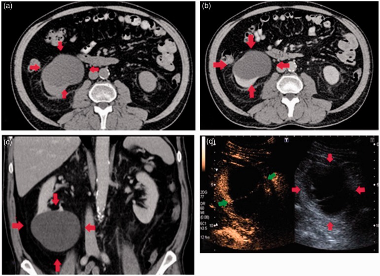
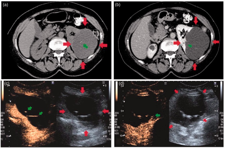
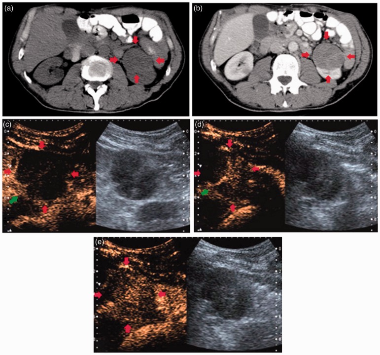
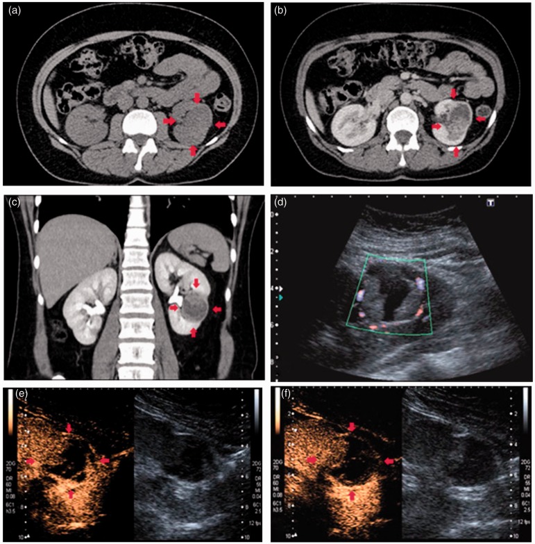
Aim: To compare contrast-enhanced ultrasound and contrast-enhanced computed tomography in the evaluation of complex renal cysts using the Bosniak classification.
Methods: Forty-six patients with 51 complex renal cysts were prospectively examined using contrast-enhanced ultrasound and contrast-enhanced computed tomography and images analysed by two observers using the Bosniak classification. Adverse effects and patients' preference were assessed for both modalities.
Results: There was complete agreement in Bosniak classification between both modalities and both observers in six cysts (11.8%). There was agreement of Bosniak classification on both modalities in 21 of 51 cysts (41.2%) for observer 1 and in 17 of 51 cysts (33.3%) for observer 2. Contrast-enhanced ultrasound gave a higher Bosniak classification than corresponding contrast-enhanced computed tomography in 31 % of cysts by both observers. Histological correlation was available in three lesions, all of which were malignant and classified as such simultaneously on both modalities by at least one observer, with remaining patients followed up with US or CT for 6-24 months. No adverse or side effects were reported following the use of US contrast, whilst 63.6% of patients suffered minor side effects following the use of CT contrast. 81.8% of the surveyed patients preferred contrast-enhanced ultrasound to contrast-enhanced computed tomography.
Conclusion: Contrast-enhanced ultrasound is a feasible tool in the evaluation of complex renal cysts in a non-specialist setting. Increased contrast-enhanced ultrasound sensitivity to enhancement compared to contrast-enhanced computed tomography, resulting in upgrading the Bosniak classification on contrast-enhanced ultrasound, has played a role in at best moderate agreement recorded by the observers with limited experience, but this would be overcome as the experience grows. To this end, we propose a standardised proforma for the contrast-enhanced ultrasound report. The benefits of contrast-enhanced ultrasound over contrast-enhanced computed tomography include patients' preference and avoidance of ionising radiation or nephrotoxicity, as well as lower cost.
Keywords: Bosniak classification; Complex renal cyst; contrast enhanced computed tomography; contrast-enhanced ultrasound; renal cell carcinoma.
Figures
References
-
- Laucks SP, McLachlan MSF. Aging and simple cysts of the kidney. Brit J Radiol 1981; 54: 12–14. - PubMed
-
- Chang C, Kuo J, Chang W, et al. Prevalence and clinical characteristics of simple renal cysts. J Chin Med Assoc 2007; 70: 486–491. - PubMed
-
- Israel GM, Bosniak MA. Follow-up CT of moderately complex cystic lesions of the kidney (Bosniak Category IIF). Amer J Roentgenol 2003; 181: 627–633. - PubMed
-
- Terada N, Ichioka K, Matsuta Y, et al. The natural history of simple renal cysts. J Urol 2002; 167: 21–23. - PubMed
-
- Zeman RK, Cronan JJ, Rosenfield AT, et al. Imaging approach to the suspected renal mass. Radiol Clin North Am 1985; 2: 503–528. - PubMed
LinkOut - more resources
Full Text Sources
Other Literature Sources