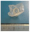
Cut endotracheal tube for endoscopic removal of an ingested push-through pack
- PMID: 27433294
- PMCID: PMC4937163
- DOI: 10.4253/wjge.v8.i13.472
Cut endotracheal tube for endoscopic removal of an ingested push-through pack
Abstract
A 52-year-old female presented to our clinic after accidentally ingesting a push-through pack (PTP). After determining that the PTP was present in the stomach, we successfully and safely removed it endoscopically by using a handmade endoscopic hood fashioned from a cut endotracheal tube. Foreign body ingestion is a common clinical problem, and most ingested foreign bodies pass spontaneously. However, the ingestion of sharp objects, such as PTPs, increases the risk of complications, and urgent endoscopy is recommended to remove such objects. Previous studies have reported the use of other devices, both commercial and handmade, for the safe endoscopic removal of foreign bodies. The novel design of our handmade hood for the removal of the PTP, which was fashioned from a cut endotracheal tube, was beneficial in terms of maintaining a wide visual field, patient safety and tolerance, and easy preparation compared to previously reported commercial and handmade devices. It may be a viable and safe device for the retrieval of PTPs and other sharp foreign bodies.
Keywords: Endoscopic removal; Foreign body ingestion; Handmade; Push-through pack; Sharp object.
Figures


Similar articles
-
Improvising in Endoscopy: Endoscopic Removal of Sharp Foreign Bodies in the Upper GI Tract, Using a Handmade Protective Device.Case Rep Gastrointest Med. 2020 Sep 9;2020:8881702. doi: 10.1155/2020/8881702. eCollection 2020. Case Rep Gastrointest Med. 2020. PMID: 32963847 Free PMC article.
-
Removal of foreign bodies in the upper gastrointestinal tract in adults: European Society of Gastrointestinal Endoscopy (ESGE) Clinical Guideline.Endoscopy. 2016 May;48(5):489-96. doi: 10.1055/s-0042-100456. Epub 2016 Feb 10. Endoscopy. 2016. PMID: 26862844
-
[Ingestion of foreign bodies in children. Recommendations of the French-Speaking Group of Pediatric Hepatology, Gastroenterology and Nutrition].Arch Pediatr. 2009 Jan;16(1):54-61. doi: 10.1016/j.arcped.2008.10.018. Epub 2008 Dec 6. Arch Pediatr. 2009. PMID: 19059766 Review. French.
-
Transparent cap-assisted endoscopic retrieval of a sharp foreign body in the esophagus.Rev Esp Enferm Dig. 2023 Apr;115(4):199. doi: 10.17235/reed.2022.9059/2022. Rev Esp Enferm Dig. 2023. PMID: 35899695
-
A double-scope technique enabled a patient with an esophageal plastic fork foreign body to avoid surgery: a case report and review of the literature.Clin J Gastroenterol. 2022 Feb;15(1):66-70. doi: 10.1007/s12328-021-01549-6. Epub 2021 Nov 5. Clin J Gastroenterol. 2022. PMID: 34741229 Review.
Cited by
-
A loop cutter is an ideal gripper for endoscopic removal of press-through-package sheets.Endoscopy. 2023 Dec;55(S 01):E889-E891. doi: 10.1055/a-2113-9265. Epub 2023 Jul 13. Endoscopy. 2023. PMID: 37442174 Free PMC article. No abstract available.
-
Improvising in Endoscopy: Endoscopic Removal of Sharp Foreign Bodies in the Upper GI Tract, Using a Handmade Protective Device.Case Rep Gastrointest Med. 2020 Sep 9;2020:8881702. doi: 10.1155/2020/8881702. eCollection 2020. Case Rep Gastrointest Med. 2020. PMID: 32963847 Free PMC article.
References
-
- Ikenberry SO, Jue TL, Anderson MA, Appalaneni V, Banerjee S, Ben-Menachem T, Decker GA, Fanelli RD, Fisher LR, Fukami N, et al. Management of ingested foreign bodies and food impactions. Gastrointest Endosc. 2011;73:1085–1091. - PubMed
-
- Kumagai M, Ikeda K, Oshima T, Nakatsuka S, Takasaka T. A press-through-pack in the larynx. Tohoku J Exp Med. 1997;183:293–295. - PubMed
Publication types
LinkOut - more resources
Full Text Sources
Other Literature Sources

