

doi: 10.1155/2016/1232594.
Epub 2016 Jun 28.
A Unique Case of an Aggressive Gangliocytic Paraganglioma of the Filum Terminale
Affiliations
- PMID: 27433367
- PMCID: PMC4940518
- DOI: 10.1155/2016/1232594
Item in Clipboard
A Unique Case of an Aggressive Gangliocytic Paraganglioma of the Filum Terminale
Case Rep Surg.
2016.
Abstract
Paragangliomas are rare neuroendocrine tumors that are mostly found in the head and neck. Even less common are gangliocytic variant paragangliomas of the spine for which there are only 7 other documented cases in the literature. We report a case of gangliocytic paraganglioma of the sacral spine in a 68-year-old man. The growth pattern is documented over three years, which to our knowledge has not previously been reported in the literature and is different from the natural history. Clinical, radiological, and pathological characteristics of the tumor are discussed in light of available reports of this rare tumor.
Figures
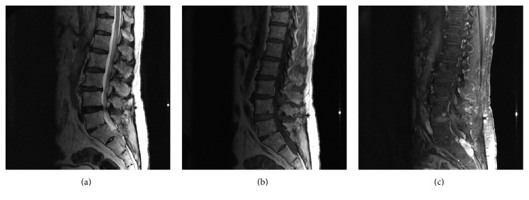
MRI of the lumbar spine with and without contrast. (a) Sagittal T2, (b) sagittal T1, and (c) sagittal T1 after fat saturation: Displays a T1 isointense, T2 hyperintense contrast enhancing mass at the S1-S2 level. The mass measures approx. 3.4 × 1.2 cm without local invasion into surrounding structures.
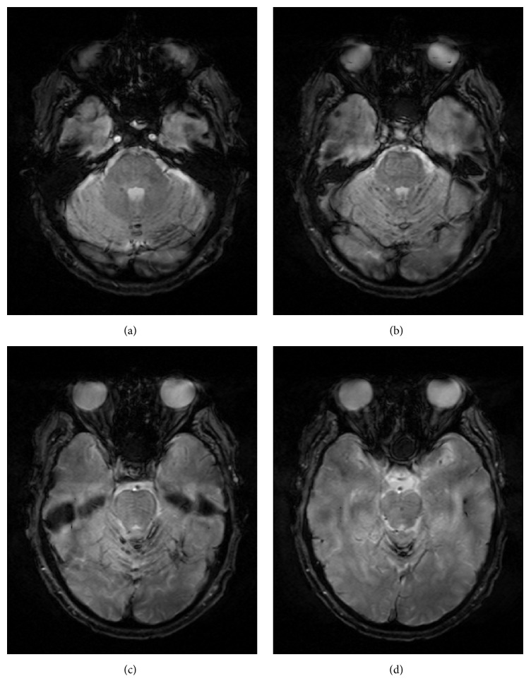
T2-weighted gradient ECHO MRI of the brain: superficial siderosis with hemosiderin deposition seen in the vermis and folia of the cerebellum.
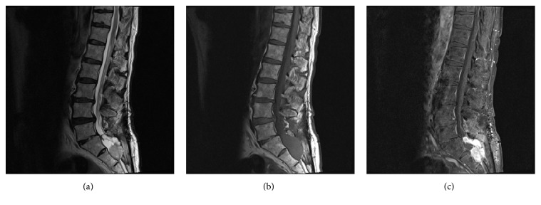
MRI of the lumbar spine with and without contrast. (a) Sagittal T2, (b) sagittal T1, and (c) sagittal T1 after fat saturation: significant enlargement of previously noted sacral mass with new bony erosion and significant mass effect within the spinal canal causing displacement of posterior spinal structures. The mass has taken a more lobulated appearance as seen on the T2 images and is vividly contrast enhancing.
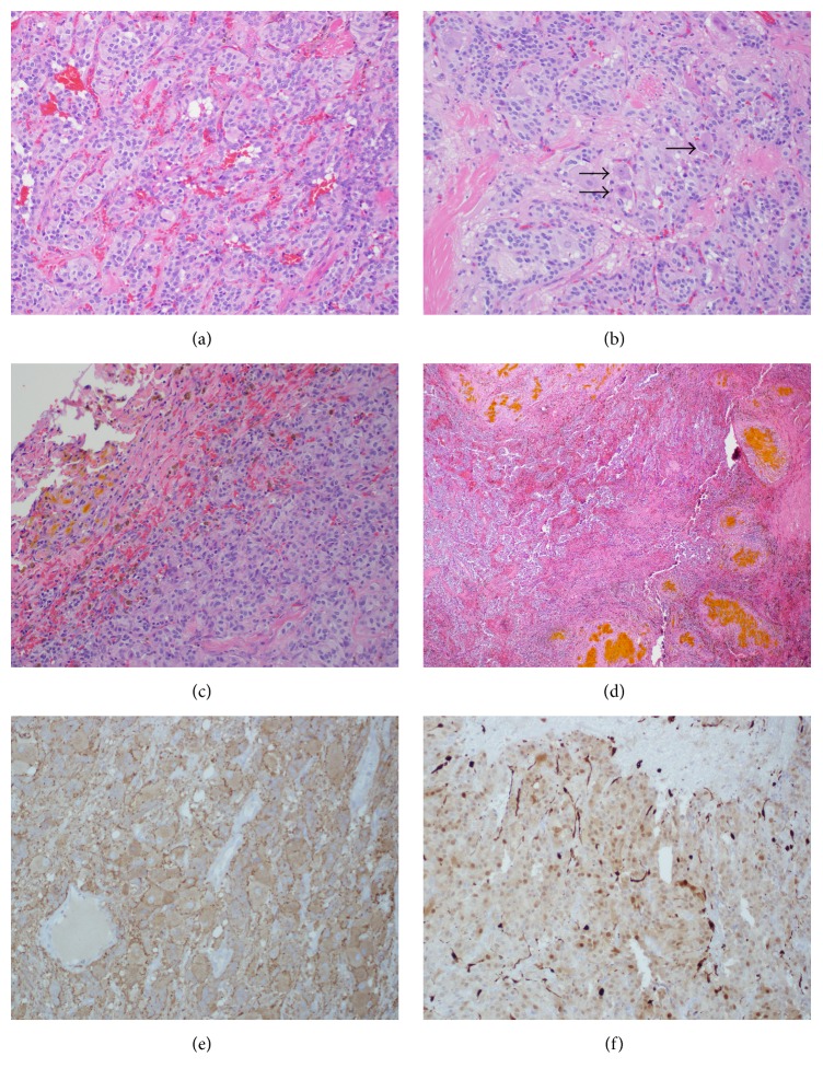
Gangliocytic paraganglioma. (a) Gangliocytic paraganglioma depicting nested arrangement of cells in a Zellballen pattern. H&E stain, 20x. (b) Gangliocytic paraganglioma with ganglion cells (arrow head). H&E stain, 40x. (c) Section of the tumor capsule showing pigmented macrophages (brown) and extracellular hemoglobin breakdown product (yellow). H&E stain, 4x. (d) Focus of remote hemorrhage toward the periphery of the tumor. H&E stain, 20x. (e) Synaptophysin stain positive (brown): positive for synaptic vesicle protein, 20x. (f) S100 stain positive for sustentacular cells (dark brown along edges of lobules), 20x.
References
-
- Burger P. C., Scheithauer B. W. Atlas of Tumor Pathology. Washington, DC, USA: Armed Forces Institute of Pathology; 1994. Tumors of paraganglionic tissue: tumors of the central nervous system; pp. 317–320.
LinkOut - more resources
Full Text Sources
Other Literature Sources
