

Studies in genera similar to Torula: Bahusaganda, Bahusandhika, Pseudotorula, and Simmonsiella gen. nov
- PMID: 27433439
- PMCID: PMC4941686
- DOI: 10.5598/imafungus.2016.07.01.03
Studies in genera similar to Torula: Bahusaganda, Bahusandhika, Pseudotorula, and Simmonsiella gen. nov
Abstract
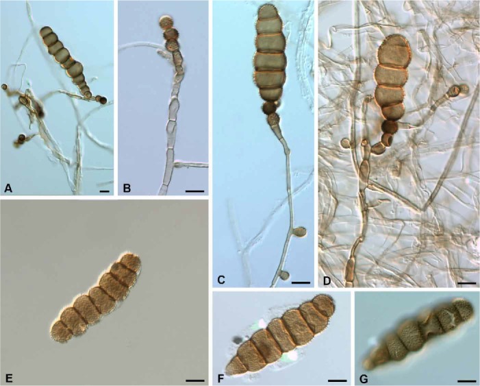
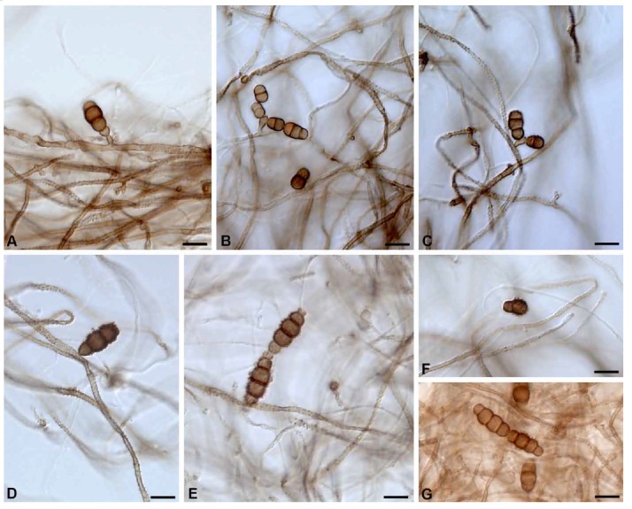
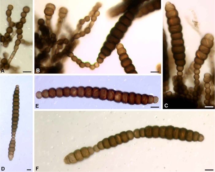
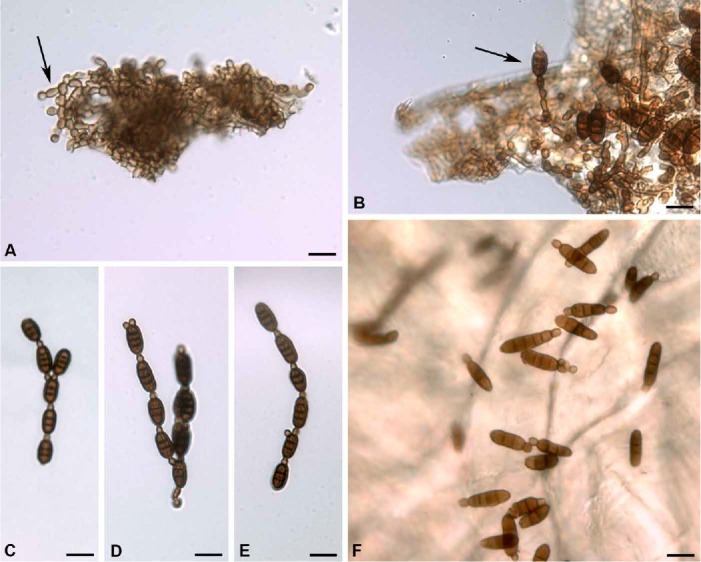
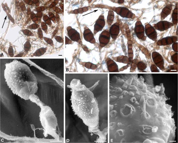
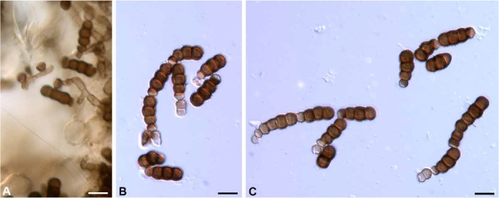
A generic key is presented to delimit Torula from seven hyphomycete genera whose type species were at one time included in the genus or whose conidium ontogeny and conidium development appear similar or superficially similar to that of Torula herbarum, the type of the genus. In Bahusaganda, two new species are described (B. elliseverhartii and B. simmonsii spp. nov.) and three new combinations made (B. ambrosiae, B. elaeodes, and B. heteromorpha combs. nov.). In Bahusandhika, one new species (B. hughesii sp. nov.) is introduced, and two new combinations made (B. rhombica and B. terrestris combs. nov.), along with emendations in the circumscription of B. caligans and B. intercalaris. Latorua is considered synonymous with Bahusandhika. In Pseudotorula, one new combination is made (P. sundara comb. nov.), and one emendation proposed (P. helica). The transfer of Dwayabeeja sundara, the type species of the genus, to Pseudotorula will require a new generic name to be introduced for D. aethiopica and D. cubensis. The new generic name Simmonsiella is established for Torula ndjilensis. Bahusandhika compacta is shown to be synonymous with Torula verrucospora.
Keywords: Bahuchashaka; Dwayabeeja; Latorua; Polyschema; corona cell.
Figures
Similar articles
-
The Luciolinae of S. E. Asia and the Australopacific region: a revisionary checklist (Coleoptera: Lampyridae) including description of three new genera and 13 new species.Zootaxa. 2019 Oct 18;4687(1):zootaxa.4687.1.1. doi: 10.11646/zootaxa.4687.1.1. Zootaxa. 2019. PMID: 31719466
-
The fine structure of conidial development in the genus Torula. II. T. caligans (Batista and Upadhyay) M. B. Ellis and T. terrestris Mistra.Can J Microbiol. 1975 Dec;21(12):1921-9. doi: 10.1139/m75-279. Can J Microbiol. 1975. PMID: 1220860
-
The Genera of Fungi - fixing the application of the type species of generic names - G 2: Allantophomopsis, Latorua, Macrodiplodiopsis, Macrohilum, Milospium, Protostegia, Pyricularia, Robillarda, Rotula, Septoriella, Torula, and Wojnowicia.IMA Fungus. 2015 Jun;6(1):163-98. doi: 10.5598/imafungus.2015.06.01.11. Epub 2015 Jun 11. IMA Fungus. 2015. PMID: 26203422 Free PMC article.
-
The Australian Ciidae (Coleoptera: Tenebrionoidea): A Preliminary Revision.Zootaxa. 2016 Nov 25;4198(1):zootaxa.4198.1.1. doi: 10.11646/zootaxa.4198.1.1. Zootaxa. 2016. PMID: 27988649
-
A review of the leafhopper genus Scaphotettix Matsumura (Hemiptera: Cicadellidae: Deltocephalinae), with description of a new genus.Zoolog Sci. 2009 Sep;26(9):656-63. doi: 10.2108/zsj.26.656. Zoolog Sci. 2009. PMID: 19799517 Review.
Cited by
-
Two novel species and three new records of Torulaceae from Yunnan Province, China.MycoKeys. 2023 Aug 7;99:1-24. doi: 10.3897/mycokeys.99.106699. eCollection 2023. MycoKeys. 2023. PMID: 37588799 Free PMC article.
-
Morphology and molecular phylogeny of Dothideomycetes fungi associated with Dracaena plants.Front Cell Infect Microbiol. 2025 Aug 11;15:1550824. doi: 10.3389/fcimb.2025.1550824. eCollection 2025. Front Cell Infect Microbiol. 2025. PMID: 40861486 Free PMC article.
-
New Species, New Record, and Antagonistic Potential of Torula (Torulaceae, Pleosporales) from Jilin Province, China.Microorganisms. 2025 Jun 23;13(7):1459. doi: 10.3390/microorganisms13071459. Microorganisms. 2025. PMID: 40731969 Free PMC article.
-
Palm Fungi and Their Key Role in Biodiversity Surveys: A Review.J Fungi (Basel). 2023 Nov 19;9(11):1121. doi: 10.3390/jof9111121. J Fungi (Basel). 2023. PMID: 37998926 Free PMC article. Review.
-
Multigene phylogeny and taxonomy of Dendryphion hydei and Torula hydei spp. nov. from herbaceous litter in northern Thailand.PLoS One. 2020 Feb 5;15(2):e0228067. doi: 10.1371/journal.pone.0228067. eCollection 2020. PLoS One. 2020. PMID: 32023268 Free PMC article.
References
-
- Bhat DJ. (1985) Two new species from Ethiopia. Proceedings of the Indian National Academy of Science, B 94: 269–272.
-
- Crane JL, Schoknecht JD. (1977) Revision of Torula species. Rutola, a new name for Torula graminis. Canadian Journal of Botany 55: 3013–3019.
-
- Crous PW, Carris LM, Giraldo A, Groenewald JZ, Hawksworth DL, et al. (2015) The Genera of Fungi – fixing the application of the type species of generic names - G 2: Allantophomopsis, Latorua, Macrodiplodiopsis, Macrohilum, Milospium, Protostegia, Pyricularia, Robillarda, Rotula, Septoriella, Torula, and Wojnowicia. IMA Fungus 6: 163–198. - PMC - PubMed
-
- Ellis MB. (1971) Dematiaceous Hyphomycetes. Kew: Commonwealth Mycological Institute.
-
- Ellis MB. (1976) More Dematiaceous Hyphomycetes. Kew: Commonwealth Mycological Institute.
LinkOut - more resources
Full Text Sources
Other Literature Sources
Miscellaneous