

Endosialin expression in soft tissue sarcoma as a potential marker of undifferentiated mesenchymal cells
- PMID: 27434038
- PMCID: PMC4985356
- DOI: 10.1038/bjc.2016.214
Endosialin expression in soft tissue sarcoma as a potential marker of undifferentiated mesenchymal cells
Abstract
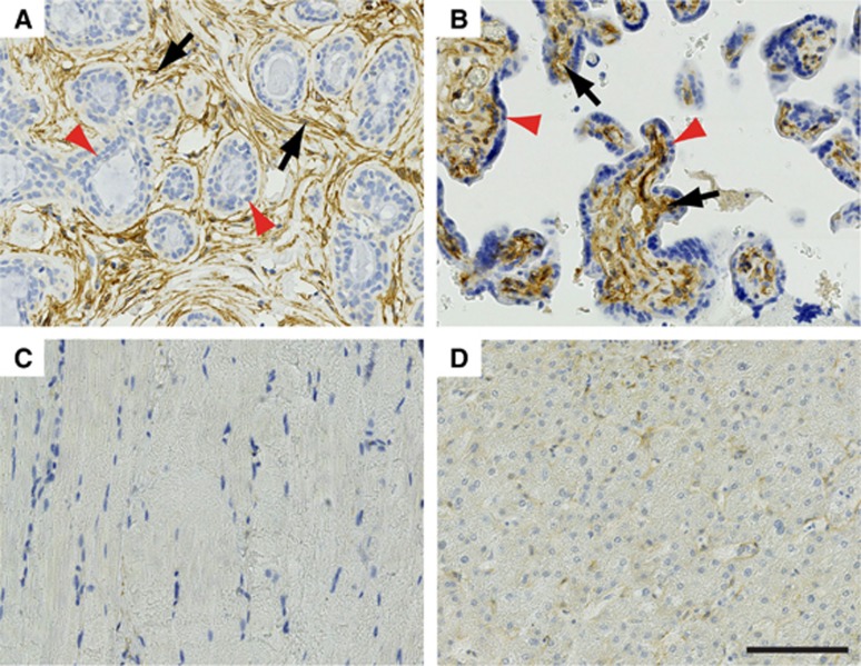
Background: Soft tissue sarcomas are a group of neoplasms with differentiation towards mesenchymal tissue, many of which are aggressive and chemotherapy resistant. Histology and immunoprofiles often overlap with neoplasms of other lineages, and establishing an accurate histopathological diagnosis is crucial for correct management, and therapeutic stratification. The endosialin cell surface glycoprotein is predominantly expressed by stromal fibroblasts and pericytes in epithelial neoplasms; however, tumour cell expression has been reported in small series of sarcomas.
Methods: We assessed endosialin expression by immunohistochemistry in a large set of 514 human soft tissue sarcomas.
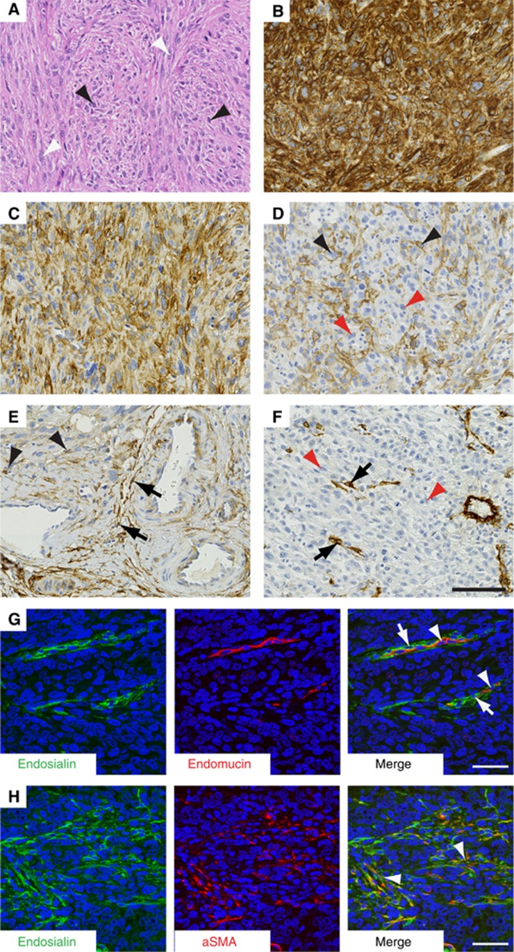
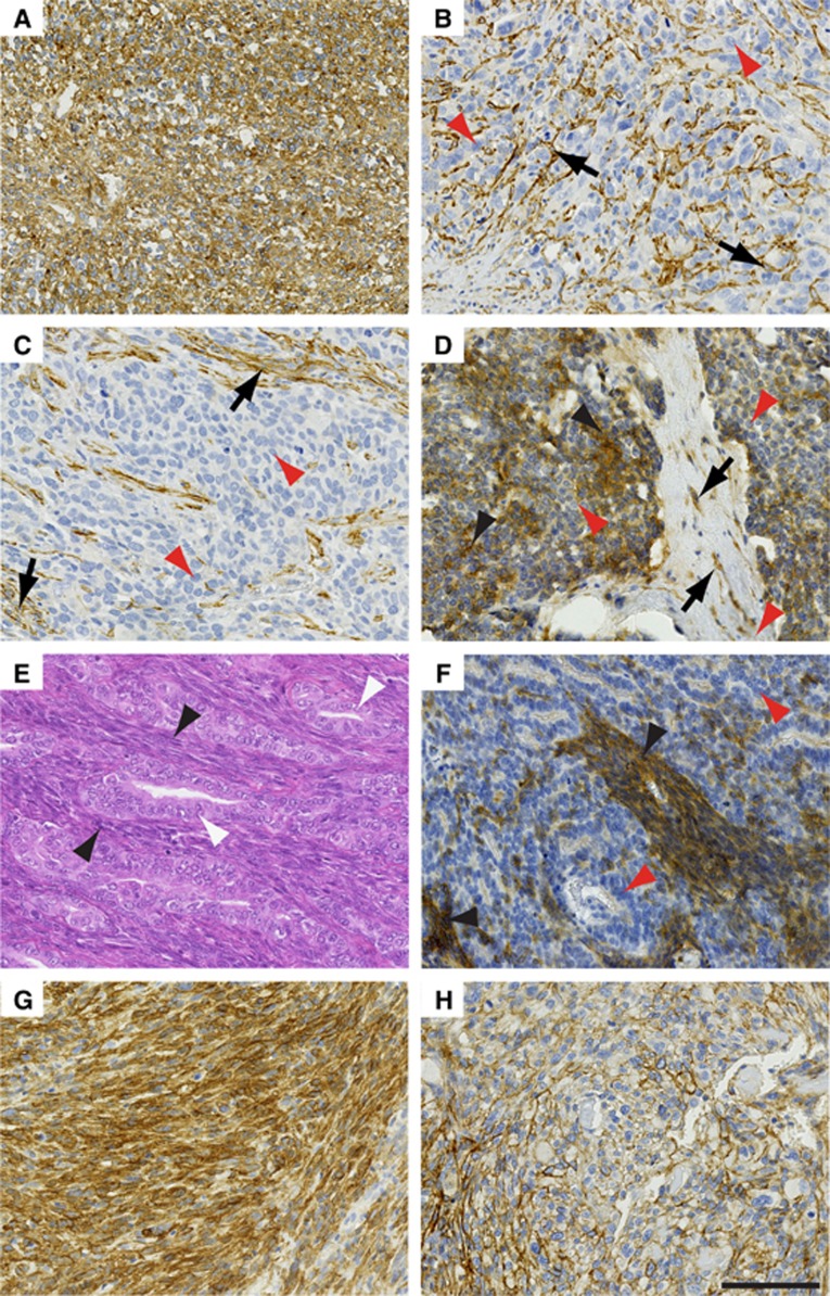
Results: Tumour cell endosialin expression was seen in 89% of undifferentiated pleomorphic sarcomas (104/117), 77% adult fibrosarcomas/spindle cell sarcomas (20/26), 62% synovial sarcomas (37/60), 51% leiomyosarcomas (94/185) and 31% rhabdomyosarcomas (39/126).
Conclusions: Endosialin immunohistochemistry has potential to distinguish undifferentiated and poorly differentiated sarcomas from other poorly differentiated, non-mesenchymal neoplasms. A Phase II trial randomising patients with advanced sarcomas to receive chemotherapy with/without an endosialin therapeutic antibody has recently completed enrolment. Endosialin expression could be used to select patients for such clinical trials. Based on our results, patients with undifferentiated pleomorphic sarcoma may be particularly suitable for such a therapeutic approach.
Conflict of interest statement
RLJ conducted a trial of a monoclonal antibody to endosialin sponsored by Morphotek. All other authors declare no conflict of interest.
Figures
Similar articles
-
Glypican-3 is expressed in rhabdomyosarcomas but not adult spindle cell and pleomorphic sarcomas.J Clin Pathol. 2011 Jul;64(7):587-91. doi: 10.1136/jclinpath-2011-200071. Epub 2011 Apr 14. J Clin Pathol. 2011. PMID: 21493758
-
Keratin subsets in spindle cell sarcomas. Keratins are widespread but synovial sarcoma contains a distinctive keratin polypeptide pattern and desmoplakins.Am J Pathol. 1991 Feb;138(2):505-13. Am J Pathol. 1991. PMID: 1704194 Free PMC article.
-
[Pathology of soft tissue sarcomas: 238 cases of the childhood tumor registry].Klin Padiatr. 1982 Jul-Aug;194(4):275-80. doi: 10.1055/s-2008-1033817. Klin Padiatr. 1982. PMID: 6290752 German.
-
Heterologous and rare homologous sarcomas of the uterine corpus: a clinicopathologic review.Adv Anat Pathol. 2011 Jan;18(1):60-74. doi: 10.1097/PAP.0b013e3182026be7. Adv Anat Pathol. 2011. PMID: 21169739 Review.
-
MicroRNAs in Different Histologies of Soft Tissue Sarcoma: A Comprehensive Review.Int J Mol Sci. 2017 Sep 12;18(9):1960. doi: 10.3390/ijms18091960. Int J Mol Sci. 2017. PMID: 28895916 Free PMC article. Review.
Cited by
-
Endosialin in Cancer: Expression Patterns, Mechanistic Insights, and Therapeutic Approaches.Theranostics. 2024 Jan 1;14(1):379-391. doi: 10.7150/thno.89495. eCollection 2024. Theranostics. 2024. PMID: 38164138 Free PMC article. Review.
-
CD248 promotes migration and metastasis of osteosarcoma through ITGB1-mediated FAK-paxillin pathway activation.BMC Cancer. 2023 Mar 30;23(1):290. doi: 10.1186/s12885-023-10731-7. BMC Cancer. 2023. PMID: 36997926 Free PMC article.
-
Endosialin (CD248) Expression in Fibromas and Soft-tissue Fibrosarcomas in Dogs.In Vivo. 2021 May-Jun;35(3):1467-1472. doi: 10.21873/invivo.12399. In Vivo. 2021. PMID: 33910824 Free PMC article.
-
Endo180 (MRC2) Antibody-Drug Conjugate for the Treatment of Sarcoma.Mol Cancer Ther. 2023 Feb 1;22(2):240-253. doi: 10.1158/1535-7163.MCT-22-0312. Mol Cancer Ther. 2023. PMID: 36399638 Free PMC article.
-
Current research and management of undifferentiated pleomorphic sarcoma/myofibrosarcoma.Front Genet. 2023 Feb 16;14:1109491. doi: 10.3389/fgene.2023.1109491. eCollection 2023. Front Genet. 2023. PMID: 36873946 Free PMC article. Review.
References
-
- Bagley R, Honma N, Weber W, Boutin P, Rouleau C, Shankara S, Kataoka S, Ishida I, Roberts B, Teicher B (2008) Endosialin/TEM 1/CD248 is a pericyte marker of embryonic and tumor neovascularization. Microvasc Res 76: 180–188. - PubMed
-
- Brady J, Neal J, Sadakar N, Gasque P (2004) Human endosialin (tumor endothelial marker 1) is abundantly expressed in highly malignant and invasive brain tumors. J Neuropathol Exp Neurol 63: 1274–1283. - PubMed
-
- Diaz LA Jr, Coughlin CM, Weil SC, Fishel J, Gounder MM, Lawrence S, Azad N, O'Shannessy DJ, Grasso L, Wustner J, Ebel W, Carvajal RD (2015) A first-in-human phase I study of MORAb-004, a monoclonal antibody to endosialin in patients with advanced solid tumors. Clin Cancer Res 21: 1281–1288. - PMC - PubMed
-
- Dolznig H, Schweifer N, Puri C, Kraut N, Rettig WJ, Kerjaschki D, Garin-Chesa P (2005) Characterization of cancer stroma markers: in silico analysis of an mRNA expression database for fibroblast activation protein and endosialin. Cancer Immun 5: 10. - PubMed
Publication types
MeSH terms
Substances
LinkOut - more resources
Full Text Sources
Other Literature Sources
Miscellaneous
