. 2016 Nov;23(11):775-784.
doi: 10.1038/gt.2016.57. Epub 2016 Aug 11.

Construction and validation of an RNA trans-splicing molecule suitable to repair a large number of COL7A1 mutations

Affiliations

Construction and validation of an RNA trans-splicing molecule suitable to repair a large number of COL7A1 mutations

B Tockner et al. Gene Ther. 2016 Nov.

Abstract

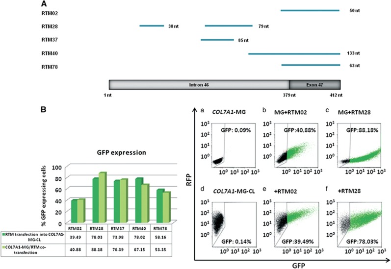
RNA trans-splicing has become a versatile tool in the gene therapy of monogenetic diseases. This technique is especially valuable for the correction of mutations in large genes such as COL7A1, which underlie the dystrophic subtype of the skin blistering disease epidermolysis bullosa. Over 800 mutations spanning the entire length of the COL7A1 gene have been associated with defects in type VII collagen, leading to excessive fragility of epithelial tissues, the hallmark of dystrophic epidermolysis bullosa (DEB). In the present study, we designed an RNA trans-splicing molecule (RTM) that is capable of repairing any given mutation within a 4200 nucleotide region spanning the 3' half of COL7A1. The selected RTM, RTM28, was able to induce accurate trans-splicing into endogenous COL7A1 pre-mRNA transcripts in a type VII collagen-deficient DEB patient-derived cell line. Correct trans-splicing was detected at the RNA level by semiquantitative RT-PCR and correction of full-length type VII collagen was confirmed at the protein level by immunofluorescence and western blot analyses. Our results demonstrate that RTM28, which covers >60% of all mutations reported in DEB and is thus the longest RTM described so far for the repair of COL7A1, represents a promising candidate for therapeutic applications.


Conflict of interest statement

JWB is an inventor on US (US8735366) and European (EP2320952) patent for ‘Improved pre-mRNA trans-splicing molecules (RTM) and their uses'. The other authors declare no conflict of interest.

Figures

Figure 1
Figure 1
A fluorescence-based screening system for RTM selection. (a) The screening system is built up by a COL7A1-MG, containing the 5′ portion of GFP and the selected COL7A1 pre-mRNA region of intron 46/exon 47, and an RTM, carrying a randomly cloned BD, splicing elements for efficient trans-splicing, the 3′ portion of GFP, an IRES and dsRED. Co-expression of both screening molecules in HEK293 cells leads to the restoration and expression of GFP upon accurate trans-splicing. (bd) The transfection of the RTM library into HEK293 cells, stably expressing the COL7A1-MG (COL7A1-MG-CL) and the red fluorescence reporter molecule mRuby, results in a diverse expression of the red and green reporter molecules. This is dependent on the levels of RTM and COL7A1-MG expression and more importantly on the functionality of the introduced RTM. A high GFP expression indicates trans-splicing through highly efficient RTMs in the cell, detected by microscopic (b, c) and FACS analysis (d). Overlay of the different fluorochromes in trans-spliced cells results in a yellow to orange colour, depending on the strength of green GFP expression together with red target/RTM expression (c). The cell nuclei were stained with 4′,6-diamidino-2-phenylindole (DAPI) (blue fluorescence). Scale bars: 50 μm (b); 20 μm (c). PPT, polypyrimidine tract.
Figure 2
Figure 2
Investigation of RTM binding regions within the target intron 46/exon 47 of COL7A1 and their trans-splicing efficiencies analysed by flow cytometry. Eighty individual clones of the RTM library were analysed for the presence of a BD sequence by sequence analysis. (A) Five single RTMs, termed RTM02, RTM28, RTM37, RTM40 and RTM78, contained one or more specific BD(s) complementary to the selected COL7A1 target region intron 46/exon 47. These RTMs were included in initial experiments to investigate their potential to induce accurate trans-splicing into COL7A1. (B) Co-transfection of COL7A1-MG together with a representative low-efficiency (RTM02) (b) or high-efficiency RTM (RTM28) (c) into HEK293 cells resulted in the expression of GFP in ~40% or ~88% of all analysed cells, respectively, upon accurate 3′ RNA trans-splicing. Similar results were achieved after RTM02 (e) and RTM28 (f) transfection into the COL7A1-MG-CL-expressing cell line, in which ~40% and ~78% GFP expression, respectively, were detectable by flow cytometric analysis. GFP expression was undetectable in HEK293 cells exclusively transfected with RTM28 (a) and in the COL7A1-MG-CL-expressing cell line (d). Regardless of whether the COL7A1-MG was transiently or stably expressed in HEK293 cells, the RTM efficiencies were comparable. The most efficient RTMs, RTM28, RTM37 and RTM40, showed higher trans-splicing efficiencies than RTM02 and RTM78 in both screening settings, measured by flow cytometric analysis.
Figure 3
Figure 3
Validation of FACS analysis by fluorescence-based microscopy. Co-transfection of COL7A1-MG together with high-efficiency RTM (RTM28) (a) into HEK293 cells resulted in the expression of GFP upon accurate 3′ RNA trans-splicing. Similar results were achieved after RTM28 (b) transfection into the COL7A1-MG-CL-expressing cell line. GFP expression was undetectable in HEK293 cells exclusively transfected with RTM28 (a, upper panel) and the COL7A1-MG-CL-expressing cell line (b, upper panel). HEK293 cells transfected with RTM28 containing dsRED and the 3′ half of GFP (a, upper panel) and stably expressing COL7A1-MG cells (COL7A1-MG-CL) containing mRuby (b, upper panel) showed red fluorescence upon reporter molecule expression. Overlay of the different flurochromes in trans-spliced cells resulted in a yellow to orange colour, depending on the strength of the merged green GFP expression and the red target expression (a, b, lower panel). Scale bars: 50 μm.
Figure 4
Figure 4
Detection of COL7A1-RTM fusion transcripts by sqRT-PCR. (a) Schematic depiction of the endogenous RTM28, containing a FLAG tag and silent mutations within the wild-type coding domain of COL7A1. (b) SqRT-PCR analysis, performed on total RNA isolated from RTM28-transduced RDEB keratinocytes using primers spanning exons 46–49, revealed the expression of COL7A1-RTM fusion transcripts at the RNA level. The respective PCR product with a size of 205 bp was verified by sequence analysis. (c) SqRT-PCR was performed to quantify total COL7A1 transcripts present in RTM-treated keratinocytes. As a result, a fivefold increase of COL7A1 expression was obtained after RTM28 treatment in comparison with RDEB patient cells transduced with an empty retroviral vector (RDEB-mock).
Figure 5
Figure 5
Correction of type VII collagen in RTM28-transduced patient keratinocytes. (a) Immunofluorescene staining of a wild-type human keratinocyte line (1, 4), an RDEB keratinocyte line (2, 5) and an RTM28-transduced RDEB keratinocyte line (3, 6). The upper panel shows type VII collagen staining using an antibody against the NC1 domain of type VII collagen, whereas staining against the FLAG tag is shown in the lower panel. Staining with the type VII collagen-specific antibody resulted in a strong signal in wild-type human keratinocytes (1), while the staining was absent in the untransduced RDEB keratinocyes (2). RTM28-transduced RDEB keratinocytes displayed a specific staining with the anti-type VII collagen antibody, corresponding to corrected type VII collagen expression upon trans-splicing (3). As expected, wild-type keratinocytes (4) and RDEB keratinocytes (5) did not show a specific signal using an anti-FLAG tag antibody, whereas staining of RTM28-transduced RDEB keratinocytes (6) resulted in a specific signal (green) in the cytoplasm of the cells. The cell nuclei were counterstained with DAPI (blue). Scale bar: 50 μm. (b) Western blot analysis of cell lysates using either an antibody against type VII collagen (upper panel) or the FLAG tag (lower panel). Wild-type human keratinocytes showed a strong type VII collagen band (1), whereas there was no band detectable in untransduced RDEB keratinocytes (2). RTM28-transduced RDEB keratinocytes (3) showed a weak band at the correct size of ~290 kDa, indicating accurate trans-splicing. Further, no band was detectable in wild-type keratinocytes (4) and untransduced RDEB keratinocytes (5) using a FLAG-specific antibody, whereas a specific ~293 kDa FLAG-tagged trans-spliced type VII collagen was visible in lysates of RTM28-transduced RDEB keratinocytes (6). Actinin staining served as loading control.

Similar articles

Cited by

References

    1. Fine JD, Hintner H (eds). Life with Epidermolysis Bullosa: Etiology, Diagnosis, and Multidisciplinary Care and Therapy. Springer: Wien, Germany and New York, NY, USA, 2009.
    1. Fine JD, Bruckner-Tuderman L, Eady RA, Bauer EA, Bauer JW, Has C et al. Inherited epidermolysis bullosa: updated recommendations on diagnosis and classification. J Am Acad Dermatol 2014; 70: 1103–1126. - PubMed
    1. Lin AN, Carter DM. Epidermolysis bullosa. Annu Rev Med 1993; 44: 189–199. - PubMed
    1. Wertheim-Tysarowka K, Sobczyńska-Tomaszewska A, Kowalewski C, Skroński M, Swięćkowski G, Kutkowska-Kaźmierczak A et al. The COL7A1 mutation database. Hum Mutat 2012; 33: 327–331. - PubMed
    1. Wagner JE, Ishida-Yamamoto A, McGrath JA, Hordinsky M, Keene DR, Woodley DT et al. Bone marrow transplantation for recessive dystrophic epidermolysis bullosa. N Engl J Med 2010; 363: 629–639. - PMC - PubMed

Publication types