. 2016 Sep;24(9):1550-60.
doi: 10.1038/mt.2016.125. Epub 2016 Jun 23.

Anti-apoptotic Effects of Human Wharton's Jelly-derived Mesenchymal Stem Cells on Skeletal Muscle Cells Mediated via Secretion of XCL1

Affiliations

Anti-apoptotic Effects of Human Wharton's Jelly-derived Mesenchymal Stem Cells on Skeletal Muscle Cells Mediated via Secretion of XCL1

SooJin Kwon et al. Mol Ther. 2016 Sep.

Abstract

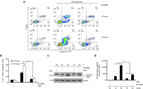
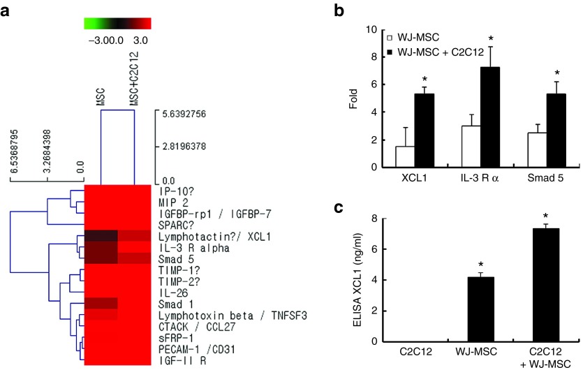
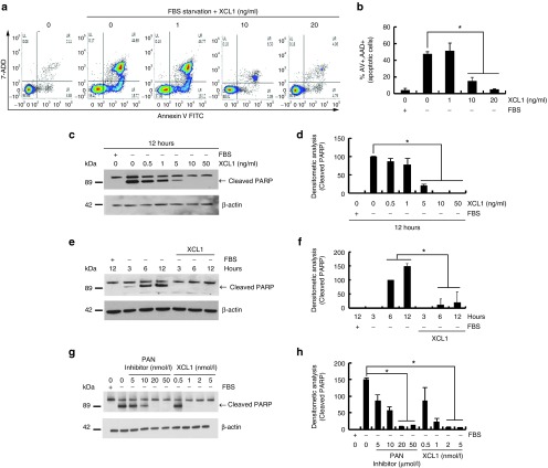
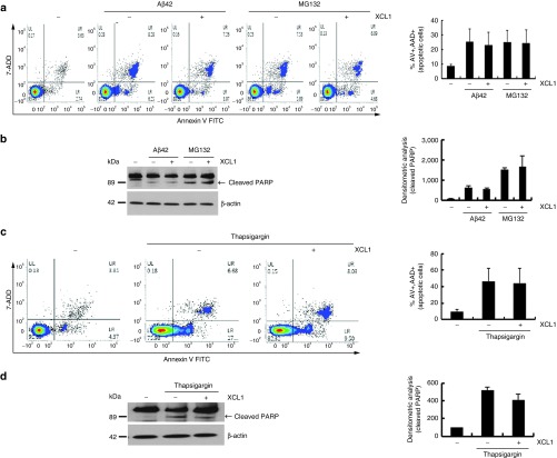
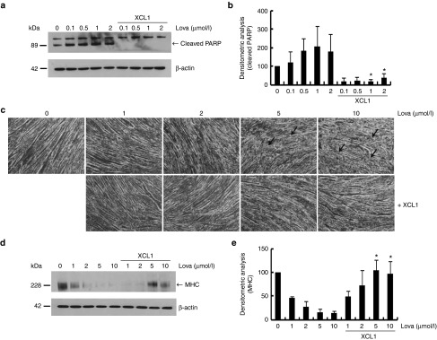
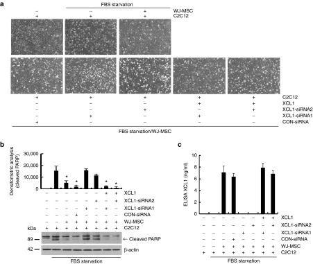
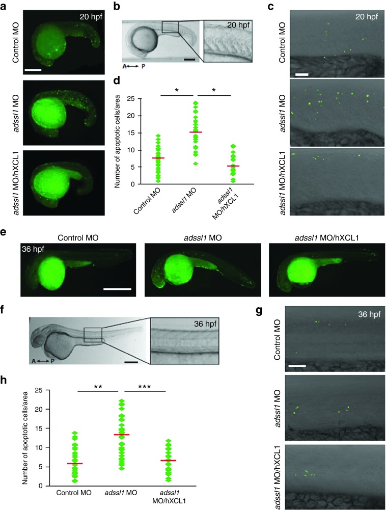
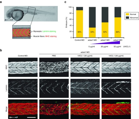
The role of Wharton's jelly-derived human mesenchymal stem cells (WJ-MSCs) in inhibiting muscle cell death has been elucidated in this study. Apoptosis induced by serum deprivation in mouse skeletal myoblast cell lines (C2C12) was significantly reduced when the cell lines were cocultured with WJ-MSCs. Antibody arrays indicated high levels of chemokine (C motif) ligand (XCL1) secretion by cocultured WJ-MSCs and XCL1 protein treatment resulted in complete inhibition of apoptosis in serum-starved C2C12 cells. Apoptosis of C2C12 cells and loss of differentiated C2C12 myotubes induced by lovastatin, another muscle cell death inducer, was also inhibited by XCL1 treatment. However, XCL1 treatment did not inhibit apoptosis of cell lines other than C2C12. When XCL1-siRNA pretreated WJ-MSCs were cocultured with serum-starved C2C12 cells, apoptosis was not inhibited, thus confirming that XCL1 is a key factor in preventing C2C12 cell apoptosis. We demonstrated the therapeutic effect of XCL1 on the zebrafish myopathy model, generated by knock down of a causative gene ADSSL1. Furthermore, the treatment of XCL1 resulted in significant recovery of the zebrafish skeletal muscle defects. These results suggest that human WJ-MSCs and XCL1 protein may act as promising and novel therapeutic agents for treatment of myopathies and other skeletal muscle diseases.


Figures

Figure 1
Figure 1
Coculturing with human Wharton's jelly-derived human mesenchymal stem cells (WJ-MSCs) reduced serum starvation-induced C2C12 cell death via secretion of soluble factors. C2C12 cells were serum-deprived during culture both in the absence and presence of human WJ-MSC (1 × 105 cells/well) for 12 and 24 hours in a transwell chamber. C2C12 cells alone and C2C12 cells cocultured with human WJ-MSC in the absence of serum were analyzed by fluorescence-activated cell-sorting (FACS) analysis of cells stained with annexin V/7-AAD (a,b) or Western blot analysis with anti-poly ADP-ribose polymerase (PARP) antibody (c,d). The arrow indicates cleaved PARP during apoptosis. (b) Percentage of apoptotic cells by FACS analysis (*P < 0.05, n = 3). (d) The cleaved PARP bands were analyzed through densitometry (*P < 0.05, n = 3)
Figure 2
Figure 2
Identification of XCL1 as a paracrine factor of Wharton's jelly-derived human mesenchymal stem cell (WJ-MSC) during coculture. Secreted proteins in the media collected from the experiments were measured using the RayBio Biotin Label-based Human Antibody. (a,b) Increased fold of spot intensity of XCL1, IL-3 R, and smad5 in cocultured cells as compared to WJ-MSC alone. (c) The conditioned media was collected from C2C12, WJ-MSC alone, and cocultured cells. Concentration of secreted XCL1 in each conditioned media was measured by XCL1 ELISA kit (*P < 0.05, n = 3).
Figure 3
Figure 3
Recombinant XCL1 protein inhibited serum starvation-induced apoptosis of C2C12 cells. C2C12 cell was serum-deprived during culture both in the absence or presence of recombinant XCL1 proteins (1, 10, and 20 ng/ml) for 12 hours. (a) Fluorescence-activated cell-sorting (FACS) analysis of cells stained with annexin V/7-AAD. (b) Percentage of apoptotic cells by FACS analysis (*P < 0.05, n = 3). (c) Harvested cells were analyzed by Western blot analysis with anti-poly ADP-ribose polymerase (PARP) antibody. (d) PARP cleaved bands were analyzed by densitometry (*P < 0.05, n = 3). (e) Serum-deprived C2C12 cells were treated with recombinant XCL1 (8 ng/ml) in a time-dependent manner. (f) PARP cleavage was monitored by Western blot analysis (*P < 0.05, n = 3). (g) Serum-deprived C2C12 cells were treated with either PAN caspase inhibitor (z-VAD-FMK) or XCL1 protein in a dose-dependent manner for 24 hours. (h) Apoptotic cells were analyzed by Western blot with anti-PARP antibody. (*P < 0.05, n = 3).
Figure 4
Figure 4
Absence of anti-apoptotic effect of XCL1 in other types of cells. The HT22, mouse hippocampal neuron cells were treated with amyloid-β42 (1 μmol/l for 48 hours) or MG132 (5 μmol/l for 24 hours) in the absence or presence of XCL1. (a) Fluorescence-activated cell-sorting (FACS) analysis of cells stained with annexin V/7-AAD. (b) Poly ADP-ribose polymerase (PARP)-cleaved bands were analyzed by densitometry. S16 rat Schwann cells were treated with thapsigargin (0.1 μmol/l for 12 hours) in the absence or presence of XCL1. The thapsigargin (0.1 μmol/l for 12 hours) treated S16 cells were analyzed by (c) FACS analysis and (d) Western blot analysis with anti-PARP antibody.
Figure 5
Figure 5
XCL1 treatment inhibited lovastatin-induced apoptosis of C2C12 cells and reduction of myotube formation. (a) C2C12 cells were treated with lovastatin (0.1, 0.5, 1, and 2 μmol/l) in a dose-dependent manner in the absence or presence of XCL1 and protein lysates were analyzed by Western blot analysis using anti-poly ADP-ribose polymerase (PARP) antibody. (b) Cleaved PARP fragments were monitored by densitometric analysis (*P < 0.05, n = 3). (c) The 7-day differentiated myotubes from C2C12 cells were treated with lovastatin (1, 2.5, 5, and 10 μmol/l) in the absence or presence of XCL1. Each arrow indicates defects in the myotubes. (d,e) Protein extracts of myotubes were analyzed by Western blot analysis using anti-myosin heavy chain (MHC) antibody. Bands of MHC were monitored by densitometric analysis (*P < 0.05, n = 3).
Figure 6
Figure 6
XCL1 siRNA treatment abolished anti-apoptotic effect of human Wharton's jelly-derived human mesenchymal stem cell (WJ-MSC). (a) Before coculturing with serum-starved C2C12 cells, WJ-MSCs were separately pretreated with two siRNAs of human XCL1 containing different sequences for 24 hours. Each siRNA-treated WJ-MSC was cocultured with serum-deprived C2C12 cells for 12 hours. Human-recombinant XCL1 was added to cocultured C2C12 cells with siRNA-treated WJ-MSC to monitor gain of function of XCL1. C2C12 cell images were taken at each condition. (b) Cell lysates were analyzed by Western blot using anti-poly ADP-ribose polymerase (PARP) antibody. Cleaved PARP fragments were monitored by densitometric analysis (*P < 0.05, n = 3). (c) Secreted XCL1 concentration in each conditioned media was measured by XCL1 ELISA kit (*P < 0.05, n = 3).
Figure 7
Figure 7
Induction of human XCL1 reduces apoptosis of the skeletal muscles in the zebrafish myopathy model. Twenty hours post fertilization (hpf) zebrafish embryos (a–d) and 36 hpf embryos were observed (e–h). (a,e) The apoptotic cells of control and adssl1 morphants or human XCL1-induced adssl1 morphants were detected by TUNEL assay, and the whole-mounted embryos were observed by fluorescent microscopy. (b,f) The boxed region of the embryos was photographed using confocal microscopy. (c,g) The representative confocal images show that the number of dead cells in adssl1 morphants was more than in control morphants, and induction of human XCL1 inhibited the increase of cell death in adssl1 morphants. (d,h) The cell death in the selected area (within five somites in the trunk) was quantified and presented in the scattered chart. A, anterior; P, posterior. a,e, bar = 100 µm; b,c,f,g, bar = 50 µm. d,h, Student's t-test: *P < 0.05, **P < 0.005, ***P < 0.001.
Figure 8
Figure 8
The rescue of defective skeletal muscles by human XCL1 in the zebrafish model. (a) The boxed region indicates the observed area to determine the effect of human XCL1 on skeletal muscles in zebrafish at 36 hpf. Myosepta and muscle fibers were stained with anti-Laminin and myosin heavy chain (MHC) antibodies, respectively. (b) The whole-mounted embryos after double staining with anti-Laminin and MHC antibodies were observed by confocal microscopy. The adssl1 morphants (phosphate-buffered saline injected embryos) showed disrupted muscle fibers and myosepta, whereas the morphants injected with human XCL1 showed recovery of muscle defects. (c) The zebrafish embryos demonstrating skeletal muscle phenotype were counted and quantified data were presented graphically. The data indicates that human XCL1 restores muscle abnormality of adssl1 morphants in a dose-dependent manner. A, anterior; P, posterior. Scale bar = 50 µm.

Similar articles

Cited by

References

    1. Danial, NN and Korsmeyer, SJ (2004). Cell death: critical control points. Cell 116: 205–219. - PubMed
    1. Portt, L, Norman, G, Clapp, C, Greenwood, M and Greenwood, MT (2011). Anti-apoptosis and cell survival: a review. Biochim Biophys Acta 1813: 238–259. - PubMed
    1. Li, J and Yuan, J (2008). Caspases in apoptosis and beyond. Oncogene 27: 6194–6206. - PubMed
    1. Taylor, RC, Cullen, SP and Martin, SJ (2008). Apoptosis: controlled demolition at the cellular level. Nat Rev Mol Cell Biol 9: 231–241. - PubMed
    1. Adhihetty, PJ and Hood, DA (2003). Mechanisms of apoptosis in skeletal muscle. Basic Appl Myol 13: 171–179.