. 2016 Sep:270:115-123.
doi: 10.1016/j.jmr.2016.07.002. Epub 2016 Jul 9.

Separation of extra- and intracellular metabolites using hyperpolarized (13)C diffusion weighted MR

Affiliations

Separation of extra- and intracellular metabolites using hyperpolarized (13)C diffusion weighted MR

Bertram L Koelsch et al. J Magn Reson. 2016 Sep.

Abstract

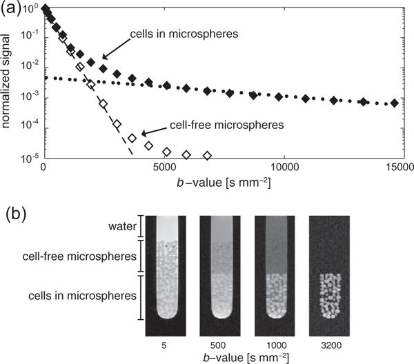
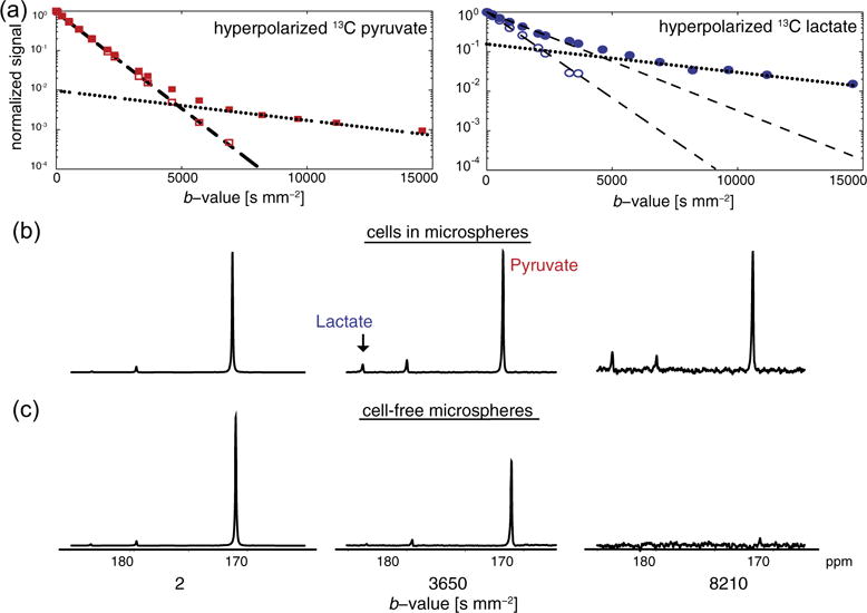
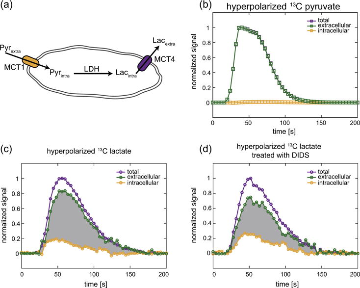
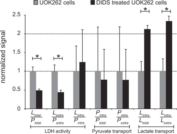
This work demonstrates the separation of extra- and intracellular components of glycolytic metabolites with diffusion weighted hyperpolarized (13)C magnetic resonance spectroscopy. Using b-values of up to 15,000smm(-2), a multi-exponential signal response was measured for hyperpolarized [1-(13)C] pyruvate and lactate. By fitting the fast and slow asymptotes of these curves, their extra- and intracellular weighted diffusion coefficients were determined in cells perfused in a MR compatible bioreactor. In addition to measuring intracellular weighted diffusion, extra- and intracellular weighted hyperpolarized (13)C metabolites pools are assessed in real-time, including their modulation with inhibition of monocarboxylate transporters. These studies demonstrate the ability to simultaneously assess membrane transport in addition to enzymatic activity with the use of diffusion weighted hyperpolarized (13)C MR. This technique could be an indispensible tool to evaluate the impact of microenvironment on the presence, aggressiveness and metastatic potential of a variety of cancers.

Keywords: Aerobic glycolysis; Cancer; Cancer aggressiveness; Cellular transport; Diffusion weighted magnetic resonance; Dynamic nuclear polarization (DNP); Hyperpolarized (13)C magnetic resonance (HP (13)C MR); Lactate; Lactate efflux; Pyruvate; Renal cell carcinoma (RCC).


Figures

Fig. 1
Fig. 1
(a) The pulsed gradient double spin echo sequence used for diffusion weighting of hyperpolarized 13C metabolites. Diffusion gradients (G) are placed symmetrically around the adiabatic 180° refocusing pulses, with a duration δ and a separation Δ. A crusher gradient after the readout ensures no transverse magnetization carries-over to subsequent scans. The excitation flip angle θ was either 15° or 30°. (b) The diffusion gradient array used to measure the extra- and intracellular weighted diffusion coefficients of hyperpolarized 13C metabolites. Every third scan (small boxes) was used to normalize adjacent diffusion weighted scans (large boxes), thereby removing the effects of T1 relaxation and metabolism on the signal change from that due to the diffusion weighting. (c) The gradient array used to measure the change in the total (small boxes) and the intracellular weighted (large boxes) hyperpolarized 13C metabolite pools over time. See methods for a more detailed description of these two acquisition schemas.
Fig. 2
Fig. 2
(a) The water signal response with increasing diffusion weighting in the bioreactor. For cells encapsulated in the alginate microspheres (◆), the water signal response reveals the presence of multiple environments with different water diffusion coefficients. The fast and slow asymptotes (dotted lines) were used to determine extra- and intracellular weighted diffusion coefficients, respectively. The water signal response in cell-free microspheres (◊) reflects water diffusion in solution and in the microspheres. (b) A diffusion weighted imaging experiment confirms the presence of the different diffusion environments. At low b-values, water signal is present in all three environments. As the b-values increase, signal from compartments with faster diffusion decreases while signal from highly restricted environments (i.e., intracellular) persists. At b-values above 3200 s mm−2, only signal from within the cells can be seen. The intensity of these images are scaled independently to more easily identify the various features. Only in this imaging experiment were alginate microspheres with and without cells layered for a single experiment; all other experiments acquired either with one or the other.
Fig. 3
Fig. 3
(a) Hyperpolarized 13C metabolite signal response with increasing diffusion weighting in the bioreactor. The multi-exponential signal response of hyperpolarized 13C pyruvate and lactate reveals multiple diffusion environments in experiments with UOK262 cells encapsulated in alginate microspheres (filled symbols). The fast and slow asymptotes were used to determine the diffusion coefficients in the extra- and intracellular weighted environments, respectively similar to that of proton in Fig. 2. Diffusion weighting of the injected hyperpolarized 13C substrates in cell-free microspheres are also shown (empty symbols). Hyperpolarized 13C lactate in cell experiments was produced by conversion from pyruvate while in the cell-free microsphere experiment 13C lactate was polarized and injected. Spectra of the hyperpolarized 13C pyruvate and lactate with increasing diffusion weighting (b-value) shows the suppression of extracellular signals at higher b-values in cells encapsulated in alginate (b) and in cell-free microspheres (c). For (b) and (c) spectra at a single b-value are scaled to the same SNR.
Fig. 4
Fig. 4
(a) A schematic of a cell showing the transport of hyperpolarized 13C pyruvate into the cell via the monocarboxylate transporter 1 (MCT1), its conversion to hyperpolarized 13C lactate by the enzyme lactate dehydrogenase (LDH) and the transport of 13C lactate out of the cell via MCT4. The dynamic signals of hyperpolarized 13C pyruvate (b) and lactate (c) in UOK262 cells perfused in the bioreactor, showing the total, extra- and intracellular metabolite pools. The total and intracellular signals are acquired at intermittent low and high b-values, namely 2.4 and 3863 s mm−2 respectively. (d) UOK262 cells treated with the high-affinity MCT4 inhibitor DIDS show an increase in the fraction of intracellular hyperpolarized 13C lactate relative to the extracellular hyperpolarized 13C lactate. The change in shaded area in (c) and (d) between the extra- and intracellular hyperpolarized 13C lactate curves highlights the relative changes in these lactate pools due to the treatment with DIDS. All plots are normalized to the maximum total signal of the respective hyperpolarized metabolite.
Fig. 5
Fig. 5
The relative changes in metabolism and membrane transport of hyperpolarized 13C metabolites in UOK262 cells, without and with treatment with DIDS, a high-affinity inhibitor of MCT4. The ratios that reflect LDH activity, or conversion of hyperpolarized 13C pyruvate to 13C lactate, show a significant decrease with DIDS treatment. The membrane transport ratios for hyperpolarized 13C pyruvate do not significantly change with DIDS treatment. The increase in the lactate transport ratios shows that DIDS inhibition of MCT4 leads to a higher intracellular fraction of hyperpolarized 13C lactate. Significant differences with p-value < 0.05 are represented by*.

References

    1. Nelson SJ, Kurhanewicz J, Vigneron DB, Larson PEZ, Harzstark AL, Ferrone M, et al. Metabolic imaging of patients with prostate cancer using hyperpolarized [1-13C]pyruvate. Sci Transl Med. 2013;5:198ra108. http://dx.doi.org/10.1126/scitranslmed.3006070. - DOI - PMC - PubMed
    1. Warburg O. On the origin of cancer cells. Science. 1956;123:309–314. 1956 ed. - PubMed
    1. Albers MJ, Bok R, Chen AP, Cunningham CH, Zierhut ML, Zhang VY, et al. Hyperpolarized 13C lactate, pyruvate, and alanine: noninvasive biomarkers for prostate cancer detection and grading. Cancer Res. 2008;68:8607–8615. http://dx.doi.org/10.1158/0008-5472.CAN-08-0749. - DOI - PMC - PubMed
    1. Kroemer G, Pouysségur J. Tumor cell metabolism: cancer’s Achilles’ heel. Cancer Cell. 2008;13:472–482. http://dx.doi.org/10.1016/j.ccr.2008.05.005. - DOI - PubMed
    1. Gallagher SM, Castorino JJ, Wang D, Philp NJ. Monocarboxylate transporter 4 regulates maturation and trafficking of CD147 to the plasma membrane in the metastatic breast cancer cell line MDA-MB-231. Cancer Res. 2007;67:4182–4189. http://dx.doi.org/10.1158/0008-5472.CAN-06-3184. - DOI - PubMed