. 2017 Mar;10(2):341-351.
doi: 10.1038/mi.2016.61. Epub 2016 Jul 20.

Periostin regulates fibrocyte function to promote myofibroblast differentiation and lung fibrosis

Affiliations

Periostin regulates fibrocyte function to promote myofibroblast differentiation and lung fibrosis

S L Ashley et al. Mucosal Immunol. 2017 Mar.

Abstract

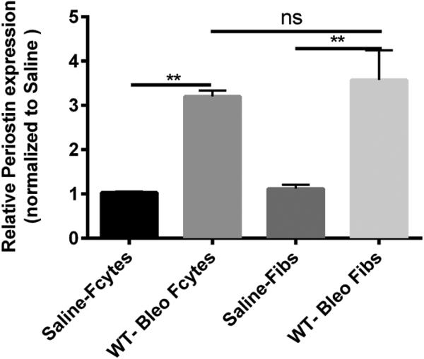
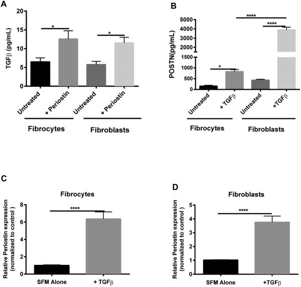
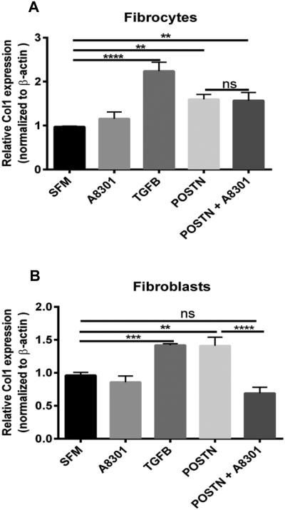
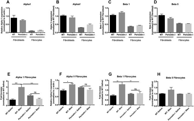
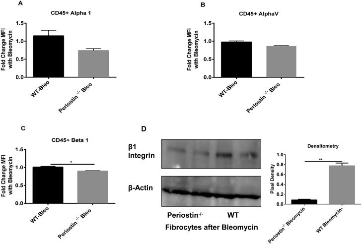
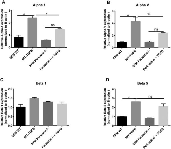
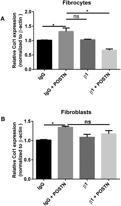
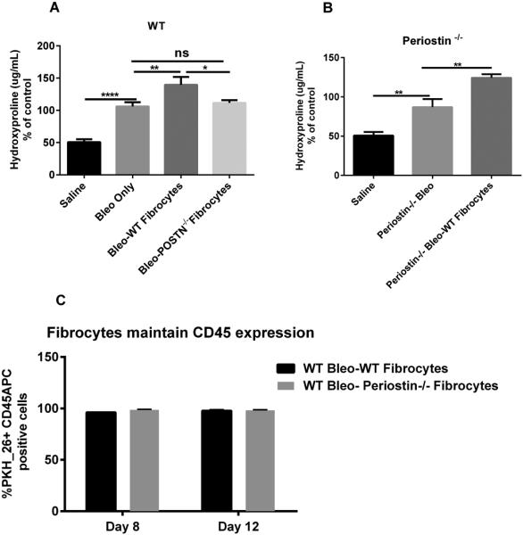
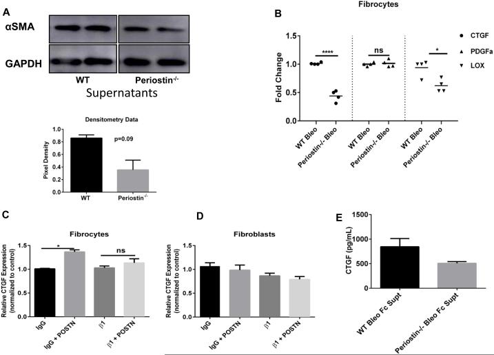
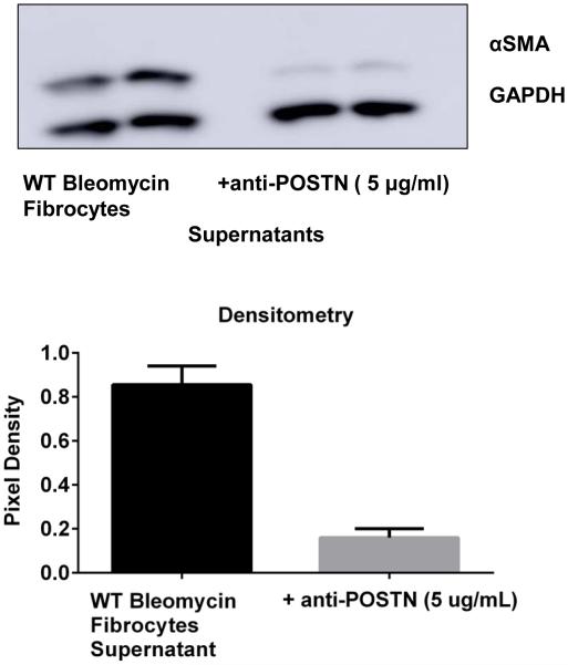
Fibrocytes are circulating mesenchymal precursors (CD45+, col 1+) recruited to fibrotic areas. Fibrocytes secrete profibrotic mediators including periostin; a matricellular protein that regulates cellular interactions with extracellular matrix (ECM) components. In bleomycin-induced fibrosis, periostin deficiency in structural or hematopoietic cells limits development of pulmonary fibrosis. To determine if hematopoietic-derived fibrocytes might secrete soluble factors to activate structural myofibroblast differentiation, wild-type (WT) fibroblasts were treated with conditioned medium from fibrocytes isolated from bleomycin-treated WT or periostin-/- mice. After 24 h we saw less α-smooth muscle actin expression in cells treated with conditioned medium from periostin-/- fibrocytes. Adoptive transfer of WT fibrocytes augmented lung fibrosis to a greater extent than transfer of fibrocytes from periostin-/- mice. In vitro analysis of fibrocytes and fibroblasts isolated from WT and periostin-/- mice treated with TGFβ1 or periostin demonstrated co-regulation of mesenchymal activation and beta 1 integrin as a potential receptor for periostin on fibrocytes. Additionally, connective tissue growth factor (CTGF) mRNA expression was increased in fibrocytes treated with periostin whereas CTGF and lysl oxidase (LOX) mRNA expression was low in bleomycin-treated periostin-/- fibrocytes. These data suggest fibrocytes may augment bleomycin-induced fibrosis via secretion of periostin and other soluble factors that promote myofibroblast differentiation.


Figures

Figure 1
Figure 1. Increased mRNA expression of periostin in lung mesenchymal cells post-bleomycin treatment
Wild type mice were given 0.025U of bleomycin or saline intratracheally on day 0. On day 14, lung mesenchymal cells were cultured and sorted by Magnetic bead separation for fibroblasts and fibrocytes. Total RNA was isolated and by real time RT-PCR we measured the expression of periostin and β-actin in fibrocytes and fibroblasts . Data represent n=3 per group pooled from multiple mice. **p < 0.01, *p< 0.05, ns=not significant.
Figure 2
Figure 2. Periostin and TGFβ regulate each other in fibroblasts and fibrocytes
Fibroblasts and fibrocytes from wild-type mice were treated with periostin (500ng/mL) or TGFβ (2ng/mL) for 48 hours. Cell-free supernatants were collected and analyzed by ELISA for TGFβ (A) and periostin (B). Total RNA was isolated and by real time RT-PCR we measured the expression of periostin and β-actin in fibrocytes (C) and fibroblasts (D). Data are representative of mean ±SEM, n=3 wells/treatment per group. *p< 0.05, **p < 0.01 ,****p<0.0001, ns=not significant
Figure 3
Figure 3. Periostin induces collagen 1 expression in fibrocytes independently of TGFβ signaling
Mesenchymal cells were cultured in complete media, sorted into fibrocytes and fibroblasts, then switched to SFM where they were treated with TGFB (2ng/mL), A8301 (ALK5 inhibitor) or periostin (500ng/mL) for 48hours. Total RNA was isolated and by real time RT-PCR we measured the expression of collagen I and β-actin in fibrocytes (A) and fibroblasts (B). Data are representative of mean ±SEM, n=3 wells/treatment per group from 2 independent experiments ****p<0.0001, ***p<0.001,**p<0.01, *p<0.05 and ns + not significant.
Figure 4
Figure 4. Loss of periostin decreases the expression of integrins post-bleomycin treatment A-D)
Wild type or periostin−/− mice were given bleomycin or saline intratracheally on day 0. On day 14 lungs were harvested and mesenchymal cells were cultured and sorted as previously described. Total RNA was isolated and by real time RT-PCR we measured the expression of alpha1, alpha V, beta 1 and beta 5 expression in saline treated fibrocytes and fibroblasts (A-D) and bleomycin treated (E-H) fibrocytes. Values are expressed as mean ± SEM, and represent n=3 animals/group from two independent experiments ns=not significant, ***p<0.001,**p<0.01 and *p<0.05.
Figure 5
Figure 5. Periostin−/− fibrocytes express significantly less Beta 1 integrin post-bleomycin treatment
(A-C) Wild type or periostin−/− mice were given bleomycin or saline intratracheally on day 0. On day 14 lungs were harvested and mesenchymal cells were incubated with a pan CD45 antibody, alpha 1, alpha V and beta 1-integrin antibodies. Cells were analyzed by flow cytometry to assess surface expression of integrins on CD45+ and CD45− cells. (D) Fibrocytes from bleomycin-treated WT and periostin−/− mice were lysed in RIPA buffer with protease inhibitor and we assessed the expression of Beta 1 and β-actin by Western blot The intensity of the bands were also quantitated using the NIH Image J software from duplicate cultures, **p<0.01 and *p<0.05.
Figure 6
Figure 6. TGFβ treatment did not induce of β1 integrin mRNA expression in fibrocytes in the absence of periostin
Fibrocytes from wild-type and periostin−/− mice were treated with recombinant TGFβ (2ng/mL) for 48 hours. Total RNA was isolated and by real time RT-PCR we measured the expression of different integrins and β-actin in fibrocytes (A) Alpha 1, (B) Alpha V, (C) Beta 1 and (D) Beta 5. Data are representative of mean ±SEM, n=3 wells/treatment per group ,**p<0.01, *p<0.05 and ns=not significant.
Figure 7
Figure 7. Beta 1 integrin blockade in WT fibrocytes caused decreased Collagen 1 expression with periostin treatment but showed no effect in fibroblasts
Mesenchymal cells were cultured in complete media, sorted into fibrocytes and fibroblasts, then switched to SFM where they were treated with purified rat anti-mouse beta 1 (β1)(HMβ1-1, rat CD29) blocking antibody or purified armenian Hamster IgG (400916) isotype control for 30mins, then incubated with or without periostin (500ng/mL) for 48hours. Total RNA was isolated and by real time RT-PCR we measured the expression of collagen 1 and β-actin in fibrocytes (A) and fibroblasts (B). Data are representative of mean ±SEM, n=3 wells/treatment per group *p<0.05. and ns=not significant.
Figure 8
Figure 8. Adoptive transfer of WT but not periostin−/− fibrocytes augments bleomycin-induced fibrosis but both maintained CD45 expression in vivo
(A) Wild type mice were given 0.025U of bleomycin or PBS intratracheally on day 0. On day 5, post-bleomycin treatment half of each group received 5×10⁵ WT or periostin−/− fibrocytes by intravenous tail vein injection. Lungs were harvested on day 21 post-bleomycin for hydroxyproline quantification. Data shown are pooled from two independent experiments, with n = 4-6 mice per group in each experiment. **p<0.01, *p<0.05 and ns=not significant (B) Periostin−/− mice were given 0.025U of bleomycin intratracheally on day 0. On day 5 post-bleomycin treatment, half of each group received 5×10⁵ WT fibrocytes, lungs were harvested and assessed collagen content on day 21 by hydroxyproline assay *p<0.05. (C) Wild type mice were given 0.025U of bleomycin or PBS intratracheally on day 0. On day 5, post-bleomycin treatment half of each group received 5×10⁵ PKH-26 labeled WT or periostin−/− fibrocytes by intravenous tail vein injection. On day 3 and 7 post-transfer, lungs were then harvested and total lung leukocytes were enumerated and then labeled with CD45 APC to determine the percentages of PKH-26+CD45+APC fibrocytes. Data shown represents mean ± SEM, n=3 animals/group, ns, not significant.
Figure 9
Figure 9. WT fibrocytes secrete CTGF in the presence of periostin and increase αSMA protein expression in fibroblasts in the presence of periostin
(A) Wild type mice were given 0.025U of bleomycin or PBS intratracheally on day 0. On day 14, post-bleomycin treatment lung mesenchymal cells were cultured for 14 days. After 14 days in culture cells were sorted and CD45 positive fibrocytes were incubated in SFM overnight. Cell-free supernatants were collected from both bleomycin-WT and periostin−/− cells and added 1:1 with SFM onto WT untreated fibroblasts (CD45 negative) cells for 24h. Cells were lysed in RIPA buffer with protease inhibitor and we assessed the expression of αSMA and GAPDH by western blot. Densitometry data is also shown. (B) Total RNA was isolated from the WT and periostin−/− fibrocytes after overnight incubation in serum-free media and by real time RT-PCR we measured the mRNA expression of CTGF, PDGFα and LOX. Data shown represents mean ± SEM, n=3 wells/group, ns=not significant, ****p<0.0001, **p<0.01 and *p<0.05. (C-D) Total RNA was isolated from WT mesenchymal cells treated with periostin (500ng/mL) and by real time RT-PCR we measured the mRNA expression of CTGF. Data shown represents mean ± SEM, n=3 wells/group, ns=not significant and *p<0.05. (E)) Cell free supernatants were analyzed by ELISA for CTGF.
Figure 10
Figure 10. Neutralization of periostin secretion by fibrocytes post-bleomycin treatment limited their ability to promote myofibrolast differentiation
WT mice were given 0.025U of bleomycin or PBS intratracheally on day 0. On day 14, post-bleomycin treatment lung mesenchymal cells were cultured for 14 days. After 14 days in culture cells were sorted and CD45 positive fibrocytes were incubated in SFM overnight in presence or absence of a mouse periostin neutralizing antibody (AF-2955, R&D systems). Cell-free supernatants were collected from both and added 1:1 with SFM onto WT untreated fibroblasts (CD45 negative) for 24h. Cells were lysed in RIPA buffer with protease inhibitor and we assessed the expression of αSMA and GAPDH by western blot. Data was quantitated using Image J software and pixel density is represented on bar graph.

References

    1. Wilson MS, Wynn TA. Pulmonary fibrosis: pathogenesis, etiology and regulation. Mucosal Immunol. 2009;2(2):103–121. - PMC - PubMed
    1. King TE, Jr., Pardo A, Selman M. Idiopathic pulmonary fibrosis. Lancet. 2011;378(9807):1949–1961. - PubMed
    1. Hutchinson JP, McKeever TM, Fogarty AW, Navaratnam V, Hubbard RB. Increasing global mortality from idiopathic pulmonary fibrosis in the twenty-first century. Ann Am Thorac Soc. 2014;11(8):1176–1185. - PubMed
    1. Martinez FJ, Safrin S, Weycker D, Starko KM, Bradford WZ, King TE, Jr., et al. The clinical course of patients with idiopathic pulmonary fibrosis. Ann Intern Med. 2005;142(12):963–967. Pt 1. - PubMed
    1. Antoniou KM, Pataka A, Bouros D, Siafakas NM. Pathogenetic pathways and novel pharmacotherapeutic targets in idiopathic pulmonary fibrosis. Pulm Pharmacol Ther. 2007;20(5):453–461. - PubMed

MeSH terms