. 2016 Jul 19:18:172.
doi: 10.1186/s13075-016-1070-6.

The unfolded protein response genes in human osteoarthritic chondrocytes: PERK emerges as a potential therapeutic target

Affiliations

The unfolded protein response genes in human osteoarthritic chondrocytes: PERK emerges as a potential therapeutic target

Ying-Hua Li et al. Arthritis Res Ther. .

Abstract

Background: The unfolded protein response (UPR) is activated following an endoplasmic reticulum (ER) stress. The aim of this study was to investigate the global expression of UPR genes in human OA chondrocytes in induced (I)-UPR conditions, and to explore the regulation and role of the UPR genes in homeostatic (H)-UPR conditions in human normal and OA chondrocytes.

Methods: Gene expression was determined by PCR array and qPCR. Protein production in cartilage was determined by immunohistochemistry, gene silencing by specific siRNAs, and gene regulation by treating chondrocytes with cytokines and growth factors associated with cartilage pathobiology.

Results: Several UPR genes, among them ERN1, PERK, and CREB3L2 were downregulated in OA compared to normal chondrocytes at both the mRNA and protein levels, but the ER stress response triggered by thapsigargin or tunicamycin treatment was similar in normal and OA chondrocytes. The activation of ER stress sensors (phosphorylated PERK, cleavage of ATF6B, and the spliced mRNA forms of XBP1) was not significantly increased in OA chondrocytes/cartilage. PDGF-BB and IL-6 significantly downregulated the expression of ERN1, PERK, and CREB3L2, but not that of ATF6B. Silencing experiments done under conditions of no ER stress (physiological conditions) revealed that decreasing ERN1 expression led to decreased COL2a1, MMP-13, ADAMTS4 and ADAMTS5 expression, while decreasing CREB3L2 and ATF6B led to decreased ADAMTS5 and ADAMTS4 expression, respectively. Importantly, the downregulation of PERK expression increased COL1a1 and suppressed COL2a1 expression.

Conclusions: Although the level of ER stress is not significantly increased in OA chondrocytes, these cells respond strongly to an acute ER stress despite the decreased expression of ERN1, PERK, and CREB3L2. Emerging findings revealed for the first time that these genes play a role in cartilage biology in conditions where an acute ER stress response is not triggered and OA is not characterized by an overall basal activation of the ER stress response. Importantly, these findings identify PERK as a potential target for new OA treatment avenues.

Keywords: Chondrocyte; ER stress; Osteoarthritis; PERK; Unfolded protein response.


Figures
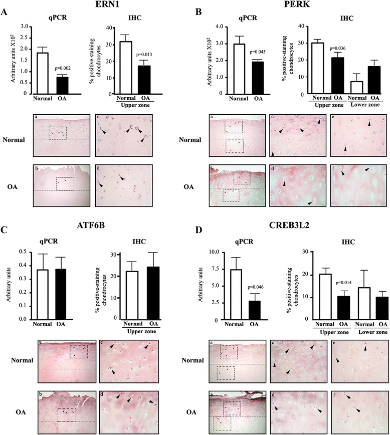
Fig. 1
Fig. 1
Expression/production of UPR genes in normal and osteoarthritic (OA) human chondrocytes and cartilage. mRNA levels (qPCR, normal [n = 6)] and OA [n = 6]) and protein production (immunohistochemistry [IHC], normal [n = 7] and OA [n = 7]) of (a) ERN1, (b) PERK, (c) ATF6B, and (d) CREB3L2. Illustrated are representative images of cartilage IHC. Data are expressed as mean ± SEM, and p values assessed by the unpaired t test comparing OA to normal chondrocytes. Magnification × 63 (a, b) and × 250 (c and d, upper cartilage zone; e and f, lower zone); boxes indicate where the magnifications were taken, and arrows indicate positively stained chondrocytes
Fig. 2
Fig. 2
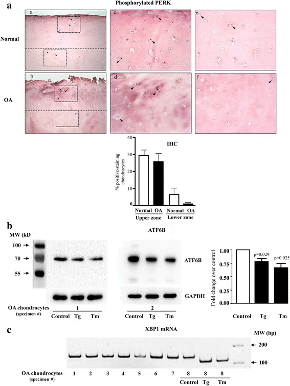
Basal activation status of the ER stress sensors in osteoarthritic (OA) chondrocytes. a The phosphorylation of PERK was determined by immunohistochemistry (IHC) in normal (n = 7) and OA (n = 7) cartilage. Illustrated are representative images of cartilage IHC and a graph of the data expressed as mean ± SEM. Statistical significance assessed by the unpaired t test comparing OA to normal chondrocytes showed no difference. Magnification × 63 (a, b) and × 250 (c and d, upper cartilage zone; e and f, lower zone); boxes indicate where the magnifications were taken, and arrows, the positively stained chondrocytes. b The uncleaved form (approximately 75 kDa) of ATF6B in OA chondrocytes (n = 6) treated or not (control) for 20 minutes with thapsigargin (Tg; 50 nM) and tunicamycin (Tm; 500 ng/ml) assessed by Western blot using an antibody that recognizes the C-terminus of the protein. Illustrated are representative Western blots with GAPDH as loading control and a graph of the densitometry analysis of the uncleaved ATF6B. Data are expressed as mean ± SEM, and p values assessed by the unpaired t test comparing OA to normal chondrocytes. c The presence of the spliced form of XBP1 mRNA was determined by PCR followed by gel electrophoresis (n = 8). In addition, OA chondrocyte sample #8 treated for 20 minutes with Tg (50 nM) and Tm (500 ng/ml) is shown as control for the ER stress activation
Fig. 3
Fig. 3
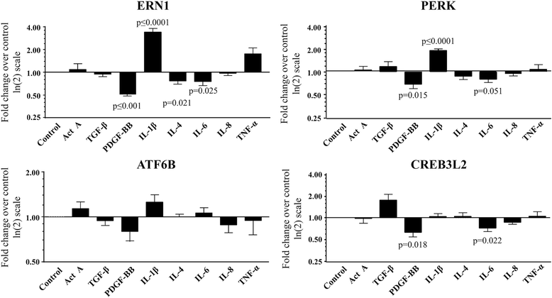
Regulation of UPR genes by cytokines/growth factors in human osteoarthritic chondrocytes under H-UPR conditions. Chondrocytes (n = 5–7) were treated with the following factors: Activin A (ActA), TGF-β, PDGF-BB, IL-4, IL-6, and IL-8 (all at 10 ng/ml), IL-1β (100 pg/ml), and TNF-α (5 ng/ml) and processed for RNA extraction and qPCR. Each control (untreated cells) was assigned an arbitrary value of 1 and the effect of the treatment evaluated as fold change over control. Data are expressed as mean ± SEM, and p values assessed by the one-sample t test, comparing the treated chondrocytes to the control
Fig. 4
Fig. 4
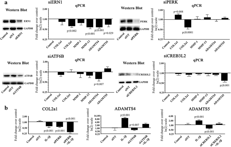
Effect of silencing UPR genes under H-UPR conditions in human osteoarthritic chondrocytes. a Silencing (si) ERN1, PERK, ATF6B, and CREB3L2 (Western blots), and the resulting expression of COL1a1, COL2a1, MMP-1, MMP-13, ADAMTS4, and ADAMTS5 (qPCR) (n = 7–10). The non-targeting or random siRNA (siNT) values were calculated for each primer as fold change over the control, which was assigned an arbitrary value of 1, and the mean ± SEM was 1.02 ± 0.03 for all the primers grouped. b Effect of IL-1β (100 pg/ml) with or without (control) siPERK, siATF6B, and siCREB3L2 on COL2a1, ADAMTS4, and ADAMTS5 levels (n = 5–7). Each control was assigned an arbitrary value of 1 and the effect of the treatment evaluated as fold change over control. Also represented are the siNTs for COL2a1, ADAMTS4, and ADAMTS5 respectively, showing values similar to those of the control. Data are expressed as mean ± SEM, and p values were assessed by the one-sample t test, comparing the treated chondrocytes to the control

References

    1. Wictome M, Henderson I, Lee AG, East JM. Mechanism of inhibition of the calcium pump of sarcoplasmic reticulum by thapsigargin. Biochem J. 1992;283(Pt 2):525–9. doi: 10.1042/bj2830525. - DOI - PMC - PubMed
    1. Reiling JH, Clish CB, Carette JE, Varadarajan M, Brummelkamp TR, Sabatini DM. A haploid genetic screen identifies the major facilitator domain containing 2A (MFSD2A) transporter as a key mediator in the response to tunicamycin. Proc Natl Acad Sci U S A. 2011;108(29):11756–65. doi: 10.1073/pnas.1018098108. - DOI - PMC - PubMed
    1. Bukau B, Weissman J, Horwich A. Molecular chaperones and protein quality control. Cell. 2006;125(3):443–51. doi: 10.1016/j.cell.2006.04.014. - DOI - PubMed
    1. Bernales S, Papa FR, Walter P. Intracellular signaling by the unfolded protein response. Annu Rev Cell Dev Biol. 2006;22:487–508. doi: 10.1146/annurev.cellbio.21.122303.120200. - DOI - PubMed
    1. Malhotra JD, Kaufman RJ. The endoplasmic reticulum and the unfolded protein response. Semin Cell Dev Biol. 2007;18(6):716–31. doi: 10.1016/j.semcdb.2007.09.003. - DOI - PMC - PubMed

Publication types