. 2016 Jul 19;7(4):e00699-16.
doi: 10.1128/mBio.00699-16.

Archaeal Haloarcula californiae Icosahedral Virus 1 Highlights Conserved Elements in Icosahedral Membrane-Containing DNA Viruses from Extreme Environments

Affiliations

Archaeal Haloarcula californiae Icosahedral Virus 1 Highlights Conserved Elements in Icosahedral Membrane-Containing DNA Viruses from Extreme Environments

Tatiana A Demina et al. mBio. .

Abstract

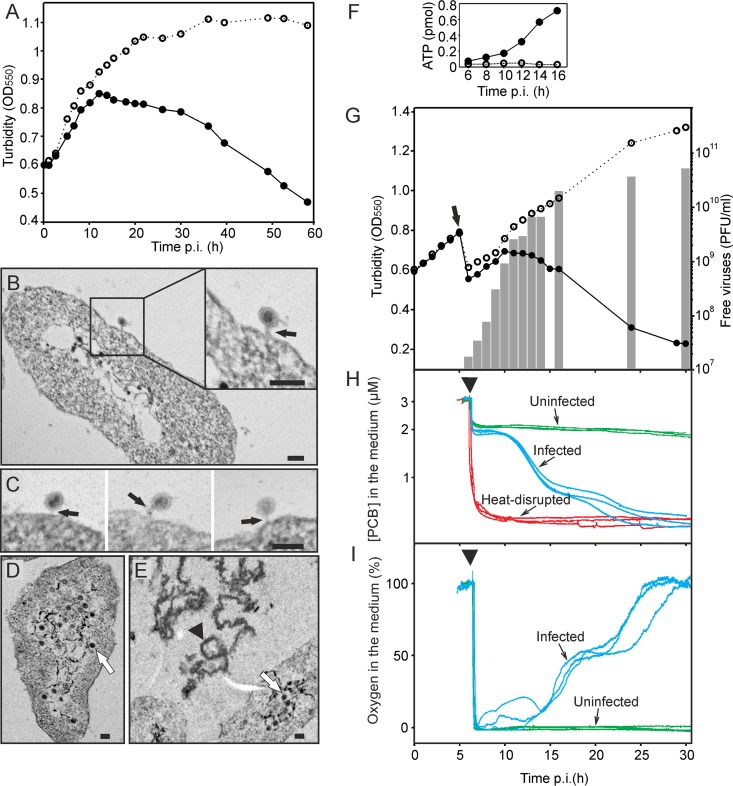
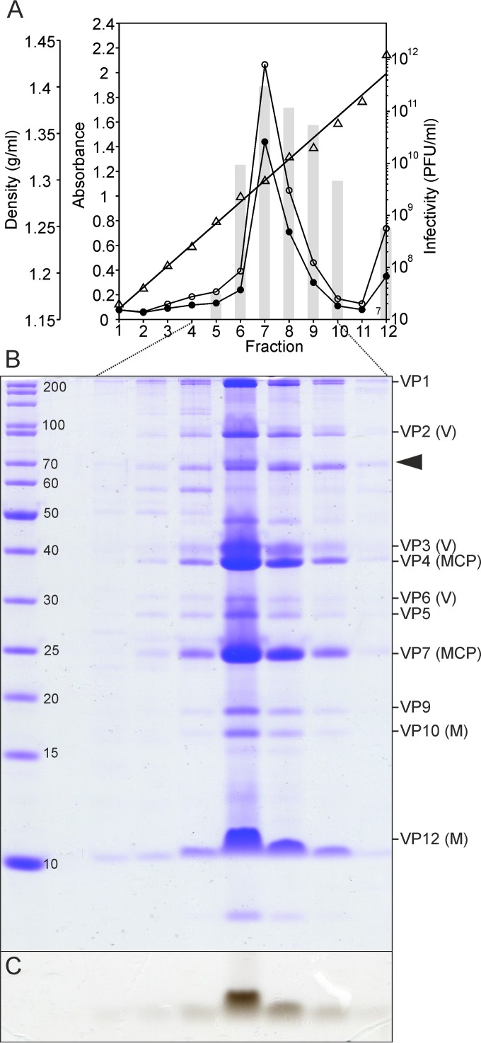
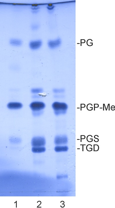
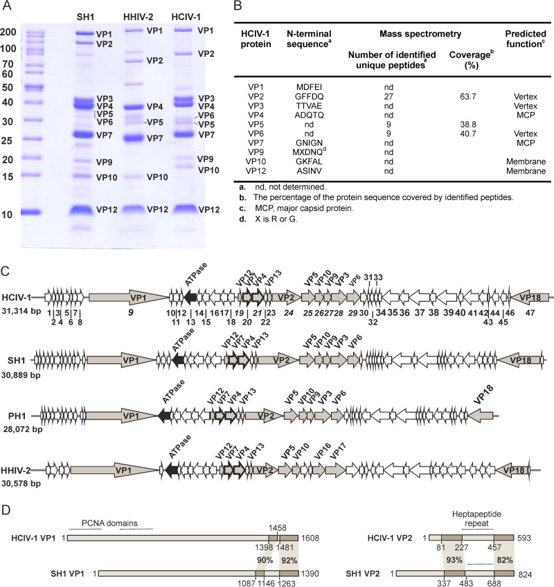
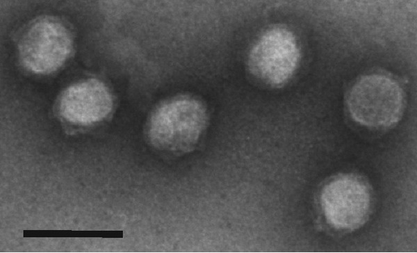
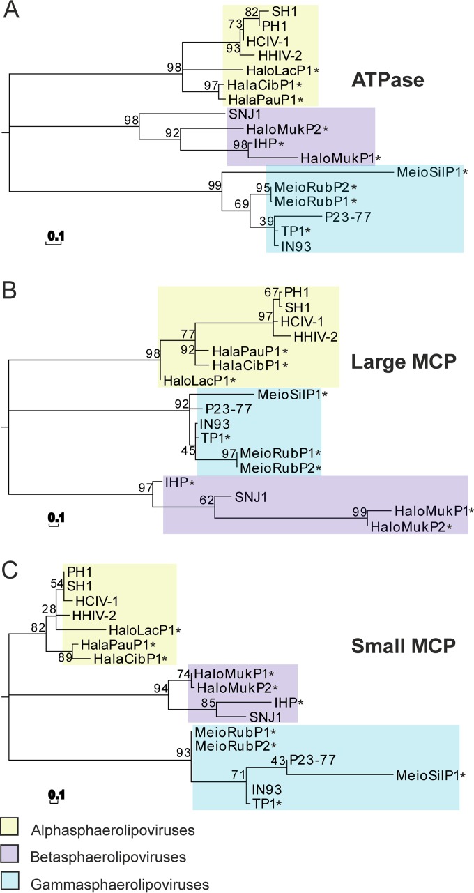
Despite their high genomic diversity, all known viruses are structurally constrained to a limited number of virion morphotypes. One morphotype of viruses infecting bacteria, archaea, and eukaryotes is the tailless icosahedral morphotype with an internal membrane. Although it is considered an abundant morphotype in extreme environments, only seven such archaeal viruses are known. Here, we introduce Haloarcula californiae icosahedral virus 1 (HCIV-1), a halophilic euryarchaeal virus originating from salt crystals. HCIV-1 also retains its infectivity under low-salinity conditions, showing that it is able to adapt to environmental changes. The release of progeny virions resulting from cell lysis was evidenced by reduced cellular oxygen consumption, leakage of intracellular ATP, and binding of an indicator ion to ruptured cell membranes. The virion contains at least 12 different protein species, lipids selectively acquired from the host cell membrane, and a 31,314-bp-long linear double-stranded DNA (dsDNA). The overall genome organization and sequence show high similarity to the genomes of archaeal viruses in the Sphaerolipoviridae family. Phylogenetic analysis based on the major conserved components needed for virion assembly-the major capsid proteins and the packaging ATPase-placed HCIV-1 along with the alphasphaerolipoviruses in a distinct, well-supported clade. On the basis of its virion morphology and sequence similarities, most notably, those of its core virion components, we propose that HCIV-1 is a member of the PRD1-adenovirus structure-based lineage together with other sphaerolipoviruses. This addition to the lineage reinforces the notion of the ancient evolutionary links observed between the viruses and further highlights the limits of the choices found in nature for formation of a virion.

Importance: Under conditions of extreme salinity, the majority of the organisms present are archaea, which encounter substantial selective pressure, being constantly attacked by viruses. Regardless of the enormous viral sequence diversity, all known viruses can be clustered into a few structure-based viral lineages based on their core virion components. Our description of a new halophilic virus-host system adds significant insights into the largely unstudied field of archaeal viruses and, in general, of life under extreme conditions. Comprehensive molecular characterization of HCIV-1 shows that this icosahedral internal membrane-containing virus exhibits conserved elements responsible for virion organization. This places the virus neatly in the PRD1-adenovirus structure-based lineage. HCIV-1 further highlights the limited diversity of virus morphotypes despite the astronomical number of viruses in the biosphere. The observed high conservation in the core virion elements should be considered in addressing such fundamental issues as the origin and evolution of viruses and their interplay with their hosts.


Figures

FIG 1
FIG 1
HCIV-1 infection cycle. (A) Growth curves of uninfected (open circles) and HCIV-1-infected (closed circles) Haloarcula californiae cultures. (B to E) Thin-section electron micrographs of HCIV-1-infected cells. (B and C) Virus particles attached to the cells at 5 h p.i. (black arrows) with tube-like structures between the viral particles and cells. (D) Intracellular virus particles at 8 h p.i. (white arrow). (E) Intracellular virus particles (white arrow) and cell debris (black arrowhead) indicating cell lysis at 12 h p.i. Scale bars in panels B to E, 100 nm. (F) Amount of extracellular ATP in the infected (closed circles) and uninfected (open circles) Haloarcula californiae cultures. (G) Growth curves of uninfected (open circles) and HCIV-1-infected (closed circles) Haloarcula californiae cells and the number of free progeny viruses in the infected culture (bars). The time point (5 h p.i.; arrow) when the cultures were washed to remove unadsorbed virus particles is indicated. (H) Binding of PCB to infected (blue lines), uninfected (green lines), and heat-disrupted (red lines) Haloarcula californiae cells measured starting at ~6 h p.i. (arrowhead) (n = 3) in the presence of PCB (calibration was performed with 3 μM PCB). (I) The relative concentrations (%) of dissolved oxygen in the medium of infected (blue lines) and uninfected (green lines) cultures measured starting at ~6 h p.i. (arrowhead). The MOI was 60 for the experiments represented in panels B and C; elsewhere, the MOI was 10. The cells were grown in flasks (A to E) or reaction vessels (F to I) at 37°C with aeration.
FIG 2
FIG 2
Protein and lipid content of HCIV-1 virions equilibrated in CsCl. (A) Density (triangles), infectivity (bars), A260 values (open circles), and A280 values (closed circles) of the CsCl gradient fractions. (B) Proteins from fractions 4 to 10 separated in polyacrylamide-Tricine-SDS gel stained with Coomassie blue. Molecular mass markers (in kilodaltons) (left lane) and HCIV-1 structural proteins (right lane) are shown. Virion proteins (VPs) and their predicted functions are indicated on the right (M, membrane proteins; MCP, major capsid proteins; V, vertex proteins). One host-derived impurity was identified by mass spectrometry (arrowhead). (C) Lipid signal detected below the 10-kDa protein marker after staining the separation gel with Sudan Black B.
FIG 3
FIG 3
HCIV-1 virion lipids. A thin-layer chromatogram of lipids extracted from the twice-purified HCIV-1 virions (lane 1), Haloarcula californiae cells (lane 2), and Haloarcula hispanica cells (lane 3) is shown. The major lipid species of Haloarcula hispanica are indicated on the right (7) as follows: PG, phosphatidylglycerol; PGP-Me, phosphatidylglycerophosphate methyl ester; PGS, phosphatidylglycerosulfate; TGD, triglycosyl glycerodiether.
FIG 4
FIG 4
HCIV-1 structural proteins and genome organization. (A) Virion proteins of highly purified SH1, HHIV-2, and HCIV-1 analyzed in a polyacrylamide-Tricine-SDS gel stained with Coomassie blue. Molecular mass markers are indicated in kilodaltons (left lane). (B) Identification of HCIV-1 structural proteins by N-terminal sequencing and mass spectrometry. (C) Comparison of the HCIV-1, SH1, PH1, and HHIV-2 genomes. The reading direction of genes/ORFs is indicated (arrows), and HCIV-1 gene/ORF numbers (1 to 47) are shown; also indicated are genes/ORFs encoding VPs (grey), putative ATPases (black), and MCPs VP4 and VP7 (thicker lines). Genes encoding structural proteins are labeled as VPs. (D) Comparison of proteins VP1 and VP2 of HCIV-1 and SH1. Amino acid coordinates and similarities (%) for conserved regions are shown. Proliferating cell nuclear antigen (PCNA) domains in HCIV-1 VP1, as well as heptapeptide regions in HCIV-1 and SH1 VP2 proteins, are highlighted with lines.
FIG 5
FIG 5
TEM micrographs of the twice-purified HCIV-1 virions stained with 3% (wt/vol) uranyl acetate (pH 4.5). Scale bar, 100 nm.
FIG 6
FIG 6
Phylogenetic analysis of conserved HCIV-1 proteins. Maximum likelihood phylogenetic trees of protein sequences of HCIV-1, sphaerolipoviruses, and related proviruses (marked with asterisks) are shown. (A) Putative ATPase. (B) Large MCP (VP4 in HCIV-1). (C) Small MCP (VP7 in HCIV-1). Evolutionary analysis was conducted using the JTT amino acid substitution model and 1,000 bootstrap values in MEGA 5.05. The bar (0.1) indicates the inferred number of substitutions per site.

References

    1. Pietilä MK, Demina TA, Atanasova NS, Oksanen HM, Bamford DH. 2014. Archaeal viruses and bacteriophages: comparisons and contrasts. Trends Microbiol 22:334–344. doi: 10.1016/j.tim.2014.02.007. - DOI - PubMed
    1. Benson SD, Bamford JK, Bamford DH, Burnett RM. 1999. Viral evolution revealed by bacteriophage PRD1 and human adenovirus coat protein structures. Cell 98:825–833. doi: 10.1016/S0092-8674(00)81516-0. - DOI - PubMed
    1. Maaty WS, Ortmann AC, Dlakić M, Schulstad K, Hilmer JK, Liepold L, Weidenheft B, Khayat R, Douglas T, Young MJ, Bothner B. 2006. Characterization of the archaeal thermophile Sulfolobus turreted icosahedral virus validates an evolutionary link among double-stranded DNA viruses from all domains of life. J Virol 80:7625–7635. doi: 10.1128/JVI.00522-06. - DOI - PMC - PubMed
    1. Happonen LJ, Redder P, Peng X, Reigstad LJ, Prangishvili D, Butcher SJ. 2010. Familial relationships in hyperthermo- and acidophilic archaeal viruses. J Virol 84:4747–4754. doi: 10.1128/JVI.02156-09. - DOI - PMC - PubMed
    1. Jäälinoja HT, Roine E, Laurinmäki P, Kivelä HM, Bamford DH, Butcher SJ. 2008. Structure and host-cell interaction of SH1, a membrane-containing, halophilic euryarchaeal virus. Proc Natl Acad Sci U S A 105:8008–8013. doi: 10.1073/pnas.0801758105. - DOI - PMC - PubMed

Publication types

LinkOut - more resources