. 2016 Jul 19;17(1):24.
doi: 10.1186/s12865-016-0154-z.

Cryptic protein-protein interaction motifs in the cytoplasmic domain of MHCI proteins

Affiliations

Cryptic protein-protein interaction motifs in the cytoplasmic domain of MHCI proteins

Karla K Frietze et al. BMC Immunol. .

Abstract

Background: Major histocompatibility complex class I (MHCI) proteins present antigenic peptides for immune surveillance and play critical roles in nervous system development and plasticity. Most MHCI are transmembrane proteins. The extracellular domain of MHCI interacts with immunoreceptors, peptides, and co-receptors to mediate immune signaling. While the cytoplasmic domain also plays important roles in endocytic trafficking, cross-presentation of extracellularly derived antigens, and CTL priming, the molecular mediators of cytoplasmic signaling by MHCI remain largely unknown.

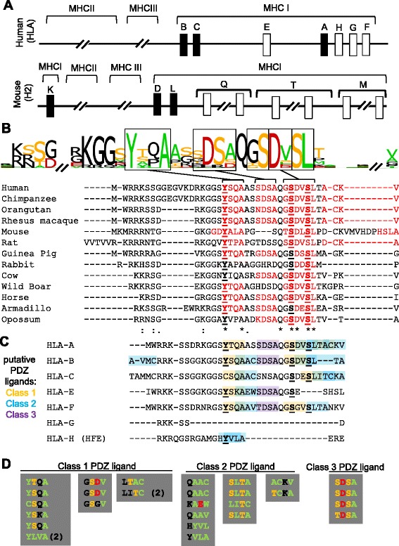
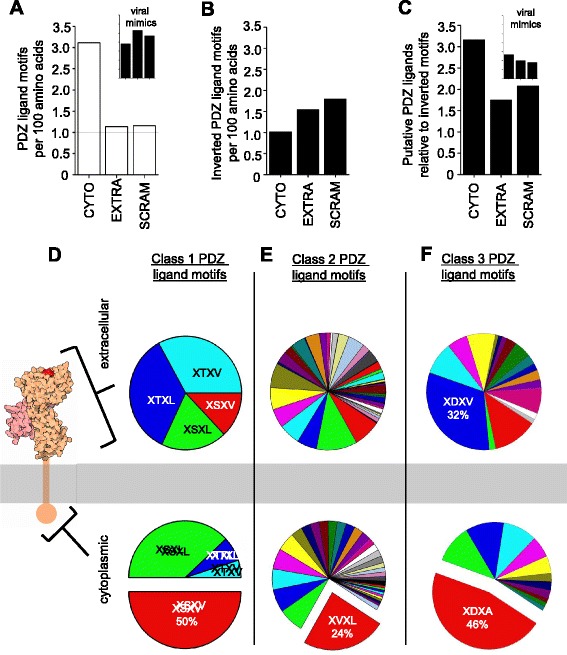
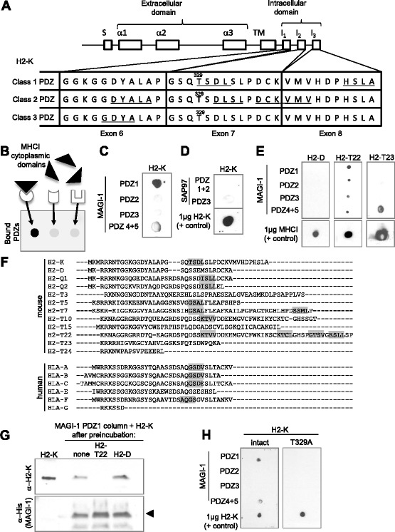
Results: Here we show that the cytoplasmic domain of MHCI contains putative protein-protein interaction domains known as PDZ (PSD95/disc large/zonula occludens-1) ligands. PDZ ligands are motifs that bind to PDZ domains to organize and mediate signaling at cell-cell contacts. PDZ ligands are short, degenerate motifs, and are therefore difficult to identify via sequence homology alone, but several lines of evidence suggest that putative PDZ ligand motifs in MHCI are under positive selective pressure. Putative PDZ ligands are found in all of the 99 MHCI proteins examined from diverse species, and are enriched in the cytoplasmic domain, where PDZ interactions occur. Both the position of the PDZ ligand and the class of ligand motif are conserved across species, as well as among genes within a species. Non-synonymous substitutions, when they occur, frequently preserve the motif. Of the many specific possible PDZ ligand motifs, a handful are strikingly and selectively overrepresented in MHCI's cytoplasmic domain, but not elsewhere in the same proteins. Putative PDZ ligands in MHCI encompass conserved serine and tyrosine residues that are targets of phosphorylation, a post-translational modification that can regulate PDZ interactions. Finally, proof-of-principle in vitro interaction assays demonstrate that the cytoplasmic domains of particular MHCI proteins can bind directly and specifically to PDZ1 and PDZ4&5 of MAGI-1, and identify a conserved PDZ ligand motif in the classical MHCI H2-K that is required for this interaction.

Conclusions: These results identify cryptic protein interaction motifs in the cytoplasmic domain of MHCI. In so doing, they suggest that the cytoplasmic domain of MHCI could participate in previously unsuspected PDZ mediated protein-protein interactions at neuronal as well as immunological synapses.

Keywords: Brain; Cytoplasmic; Immune; MHC class I; MHCI; PDZ; PDZ ligand; Scaffolding; Synapse.


Figures

Fig. 1
Fig. 1
MHCI gene family, and conservation of PDZ ligand motifs in MHCI cytoplasmic domains. a Greatly simplified schematic of the MHCI genomic region in humans (HLA, on chromosome 6, top) and mice (H2, on chromosome 17, bottom), showing the relative positions of the classical (black boxes) and non-classical (white boxes) MHCI genes or gene families. Multi-member gene clusters are indicated by breaks in the chromosome. Numerous genes and pseudogenes have been omitted for clarity, and distances are not to scale. Centromeric is to the left. Adapted from [62]. b Logo showing conservation of amino acids within closest-match homologues of the human classical MHCI HLA-A. The height of each letter corresponds to the extent of conservation across species. Color code: ST, orange; YFWCMVILA, green; DE, red; all others black. Below, individual source sequences. Putative PDZ ligand motifs are in red. Previously noted conserved serine and tyrosine residues that can be phosphorylated in some species (underlined in bold in b and c; see text) are embedded in PDZ ligand motifs in several MHCI proteins. Similar alignments have been performed previously (e.g., [15]). * = conserved; : = semi-conserved; . = similar. c Aligned amino acid sequences of the cytoplasmic domains of human MHCI proteins, with putative PDZ ligand motifs highlighted (class 1PDZ ligand, orange; class 2, blue; class 3, purple). Consensus motifs: class 1 PDZ, S/T-X-Y/F/W/C/M/V/I/L/A; class 2 PDZ, ΦXΦ (Y/F/W/C/M/V/I/L/A-X-Y/F/W/C/M/V/I/L/A); class 3 PDZ, D/E-X-Y/F/W/C/M/V/I/L/A [49]. Human reference sequences obtained from NCBI, alignments performed using T-Coffee [34]. d Conservation of PDZ ligands across human HLA genes from c, despite non-conservative substitutions. In two cases, a class 1 ligand motif is converted to class 2 by the substitution (2), but remains a putative PDZ ligand. Color code as in (b)
Fig. 2
Fig. 2
Enrichment of putative PDZ ligand motifs in the cytoplasmic domain of mouse MHCI proteins. a Frequency of the class 1 PDZ ligand consensus motif [X (S/T) X (V/L)] [51] in the cytoplasmic domain (“CYTO”), extracellular domain (“EXTRA”) or scrambled full sequence (“SCRAM”) of 20 mouse MHCI and MHCI-like proteins (see Methods for list), represented as motifs per 100 amino acids. PDZ ligand motifs are enriched in the cytoplasmic domain. Inset, frequency of PDZ ligand motifs in five viral MHCI mimics (UL-18, UL-37, UL-142, M144, and M153), which are likely to be under different selective pressures than host MHCI. Axis labels in insets match those in main graph and are omitted for clarity. b Frequency of inverted, non-functional class 1 PDZ ligand motifs [(V/L) X (S/T) X] in the same MHCI amino acid sequences. c PDZ ligand motifs in MHCI amino acid sequences, represented in relation to the number of inverted motifs in the same domain. Inset, same data from the five viral MHCI mimics used in (a). d Representation of four specific class 1 PDZ ligand consensus motifs (XSXV, XSXL, XTXV, XTXL) [51] in the extracellular (top) versus cytoplasmic (bottom) domains of 99 MHCI proteins from 21 species. Serine-containing motifs are significantly over-represented relative to threonine-containing motifs in the cytoplasmic domain. MHCI schematic, rcsb-pdb (http://pdb101.rcsb.org/motm/62). ef Representation of specific class 2 (ΦXΦ) and class 3 (X (D/E) X Φ) PDZ ligand motifs, respectively, where Φ = V/I/L/M/F/W/A/C. For a complete list of class 2 and class 3 ligands plotted and their frequencies, see Additional file 3: Figure S3. Color code is the same in both extracellular and cytoplasmic charts
Fig. 3
Fig. 3
MHCI can bind directly to PDZ domains in vitro. a Schematic showing the exons that encode the classical MHCI H2-Kb. A signal sequence (S) is followed by exons encoding the three extracellular alpha domains, a single exon encoding the transmembrane domain, and three exons (6, 7, and 8) encoding the intracellular domain (not drawn to scale). Bottom, amino acid sequence of exons 6, 7, and 8 in H2-Kb. Putative PDZ ligand motifs are underlined. Exon 6 encodes the overlapping ligand motifs GDYA, DYAL, and YALA, exon 7 encodes the overlapping ligand motifs TSDL and DLSL, and exon 8 encodes the C-terminal motif HSLA. Only the overlapping motifs DCKV and KVMV span an exon/intron boundary. Notably, KVMV is also among the least conserved putative PDZ ligand motifs in H2-Kb, since it is not detected in this position in any other mouse MHCI. Intronic structure, NCBI. Adapted from [15]. b Schematic of in vitro binding assay. Different recombinant PDZ domain peptides are bound to each membrane spot, and the membrane is panned with GST-tagged recombinant cytoplasmic domains from specific MHCI proteins. A direct interaction between the cytoplasmic domain of MHCI and a given PDZ domain will be apparent as a black spot after visualization of bound anti-GST antibodies. c The cytoplasmic domain of the classical MHCI H2-Kb can bind directly to PDZ1 and to a lesser extent PDZ4+5 of MAGI-1, but not to PDZs 2 or 3 of the same protein. See Additional file 4: Figure S4B&D for validation of the identity of H2-K peptides, and S4E-F for titration of H2-Kb binding. d The cytoplasmic domain of the classical MHCI H2-Kb does not bind to PDZ1+2 (expressed together) or PDZ 3 of SAP97. e MAGI-1 binding by recombinant cytoplasmic domains derived from different classical and nonclassical MHCI proteins. Notably, the ability to bind to PDZ1 of MAGI-1 correlates with the presence of a class 1 PDZ ligand motif, which is present in H2-Kb and H2-T22, but not H2-D or H2-T23 (see f). f Amino acid sequences of the cytoplasmic domains of mouse and human MHCIs, aligned in ClustalW. Highlighted, putative class 1 PDZ ligand motifs (consensus [X – S/T – X – V/L]) that match MAGI-1 PDZ1’s binding preferences [–82]. g Competition assays show that preincubation of column-bound MAGI-1 PDZ1 with cytoplasmic peptides derived from H2-T22, which can bind, but not H2-D, which cannot, precludes subsequent binding by peptides derived from H2-K. Left, pure H2-K not applied to a column; “none”, preincubation in buffer alone. H2-K was detected using an antibody against the cytoplasmic domain of H2-K (see Methods). Bottom, anti-His antibody shows eluted MAGI-1 peptide (arrow). h A point mutation in the lone class 1 PDZ ligand motif, TSDL, in the cytoplasmic domain of H2-Kb attenuates binding to MAGI-1 PDZ1. The location of the mutated residue (T329) is shown in (a)

Similar articles

Cited by

References

    1. Boulanger LM, Shatz CJ. Immune signalling in neural development, synaptic plasticity and disease. Nat Rev Neurosci. 2004;5(7):521–531. doi: 10.1038/nrn1428. - DOI - PubMed
    1. Shatz CJ. MHC class I: an unexpected role in neuronal plasticity. Neuron. 2009;64(1):40–45. doi: 10.1016/j.neuron.2009.09.044. - DOI - PMC - PubMed
    1. Boulanger LM. Immune proteins in brain development and synaptic plasticity. Neuron. 2009;64(1):93–109. doi: 10.1016/j.neuron.2009.09.001. - DOI - PubMed
    1. Elmer BM, McAllister AK. Major histocompatibility complex class I proteins in brain development and plasticity. Trends Neurosci. 2012;35(11):660–70. doi: 10.1016/j.tins.2012.08.001. - DOI - PMC - PubMed
    1. Huh GS, Boulanger LM, Du H, Riquelme PA, Brotz TM, Shatz CJ. Functional requirement for class I MHC in CNS development and plasticity. Science. 2000;290(5499):2155–2159. doi: 10.1126/science.290.5499.2155. - DOI - PMC - PubMed

Publication types

MeSH terms

LinkOut - more resources