. 2016 Jul 20:6:29589.
doi: 10.1038/srep29589.

Tert-butylhydroquinone lowers blood pressure in AngII-induced hypertension in mice via proteasome-PTEN-Akt-eNOS pathway

Affiliations

Tert-butylhydroquinone lowers blood pressure in AngII-induced hypertension in mice via proteasome-PTEN-Akt-eNOS pathway

Bing-Can Xu et al. Sci Rep. .

Retraction in

Abstract

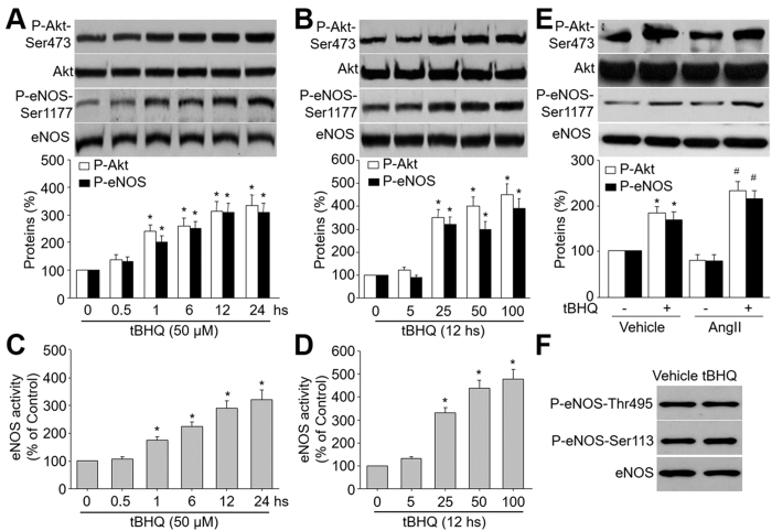
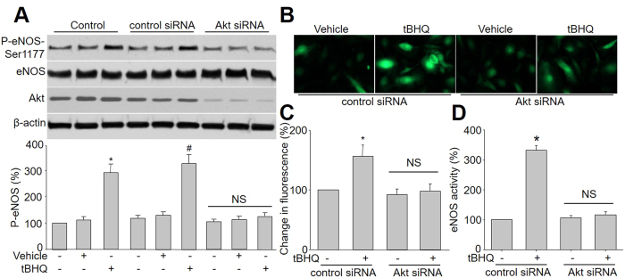
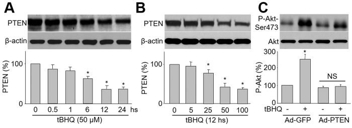
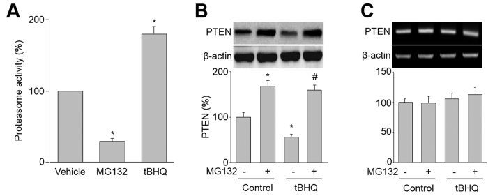
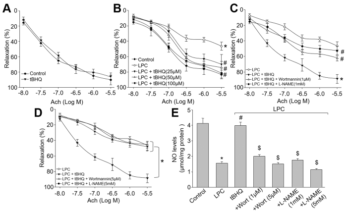
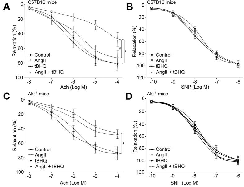
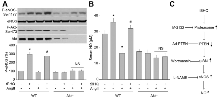
Tert-butylhydroquinone (tBHQ), as an antioxidant, has been widely used for many years to prevent oxidization of food products. The aim of this study was to investigate whether tBHQ activates endothelial nitric oxide synthase (eNOS) to prevent endothelial dysfunction and lower blood pressure. The role of Akt in tBHQ-induced eNOS phosphorylation was examined in human umbilical vein endothelial cells (HUVEC) or in mice. tBHQ treatment of HUVEC increased both Akt-Ser473 phosphorylation, accompanied with increased eNOS-Ser1177 phosphorylation and NO release. Mechanically, pharmacologic or genetic inhibition of Akt abolished tBHQ-enhanced NO release and eNOS phosphorylation in HUVEC. Gain-function of PTEN or inhibition of 26S proteasome abolished tBHQ-enhanced Akt phosphorylation in HUVEC. Ex vivo analysis indicated that tBHQ improved Ach-induced endothelium-dependent relaxation in LPC-treated mice aortic arteries, which were abolished by inhibition of Akt or eNOS. In animal study, administration of tBHQ significantly increased eNOS-Ser1177 phosphorylation and acetylcholine-induced vasorelaxation, and lowered AngII-induced hypertension in wildtype mice, but not in mice deficient of Akt or eNOS. In conclusion, tBHQ via proteasome-dependent degradation of PTEN increases Akt phosphorylation, resulting in upregulation of eNOS-derived NO production and consequent improvement of endothelial function in vivo. In this way, tBHQ lowers blood pressure in hypertensive mice.


Figures

Figure 1
Figure 1. tBHQ activates eNOS and Akt in HUVEC.
(A) HUVEC were treated with 50 μM tBHQ for the indicated times. The levels of Akt and eNOS phosphorylations in total cell lysates were analyzed by western blot. The blot is a representative of three blots obtained from separated experiments. Data presented are means ± SEM from 3 independent experiments. *P < 0.05 vs. control group (point 0). (B) HUVEC were treated with varying concentrations of tBHQ for 12 hours. The levels of Akt and eNOS phosphorylations in total cell lysates were analyzed by western blot. The blot is a representative of three blots obtained from separated experiments. Data presented are means ± SEM from 3 independent experiments. *P < 0.05 vs. control group (point 0). (C) eNOS activity was assessed by citrulline assay in (A). (D) eNOS activity was assessed by citrulline assay in (B). (E) HUVEC were pretreated with tBHQ (50 μM) for 30 minutes followed by AngII (1 μM) for 12 hours. Total cell lysates were analyzed by western blot for the indicated proteins. Data presented are means ± SEM from 3 independent experiments. *P < 0.05 vs. vehicle alone. #P < 0.05 vs. tBHQ alone. (F) HUVEC were treated with tBHQ (50 μM) for 12 hours. Total cell lysates were analyzed by western blot for the indicated proteins. The blot is a representative of three blots obtained from separated experiments.
Figure 2
Figure 2. Akt mediates tBHQ-induced eNOS phosphorylation and NO production in cultured endothelial cells.
(A) HUVEC were infected with control or Akt siRNA for 48 hours. Then cells were exposed to tBHQ at 25 μM for 6 hours. Total cell lysates were analyzed by western blot for the indicated proteins. The blot is a representative of four blots obtained from four separated experiments. Data presented are means ± SEM from 4 independent experiments. *P < 0.05 vs. control group. #P < 0.05 vs. control siRNA alone. NS indicates no significance. (B–D) HUVEC infected with control or Akt siRNA for 48 hours. The NO production was determined by measuring the intensity of DAF fluorescence in (B) and quantitative analysis of NO amount was shown in (C). Total cell lysates were subject to assay eNOS activity (D). Data presented are means ± SEM from 4 independent experiments. *P < 0.05 vs. control siRNA group. NS indicates no significance.
Figure 3
Figure 3. tBHQ induces Akt phosphorylation through PTEN reduction.
(A) HUVEC were incubated with 25 μM tBHQ for various amounts of time. After the appropriate incubation time, cells were lysed and PTEN protein level was measured by western blot. The blot is a representative of four blots obtained from four separated experiments. Data presented are means ± SE from 4 independent experiments. *P < 0.05 vs. control groups. (B) HUVEC were treated with varying concentrations of tBHQ for 6 hours. Total cell lysates were analyzed by western blot for the indicated proteins. The blot is a representative of three blots obtained from separated experiments. Data presented are means ± SEM from 3 independent experiments. *P < 0.05 vs. control groups. (C) HUVEC were infected with Ad-PTEN-CA or Ad-vector (control) prior to tBHQ stimulation. The blot is a representative of four blots obtained from four separate experiments. Results are expressed as mean ± SEM from four independent experiments. *P < 0.05 vs. control groups. NS indicates no significance.
Figure 4
Figure 4. tBHQ increases 26S-proteasome-dependent PTEN degradation in HUVEC.
(A) HUVEC were treated with tBHQ (25 μM) or MG132 (1 μM) for 6 hours. 26S proteasome activity was assayed in cell lysates. Results are expressed as mean ± SEM from four independent experiments. *P < 0.05 vs. control groups. (B,C) HUVEC were treated with tBHQ (25 μM) with or without MG132 (1 μM) for 6 hours. (B) The protein level of PTEN was assayed by western blot. (C) The mRNA level of PTEN was determined by RT-PCR. Results are expressed as mean ± SEM from four independent experiments. *P < 0.05 vs. control groups. #P < 0.05 vs. tBHQ alone. NS indicates no significance.
Figure 5
Figure 5. Inhibition of Akt or eNOS by pharmacological reagents abolishes tBHQ-prevented endothelial dysfunction ex vivo.
(A) The isolated mouse aortic rings were incubated with tBHQ (50 μM) for 12 hours and Ach-induced endothelium-dependent relaxation was assayed by organ chamber. (B) The isolated mouse aortic rings were preincubated with tBHQ (25, 50, 100 μM) for 30 minutes and then exposed to LPC (4 mg/l) for 2 hours. Ach-induced endothelium-dependent relaxation was assayed by organ chamber. *P < 0.05 vs. control, #P < 0.05 vs. LPC. (C) Isolated mouse aortic rings were preincubated with tBHQ (50 μM) for 30 minutes with or without wortmannin (1 μM) and L-NAME (1 mM) followed by LPC (4 mg/l) for 2 hours. The endothelium-dependent relaxation induced by acetylcholine was assayed by organ chamber. *P < 0.05 vs. LPC, #P < 0.05 vs. tBHQ. (D) Isolated mouse aortic rings were preincubated with tBHQ (50 μM) for 30 minutes with or without wortmannin (5 μM) and L-NAME (5 mM) followed by LPC (4 mg/l) for 2 hours. The endothelium-dependent relaxation induced by acetylcholine was assayed by organ chamber. *P < 0.05 vs. LPC, #P < 0.05 vs. tBHQ. (E) Homogenates of aortic tissues were subjected to measure NO productions by the Griess method in each group. *P < 0.05 vs. Control group. #P < 0.05 vs. LPC. $P < 0.05 vs. LPC + tBHQ.
Figure 6
Figure 6. Deficiency of Akt abrogates tBHQ-induced improvement of endothelial dysfunction in AngII-infused mice.
WT and Akt−/− mice at the age of 8–12 weeks old were fed with normal diet containing tBHQ a ratio of 1% (w/w) for 2 weeks days prior to AngII infusion for another 14 days. Aortas from mice were cut into rings and were mounted in organ chamber to detect vessel bioactivity. The relaxation was induced acetylcholine (Ach) or SNP. (A) Endothelium-dependent relaxation of the aortic rings in response to Ach from WT mice. (B) Endothelium-independent relaxation of the aortic rings in response to SNP from WT mice. (C) Ach-induced endothelium-dependent relaxation in Akt−/− mice. (D) SNP-induced endothelium-independent relaxation in Akt−/− mice. Each data point represents relaxation expressed as a percentage of the value obtained for phenylephrine-preconstricted aorta. All data were expressed as mean ± SEM. One aortic ring was isolated from each mouse. 10–15 mice in each group. *P < 0.05 vs. Control WT or Akt−/− group. #P < 0.05 vs. AngII in WT mice.
Figure 7
Figure 7. tBHQ lowers AngII-induced hypertension in wildtype (WT) mice, but not in hypertensive Akt−/− mice.
WT, Akt−/−, and eNOS−/− mice at the age of 8–12 weeks old were fed with normal diet containing tBHQ a ratio of 1% (w/w) for 2 weeks days prior to AngII infusion. Blood pressure (BP) was monitored by telemetry, as described in Materials and Methods. (A) Systolic BP and (B) diastolic BP in WT mice, (C) systolic BP and (D) diastolic BP in Akt−/− mice, and (E) systolic BP and (F) diastolic BP in eNOS−/− mice were analyzed. Quantitative results are expressed as mean ± SEM. N is 10–15 in each group. *P < 0.05 vs. Control WT or Akt−/− mice. #P < 0.05 vs. AngII mice.
Figure 8
Figure 8. tBHQ via Akt activation increases NO production in hypertensive mice.
(A,B) WT and Akt−/− mice at the age of 8–12 weeks old were fed with normal diet containing tBHQ a ratio of 1% (w/w) for 4 weeks days prior to AngII infusion for 14 days. (A) Homogenates of aortic tissues were subjected to perform western blot to assay the levels of P-eNOS and P-Akt. (B) Serum NO level was also analyzed by the Griess method. All data were expressed as mean ± SEM. 10–15 mice in each group. *P < 0.05 vs. Control WT mice. #P < 0.05 vs. AngII mice. NS indicates no significant. (C) Proposed mechanism of tBHQ on vascular function.

Similar articles

Cited by

References

    1. Wang S. et al.. Acute inhibition of guanosine triphosphate cyclohydrolase 1 uncouples endothelial nitric oxide synthase and elevates blood pressure. Hypertension 52, 484–490 (2008). - PMC - PubMed
    1. Xu J., Wang S., Wu Y., Song P. & Zou M. H. Tyrosine nitration of PA700 activates the 26S proteasome to induce endothelial dysfunction in mice with angiotensin II-induced hypertension. Hypertension 54, 625–632 (2009). - PMC - PubMed
    1. Wang S., Xu J., Song P., Viollet B. & Zou M. H. In vivo activation of AMP-activated protein kinase attenuates diabetes-enhanced degradation of GTP cyclohydrolase I. Diabetes 58, 1893–1901 (2009). - PMC - PubMed
    1. Dimmeler S. et al.. Activation of nitric oxide synthase in endothelial cells by Akt-dependent phosphorylation. Nature 399, 601–605 (1999). - PubMed
    1. Fulton D. et al.. Regulation of endothelium-derived nitric oxide production by the protein kinase Akt. Nature 399, 597–601 (1999). - PMC - PubMed

Publication types

MeSH terms