. 2016 Jul 20:6:29952.
doi: 10.1038/srep29952.

Glutathione peroxidase 3 localizes to the epithelial lining fluid and the extracellular matrix in interstitial lung disease

Affiliations

Glutathione peroxidase 3 localizes to the epithelial lining fluid and the extracellular matrix in interstitial lung disease

Andrea C Schamberger et al. Sci Rep. .

Abstract

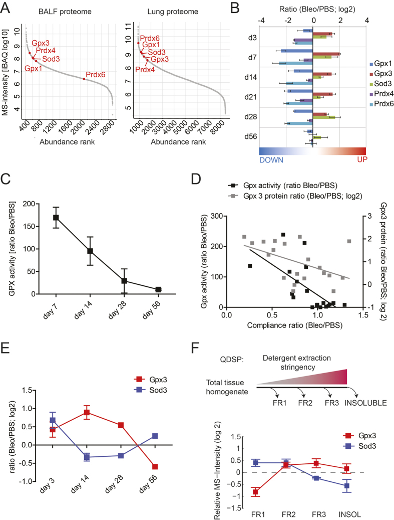
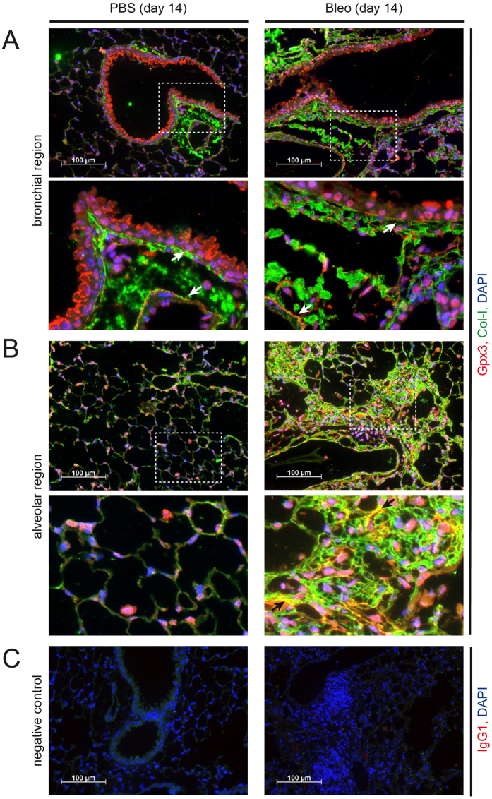
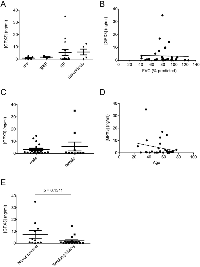
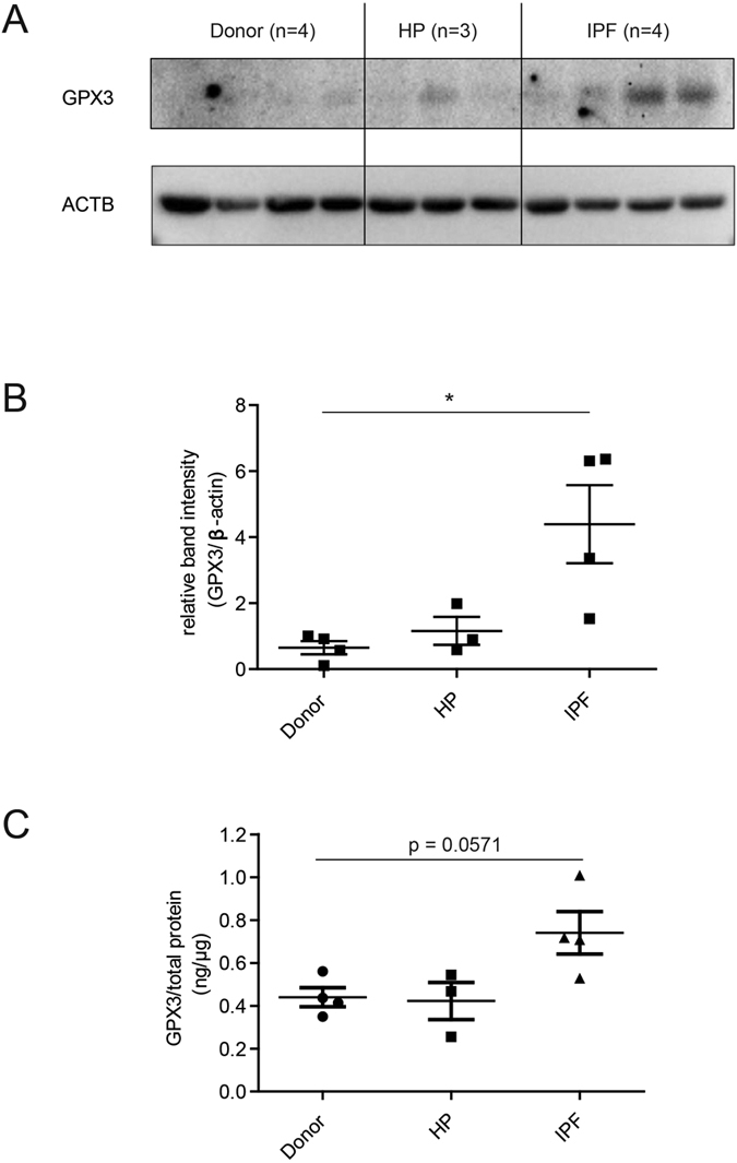
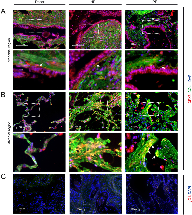
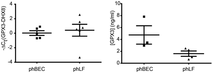
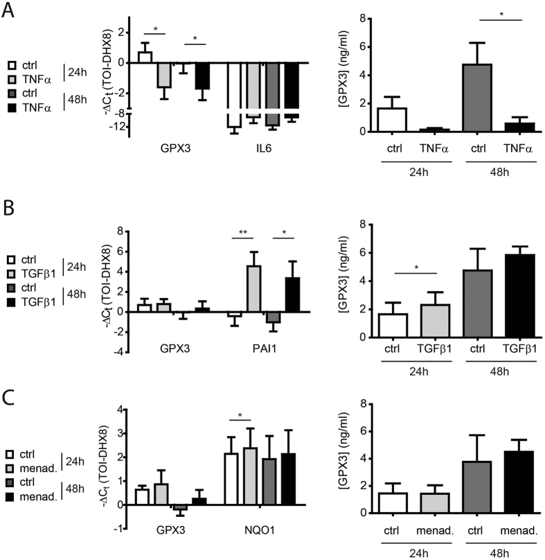
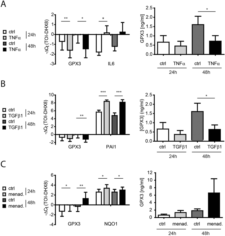
Aberrant antioxidant activity and excessive deposition of extracellular matrix (ECM) are hallmarks of interstitial lung diseases (ILD). It is known that oxidative stress alters the ECM, but extracellular antioxidant defence mechanisms in ILD are incompletely understood. Here, we extracted abundance and detergent solubility of extracellular antioxidant enzymes from a proteomic dataset of bleomycin-induced lung fibrosis in mice and assessed regulation and distribution of glutathione peroxidase 3 (GPX3) in murine and human lung fibrosis. Superoxide dismutase 3 (Sod3), Gpx3, and Gpx activity were increased in mouse BALF during bleomycin-induced lung fibrosis. In lung tissue homogenates, Gpx3, but not Sod3, was upregulated and detergent solubility profiling indicated that Gpx3 associated with ECM proteins. Immunofluorescence analysis showed that Gpx3 was expressed by bronchial epithelial cells and interstitial fibroblasts and localized to the basement membrane and interstitial ECM in lung tissue. As to human ILD samples, BALF of some patients contained high levels of GPX3, and GPX3 was upregulated in lung homogenates from IPF patients. GPX3 expression in primary human bronchial epithelial cells and lung fibroblasts was downregulated by TNF-α, but more variably regulated by TGF-β1 and menadione. In conclusion, the antioxidant enzyme GPX3 localizes to lung ECM and is variably upregulated in ILD.


Figures

Figure 1
Figure 1. Of all antioxidant proteins known to associate with the extracellular compartment, only Gpx3 is upregulated both in BALF and tissue during bleomycin-induced lung fibrosis and enriched in the matrisome fraction.
(A) Label-free proteome quantification by iBAQ shows relative abundance of the proteins in BALF (left-hand panel) and the lung proteome (right-hand panel) under basal conditions, i.e. in control mice. (B) Bar graph showing regulation of major lung antioxidant proteins during bleomycin-induced lung fibrosis. (C) Change of selenium-dependent Gpx activity in BALF during bleomycin-induced lung fibrosis. Activity ratios (i.e. Gpx activity in the Bleomycin group relative to PBS control) are shown for each time point. (D) Correlation of normalized compliance values with normalized Gpx3 protein levels as extracted from Schiller et al. (grey boxes) and with Gpx activity (black boxes, data from C) in the time course of bleomycin-induced lung fibrosis. Here, data from all time points were used and correlated. (E) Regulation of Gpx3 and Sod3 in tissue during bleomycin-induced lung fibrosis. (F) Quantitative Detergent Solubility Profiling (QDSP) shows enrichment of Gpx3, but not Sod3, in the insoluble fractions. The depicted data is derived from the bleomycin group at day 14. Data in A, B, E and F were extracted from a recently acquired proteomic data set by Schiller et al..
Figure 2
Figure 2. Gpx3 is secreted by bronchial epithelial cells and localizes to ECM structures in control (PBS) and bleomycin (Bleo) instilled mouse lung.
Immunofluorescence analysis of PBS (left-hand panels) and Bleo (right-hand panels) mouse lungs at day 14 after instillation. Representative images with Gpx3 (red), Col-I (green), and DAPI (blue) are shown from bronchial (A) and alveolar regions (B) together with an isotype control for Gpx3 staining (C). Scale bar: 100 μm. Arrows in the higher magnification inserts indicate colocalization of Gpx3 with ECM structures: Subendothelial and subepithelial basement membrane (A) and interstitial ECM (B, only right-hand panel).
Figure 3
Figure 3. Increased GPX3 levels are detected in BALF from some patients with HP and sarcoidosis, but do not significantly correlate with lung function, gender, age, or smoking history.
(A) GPX3 protein levels in BALF from patients with various ILD (n = 30) including IPF (n = 7), SRIF (n = 3), HP (n = 15), and sarcoidosis (n = 5) as measured by ELISA. GPX3 levels did not correlate with FVC (B), nor gender (C), nor age (D), nor the patients’ smoking history (E). For the latter, a non-significant trend for higher levels in never smokers is observed.
Figure 4
Figure 4. GPX3 protein is upregulated in total tissue homogenate from IPF lung.
(A) Western Blot analysis of total lung tissue homogenate showed upregulation of GPX3 in patients with IPF (n = 4), but not HP (n = 3), relative to donor samples (n = 4). (B) Densitometric analysis of the Western Blot showed that GPX3 upregulation in IPF is significant. (C) ELISA using an independent antibody confirms upregulation of GPX3 in the same samples. Data shown are mean ± SEM and two-tailed Mann-Whitney test was used for statistical analysis (*p < 0.05). ACTB, β-actin as loading control.
Figure 5
Figure 5. GPX3 is secreted by bronchial epithelial cells and localizes to ECM structures in donor, HP and IPF patients.
Representative immunofluorescence analysis of donor, HP and IPF patients tissue with GPX3 (red), COL-I (green), and DAPI (blue) are depicted from bronchial (A) and alveolar regions (B) together with an isotype control for GPX3 staining (C). Scale bar: 100 μm. Arrows indicate co-localization of GPX3 with ECM structures: Subepithelial basement membrane (A, higher magnification inserts) and fibrotic interstitial ECM (B, higher magnification inserts).
Figure 6
Figure 6. GPX3 is expressed by differentiated primary human bronchial epithelial cells (phBEC) and primary human lung fibroblasts (phLF) in culture.
Left panel: Baseline GPX3 transcript levels in phBEC and phLF normalized to DHX8 transcript levels. Right panel: GPX3 protein levels in cell culture supernatant as measured by ELISA.
Figure 7
Figure 7. Expression of glutathione peroxidase 3 (GPX3) is downregulated by TNF-α, moderately upregulated by TGF-β1, but not consistently regulated by the oxidative stress inducer menadione in differentiated primary human bronchial epithelial cells (phBEC).
(A) Left panel: GPX3 transcript levels in differentiated phBEC after treatment with 10 ng/ml TNF-α for 24 and 48 hours. Interleukin-6 (IL6) transcript is used as a positive control for TNF-α activity. Right panel: GPX3 protein levels in cell culture supernatant in response to TNF-α, as measured by ELISA. (B) Left panel: GPX3 transcript levels in differentiated phBEC after treatment with 2 ng/ml TGF-β1 for 24 and 48 hours. Plasminogen activator inhibitor 1 (PAI1) transcript is used as a positive control for TGF-β1 activity. Right panel: GPX3 protein levels in cell culture supernatant in response to TGF-β1, as measured by ELISA. (C) Left panel: GPX3 and NQO1 transcript levels in differentiated phBEC after treatment with 10 μM menadione for 24 and 48 hours. Right panel: GPX3 protein levels in cell culture supernatant in response to menadione, as measured by ELISA. The presented data are based on three independent experiments (phBEC isolated from three different donors) and given as mean ± SD. Statistical analysis was performed using paired t-test. *p < 0.1; **p < 0.01; ***p < 0.001.
Figure 8
Figure 8. Expression of glutathione peroxidase 3 (GPX3), is downregulated by TGF-β1 and TNF-α, but upregulated by the oxidative stress inducer menadione in primary human lung fibroblasts (phLF).
(A) Left panel: GPX3 transcript levels in phLF after treatment with 10 ng/ml TNF-α for 24 and 48 hours. Interleukin-6 (IL6) transcript is used as a positive control for TNF-α activity. Right panel: GPX3 protein levels in cell culture supernatant in response to TNF-α, as measured by ELISA. (B) Left panel: GPX3 transcript levels in phLF after treatment with 2 ng/ml TGF-β1 for 24 and 48 hours. Plasminogen activator inhibitor 1 (PAI1) transcript is used as a positive control for TGF-β1 activity. Right panel: GPX3 protein levels in cell culture supernatant in response to TGF-β1, as measured by ELISA. (C) Left panel: GPX3 and NQO1 transcript levels in differentiated phBEC after treatment with 10 μM menadione for 24 and 48 hours. Right panel: GPX3 protein levels in cell culture supernatant in response to menadione, as measured by ELISA. The presented data are based on four independent experiments (phLF isolated from four different donors) and given as mean ± SD. Statistical analysis was performed using paired t-test. *p < 0.1; **p < 0.01; ***p < 0.001.

Similar articles

Cited by

References

    1. Grunes D. & Beasley M. B. Hypersensitivity pneumonitis: a review and update of histologic findings. J. Clin. Pathol. 66, 888–895, doi: 10.1136/jclinpath-2012-201337 (2013). - DOI - PubMed
    1. Selman M., Pardo A. & King T. E. Jr. Hypersensitivity pneumonitis: insights in diagnosis and pathobiology. Am. J. Respir. Crit. Care Med. 186, 314–324, doi: 10.1164/rccm.201203-0513CI (2012). - DOI - PubMed
    1. Katzenstein A. L. Smoking-related interstitial fibrosis (SRIF), pathogenesis and treatment of usual interstitial pneumonia (UIP), and transbronchial biopsy in UIP. Mod. Pathol. 25 Suppl 1, S68–78, doi: 10.1038/modpathol.2011.154 (2012). - DOI - PubMed
    1. Valeyre D. et al.. Sarcoidosis. Lancet 383, 1155–1167, doi: 10.1016/S0140-6736(13)60680-7 (2014). - DOI - PubMed
    1. Patterson K. C. & Strek M. E. Pulmonary fibrosis in sarcoidosis. Clinical features and outcomes. Ann. Am. Thorac. Soc. 10, 362–370, doi: 10.1513/AnnalsATS.201303-069FR (2013). - DOI - PubMed

Publication types

MeSH terms