. 2016 Nov;151(5):923-932.
doi: 10.1053/j.gastro.2016.07.009. Epub 2016 Jul 18.

Antibiotics Suppress Activation of Intestinal Mucosal Mast Cells and Reduce Dietary Lipid Absorption in Sprague-Dawley Rats

Affiliations

Antibiotics Suppress Activation of Intestinal Mucosal Mast Cells and Reduce Dietary Lipid Absorption in Sprague-Dawley Rats

Hirokazu Sato et al. Gastroenterology. 2016 Nov.

Abstract

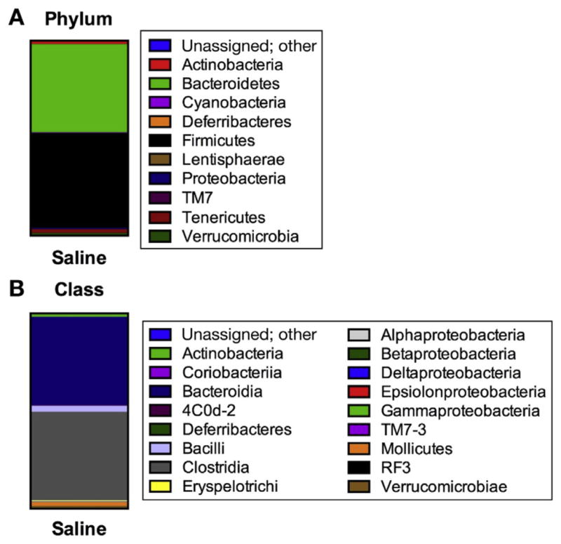
Background & aims: The gut microbiota affects intestinal permeability and mucosal mast cells (MMCs) responses. Activation of MMCs has been associated with absorption of dietary fat. We investigated whether the gut microbiota contributes to the fat-induced activation of MMCs in rats, and how antibiotics might affect this process.

Methods: Adult male Sprague-Dawley rats were given streptomycin and penicillin for 4 days (n = 6-8) to reduce the abundance of their gut flora, or normal drinking water (controls, n = 6-8). They underwent lymph fistula surgery and after an overnight recovery were given an intraduodenal bolus of intralipid. We collected intestinal tissues and lymph fluid and assessed activation of MMCs, intestinal permeability, and fat transport parameters.

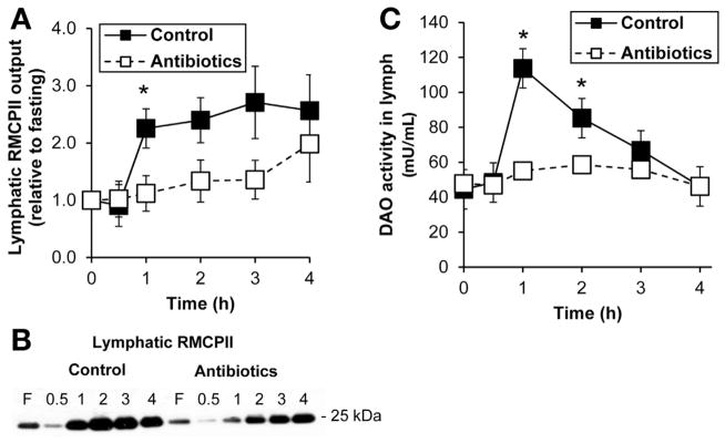
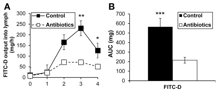
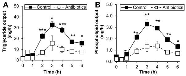
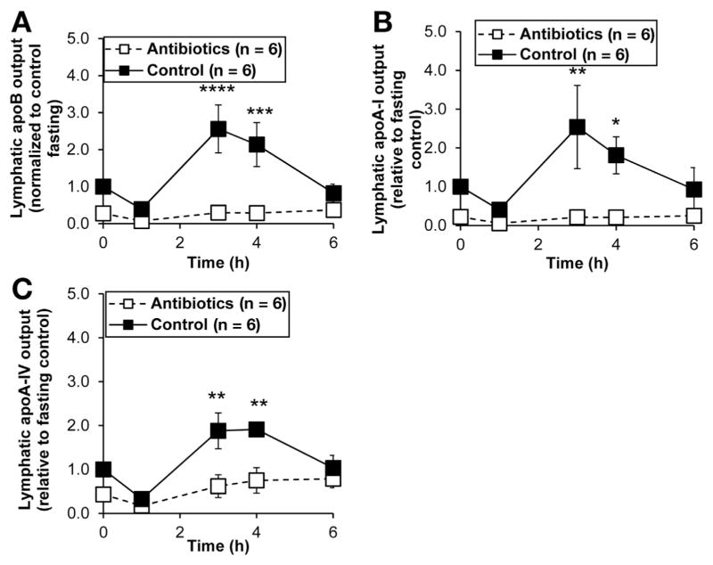
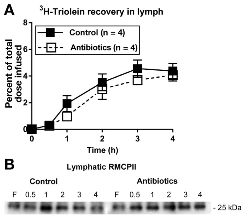
Results: Compared with controls, intestinal lymph from rats given antibiotics had reduced levels of mucosal mast cell protease II (produced by MMCs) and decreased activity of diamine oxidase (produced by enterocytes) (P < .05). Rats given antibiotics had reduced intestinal permeability in response to dietary lipid compared with controls (P < .01). Unexpectedly, antibiotics also reduced lymphatic transport of triacylglycerol and phospholipid (P < .01), concomitant with decreased levels of mucosal apolipoproteins B, A-I, and A-IV (P < .01). No differences were found in intestinal motility or luminal pancreatic lipase activity between rats given antibiotics and controls. These effects were not seen with an acute dose of antibiotics or 4 weeks after the antibiotic regimen ended.

Conclusions: The intestinal microbiota appears to activate MMCs after the ingestion of fat in rats; this contributes to fat-induced intestinal permeability. We found that the gut microbiome promotes absorption of lipid, probably by intestinal production of apolipoproteins and secretion of chylomicrons.

Keywords: APOA-I; APOA-IV; Digestion; Microbe.


Conflict of interest statement

Conflicts of interest

The authors disclose no conflicts.

Figures

Figure 1
Figure 1
Antibiotic treatment suppressed mucosal mast cell activation. (A) Lymphatic RMCPII output in saline-treated control (n = 8) and antibiotic-treated (n = 8) rats given an intraduodenal infusion of LiposynIII. (B) A representative Western blot showing RMCPII output in lymph. (C) Lymph DAO activity between the 2 groups (n = 8 each). Data are expressed as mean ± SEM; *P < .05.
Figure 2
Figure 2
Antibiotic treatment reduced intestinal permeability in response to dietary lipid. (A) Lymphatic output of FITC-labeled dextran to measure intestinal permeability in control (n = 6) and antibiotic-treated (n = 10) rats given Liposyn III. (B) Total (area under the curve [AUC]) lymph FITC levels over 4 hours. Data are expressed as mean ± SEM; *P < .05; **P < .01; ***P < .001.
Figure 3
Figure 3
Antibiotics treatment reduced the lymphatic outputs of (A) triglycerides and (B) phospholipids. Saline-treated control (n = 8) and antibiotic-treated (n = 8) rats were given an intraduodenal infusion of LiposynIII lipid. Data are expressed as mean ± SEM; *P < .05; ** P < .01; ***P < .001.
Figure 4
Figure 4
Antibiotic treatment suppressed the intestinal outputs of (A) ApoB, (B) ApoA-I, and (C) ApoA-IV into the mesenteric lymph. Saline-treated control (n = 6) and antibiotic-treated (n = 6) rats were given an intraduodenal infusion of LiposynIII. Data are expressed as mean ± SEM. *P < .05; **P < .01; ***P < .005; ****P < .001.
Figure 5
Figure 5
Antibiotic treatment did not affect (A) intestinal motility and (B) luminal fat digestion. To look at effects of antibiotics on intestinal motility, lymph fistula animals given either antibiotics (n = 7) or saline (n = 6) for 4 days were given an intraduodenal bolus of intralipid labeled with 3H triolein. Radioactivities from luminal contents of the intestinal segments (L1, duodenum; L2 and L3, jejunum; L4, ileum) were measured by liquid scintillation. To measure luminal digestion, luminal pancreatic lipase activity was assayed. Lymph-intact animals equipped with duodenal tubes were treated for 4 days with (n = 4) or without antibiotics (n = 4). On the experimental day, a lipid bolus was administered intraduodenally and, after 4 hours, the luminal contents of small intestinal segments were assayed for lipase activity.
Figure 6
Figure 6
Restoring the microbiota after antibiotic treatment restored (A) fat absorption and (B) MMC activation as measured by RMCPII output into lymph. After the 4-day antibiotic treatment regimen (n = 4) and saline as a control (n = 4), the animals were allowed to recover for 4 weeks before performing experiments.
Figure 7
Figure 7
Gut microbial composition depicting phyla (A) and class (B) of saline-treated control animals. Antibiotic treatment resulted in no amplification of the 16S rRNA gene and thus we were unable to assess fecal microbiota composition. Taxonomic structure of 16S rRNA gene was assessed using the Illumina Mi-Seq platform and results analyzed using Quantitative Insights Into Microbial Ecology software.

Similar articles

Cited by

References

    1. Savage DC. Microbial ecology of the gastrointestinal tract. Annu Rev Microbiol. 1977;31:107–133. - PubMed
    1. Forbes EE, Groschwitz K, Abonia JP, et al. IL-9- and mast cell-mediated intestinal permeability predisposes to oral antigen hypersensitivity. J Exp Med. 2008;205:897–913. - PMC - PubMed
    1. O’Sullivan M, Clayton N, Breslin NP, et al. Increased mast cells in the irritable bowel syndrome. Neurogastroenterol Motil. 2000;12:449–457. - PubMed
    1. Farhadi A, Fields JZ, Keshavarzian A. Mucosal mast cells are pivotal elements in inflammatory bowel disease that connect the dots: stress, intestinal hyperpermeability and inflammation. World J Gastroenterol. 2007;13:3027–3030. - PMC - PubMed
    1. Garrett WS, Gordon JI, Glimcher LH. Homeostasis and inflammation in the intestine. Cell. 2010;140:859–870. - PMC - PubMed

Publication types

MeSH terms