Review
. 2016 Oct;151(4):616-32.
doi: 10.1053/j.gastro.2016.07.008. Epub 2016 Jul 18.

Inflammation and the Intestinal Barrier: Leukocyte-Epithelial Cell Interactions, Cell Junction Remodeling, and Mucosal Repair

Affiliations
Review

Inflammation and the Intestinal Barrier: Leukocyte-Epithelial Cell Interactions, Cell Junction Remodeling, and Mucosal Repair

Anny-Claude Luissint et al. Gastroenterology. 2016 Oct.

Abstract

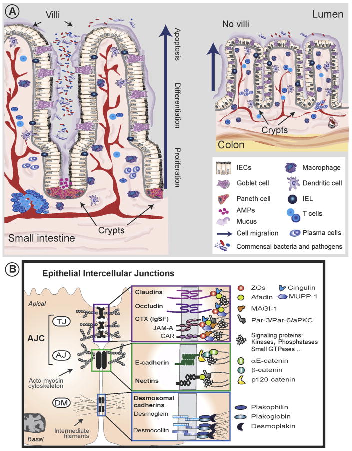
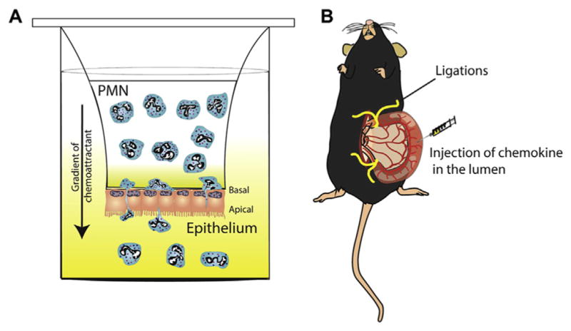
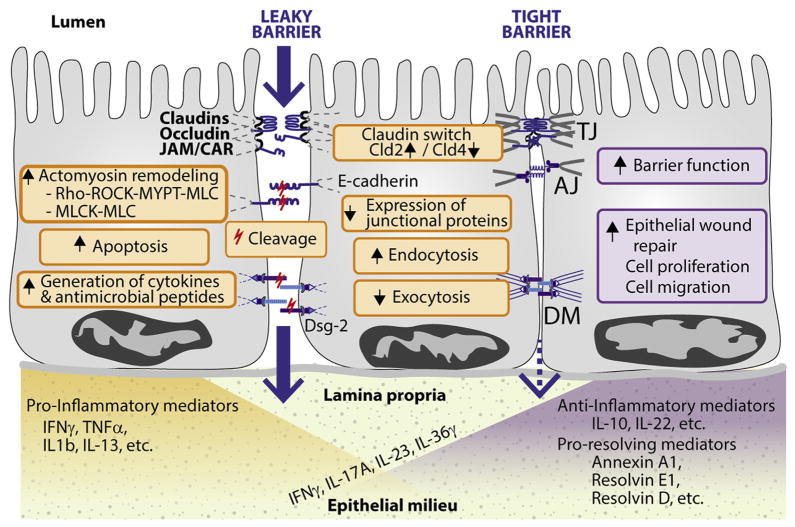
The intestinal tract is lined by a single layer of columnar epithelial cells that forms a dynamic, permeable barrier allowing for selective absorption of nutrients, while restricting access to pathogens and food-borne antigens. Precise regulation of epithelial barrier function is therefore required for maintaining mucosal homeostasis and depends, in part, on barrier-forming elements within the epithelium and a balance between pro- and anti-inflammatory factors in the mucosa. Pathologic states, such as inflammatory bowel disease, are associated with a leaky epithelial barrier, resulting in excessive exposure to microbial antigens, recruitment of leukocytes, release of soluble mediators, and ultimately mucosal damage. An inflammatory microenvironment affects epithelial barrier properties and mucosal homeostasis by altering the structure and function of epithelial intercellular junctions through direct and indirect mechanisms. We review our current understanding of complex interactions between the intestinal epithelium and immune cells, with a focus on pathologic mucosal inflammation and mechanisms of epithelial repair. We discuss leukocyte-epithelial interactions, as well as inflammatory mediators that affect the epithelial barrier and mucosal repair. Increased knowledge of communication networks between the epithelium and immune system will lead to tissue-specific strategies for treating pathologic intestinal inflammation.

Keywords: Epithelium; Inflammation; Mucosal Barrier; Mucosal Repair.


Conflict of interest statement

Conflicts of interest

The authors disclose no conflicts.

Figures

Figure 1
Figure 1
Structure of the intestinal mucosa and intercellular junction complexes. (A) The intestinal mucosal barrier is composed of a monolayer of columnar epithelial cells that is separated from luminal contents by mucus and is in close contact with a lamina propria containing immune cells, capillaries, and lymphatic vessels. In the small intestine, the epithelium is folded to form luminal structures termed villi and crypts, while the colon lacks villi and only has crypts. The epithelium is continuously renewed with progenitor stem cells in the crypt base that proliferate, differentiate, and migrate along the crypt–luminal axis followed by regulated cell death at the luminal surface. The epithelium includes absorptive enterocytes, mucus-producing goblet cells, enteroendocrine cells, and antimicrobial peptides-producing Paneth cells. AMPs, antimicrobial peptides; IEL, intraepithelial lymphocyte. (B) Epithelial intercellular junctional complexes consist of an apical junctional complex (comprising TJs and subjacent AJs) followed by DMs. The TJ and AJ is associated with an underlying perijunctional actin-myosin cytoskeleton, while DM associates with intermediate filaments. TJ, AJ, and DM complexes include transmembrane proteins that associate with TJ-associated scaffold and signaling molecules as indicated. aPKC, atypical protein kinase C; CLMP, CAR-like membrane protein; MAGI-1, membrane-associated guanylate kinase; Par-3/Par-6, partitioning defective 3/-6.
Figure 2
Figure 2
In vitro and in vivo models of neutrophil trans-epithelial migration. (A) Schematic of an in vitro model of PMN TEpM across a monolayer of intestinal epithelial cells in response to a chemoattractant (such as N-formyl-L-methionyl-L-leucyl-L-phenylalanine peptides, leukotriene B4 or CXCL1). Epithelial cells are grown on collagen-coated Transwell filters with pore sizes sufficient for PMNs to migrate through and in an inverted conformation designed to model the physiologically relevant polarity of migration, from basolateral to apical. Transmigrated PMN are collected in the bottom of the well and quantified. (B) In vivo “ileal loop” model of PMN TEpM. A vascularized 2–4 cm long ileal portion of the small intestine near the ileo–cecal junction is exteriorized and ligated at both ends without compromising the blood supply. Chemoattractant is injected into the lumen to induce PMN migration. The loop content is collected and PMN quantified by flow cytometry, while ileal loop mucosa can be analyzed histologically.
Figure 3
Figure 3
Multistep model of neutrophil transepithelial migration across intestinal mucosa. Migration of neutrophils out of blood vessels and across the epithelium during mucosal inflammation is a multistep process involving many surface adhesion molecules on the epithelium and neutrophils: (1) basolateral adhesion, (2) transmigration, and (3) apical adhesion. In addition, neutrophils secrete proteases that promote cleavage of E-cadherin, desmoglein-2, and JAML. TJs contains CAR, which binds to JAML. AJs contain E-cadherin. DMs contain desmoglein-2 (Dsg-2). ADAM10, Disintegrin and metalloproteinase domain containing protein 10; AMP, adenosine monophosphate; DC, dendritic cell; ICAM-1, intercellular adhesion molecule-1; Mϕ, macrophage; MMP, matrix metalloprotease; NETs, neutrophil extracellular traps; SIRPα, signal-regulatory protein α.
Figure 4
Figure 4
Schematic representation of the epithelial responses to pro- and anti-inflammatory cytokines. The imbalance between pro-inflammatory and anti-inflammatory cytokines is a hallmark of chronic gut inflammation, such as in inflammatory bowel disease. Overall, pro-inflammatory cytokines increase intestinal paracellular permeability, while anti-inflammatory cytokines and pro-resolving mediators stimulate epithelial repair and barrier function. Interestingly, some pro-inflammatory mediators, such as IL17A, IL23, IL36γ, and IFNγ have been reported to promote barrier protective responses (see text). Molecular mechanisms to be identified. Dsg-2, desmoglein-2.

Similar articles

Cited by

References

    1. Helander HF, Fandriks L. Surface area of the digestive tract—revisited. Scand J Gastroenterol. 2014;49:681–689. - PubMed
    1. Abraham C, Medzhitov R. Interactions between the host innate immune system and microbes in inflammatory bowel disease. Gastroenterology. 2011;140:1729–1737. - PMC - PubMed
    1. Ordas I, Eckmann L, Talamini M, et al. Ulcerative colitis. Lancet. 2012;380:1606–1619. - PubMed
    1. Katz KD, Hollander D, Vadheim CM, et al. Intestinal permeability in patients with Crohn’s disease and their healthy relatives. Gastroenterology. 1989;97:927–931. - PubMed
    1. Peeters M, Geypens B, Claus D, et al. Clustering of increased small intestinal permeability in families with Crohn’s disease. Gastroenterology. 1997;113:802–807. - PubMed

Publication types

MeSH terms