

Age-associated up-regulation of EGR1 promotes granulosa cell apoptosis during follicle atresia in mice through the NF-κB pathway
- PMID: 27436181
- PMCID: PMC5105915
- DOI: 10.1080/15384101.2016.1208873
Age-associated up-regulation of EGR1 promotes granulosa cell apoptosis during follicle atresia in mice through the NF-κB pathway
Abstract
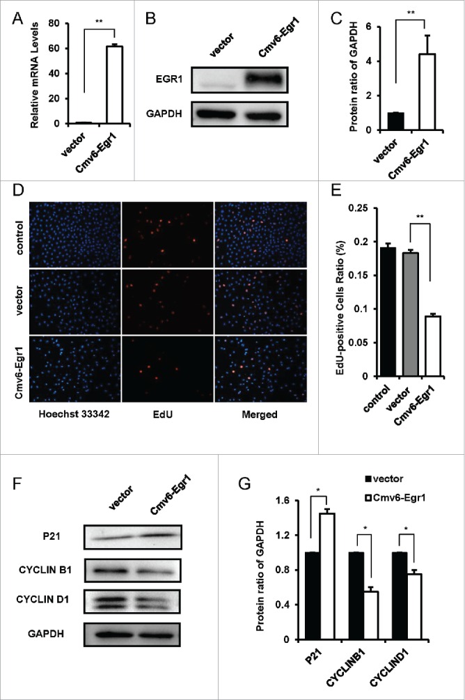
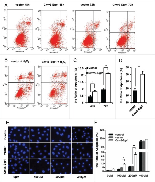
Follicular atresia is the main process responsible for the loss of follicles and oocytes from the ovary, and it is the root cause of ovarian aging. Apoptosis of granulosa cells (GCs) is the cellular mechanism responsible for follicular atresia in mammals. Recent advances have highlighted fundamental roles for EGR1 in age-related diseases via the induction of apoptosis. In the present study, we found that the expression of EGR1 was significantly increased in aged mouse ovaries compared with young ovaries. Immunohistochemical analysis revealed strongly positive EGR1 staining in atretic follicles, especially in apoptotic granulosa cells. We further showed that EGR1 up-regulation in mouse primary granulosa cells inhibited cell proliferation and promoted apoptosis. In addition, the promotion of apoptosis in GCs by EGR1 increases over time and with reactive oxygen species (ROS) stimulation. Our mechanistic study suggested that EGR1 regulates GC apoptosis in a mitochondria-dependent manner and that this mainly occurs through the NF-κB signaling pathway. In conclusion, our results suggested that age-related up-regulation of EGR1 promotes GC apoptosis in follicle atresia during ovarian aging.
Keywords: EGR1; NF-κB; apoptosis; follicular atresia; granulosa cell; ovarian aging.
Figures


Similar articles
-
Transcriptome profiling of granulosa cells from bovine ovarian follicles during atresia.BMC Genomics. 2014 Jan 18;15:40. doi: 10.1186/1471-2164-15-40. BMC Genomics. 2014. PMID: 24438529 Free PMC article.
-
Screening of key regulatory factors in apoptosis of granulosa cells in healthy and atretic pig follicles based on RNA-seq.Gene. 2025 Aug 5;959:149514. doi: 10.1016/j.gene.2025.149514. Epub 2025 Apr 17. Gene. 2025. PMID: 40250537
-
miR-361-5p Mediates SMAD4 to Promote Porcine Granulosa Cell Apoptosis through VEGFA.Biomolecules. 2020 Sep 4;10(9):1281. doi: 10.3390/biom10091281. Biomolecules. 2020. PMID: 32899767 Free PMC article.
-
Regulation of cell death and cell survival gene expression during ovarian follicular development and atresia.Front Biosci. 2003 Jan 1;8:d222-37. doi: 10.2741/949. Front Biosci. 2003. PMID: 12456353 Review.
-
Follicular atresia in pigs: measurement and physiology.J Anim Sci. 1995 Sep;73(9):2834-44. doi: 10.2527/1995.7392834x. J Anim Sci. 1995. PMID: 8582874 Review.
Cited by
-
Using bioinformatics and metabolomics to identify altered granulosa cells in patients with diminished ovarian reserve.PeerJ. 2020 Aug 28;8:e9812. doi: 10.7717/peerj.9812. eCollection 2020. PeerJ. 2020. PMID: 32923184 Free PMC article.
-
Aberrant Transferrin and Ferritin Upregulation Elicits Iron Accumulation and Oxidative Inflammaging Causing Ferroptosis and Undermines Estradiol Biosynthesis in Aging Rat Ovaries by Upregulating NF-Κb-Activated Inducible Nitric Oxide Synthase: First Demonstration of an Intricate Mechanism.Int J Mol Sci. 2022 Oct 21;23(20):12689. doi: 10.3390/ijms232012689. Int J Mol Sci. 2022. PMID: 36293552 Free PMC article.
-
Environmental toxicant induced epigenetic transgenerational inheritance of ovarian pathology and granulosa cell epigenome and transcriptome alterations: ancestral origins of polycystic ovarian syndrome and primary ovarian insufiency.Epigenetics. 2018;13(8):875-895. doi: 10.1080/15592294.2018.1521223. Epub 2018 Oct 2. Epigenetics. 2018. PMID: 30207508 Free PMC article.
-
α-SNAP is expressed in mouse ovarian granulosa cells and plays a key role in folliculogenesis and female fertility.Sci Rep. 2017 Sep 18;7(1):11765. doi: 10.1038/s41598-017-12292-9. Sci Rep. 2017. PMID: 28924180 Free PMC article.
-
Egr-1 is a key regulator of the blood-brain barrier damage induced by meningitic Escherichia coli.Cell Commun Signal. 2024 Jan 17;22(1):44. doi: 10.1186/s12964-024-01488-y. Cell Commun Signal. 2024. PMID: 38233877 Free PMC article.
References
-
- Kaipia A, Hsueh AJ. Regulation of ovarian follicle atresia. Ann Rev Physiol 1997; 59:349-63; PMID:9074768; http://dx.doi.org/10.1146/annurev.physiol.59.1.349 - DOI - PubMed
-
- Persani L, Rossetti R, Cacciatore C. Genes involved in human premature ovarian failure. J Mol Endocrinol 2010; 45:257-79; PMID:20668067; http://dx.doi.org/10.1677/JME-10-0070 - DOI - PubMed
-
- Faddy MJ. Follicle dynamics during ovarian ageing. Mol Cell Endocrinol 2000; 163:43-8; PMID:10963872; http://dx.doi.org/10.1016/S0303-7207(99)00238-5 - DOI - PubMed
-
- Tilly JL, Kowalski KI, Johnson AL, Hsueh AJ. Involvement of apoptosis in ovarian follicular atresia and postovulatory regression. Endocrinology 1991; 129:2799-801; PMID:1718732; http://dx.doi.org/10.1210/endo-129-5-2799 - DOI - PubMed
-
- Shen M, Lin F, Zhang J, Tang Y, Chen WK, Liu H. Involvement of the up-regulated FoxO1 expression in follicular granulosa cell apoptosis induced by oxidative stress. J Biol Chem 2012; 287:25727-40; PMID:22669940; http://dx.doi.org/10.1074/jbc.M112.349902 - DOI - PMC - PubMed
MeSH terms
Substances
LinkOut - more resources
Full Text Sources
Other Literature Sources
Medical
Miscellaneous