. 2016 Jul 20:7:12235.
doi: 10.1038/ncomms12235.

SIRT7 is a histone desuccinylase that functionally links to chromatin compaction and genome stability

Affiliations

SIRT7 is a histone desuccinylase that functionally links to chromatin compaction and genome stability

Lei Li et al. Nat Commun. .

Abstract

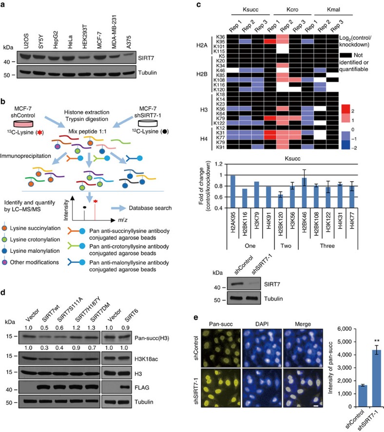
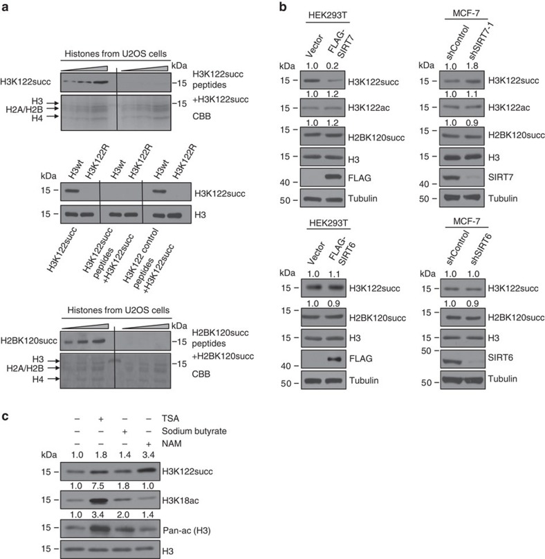
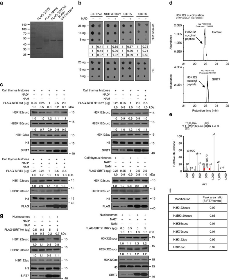
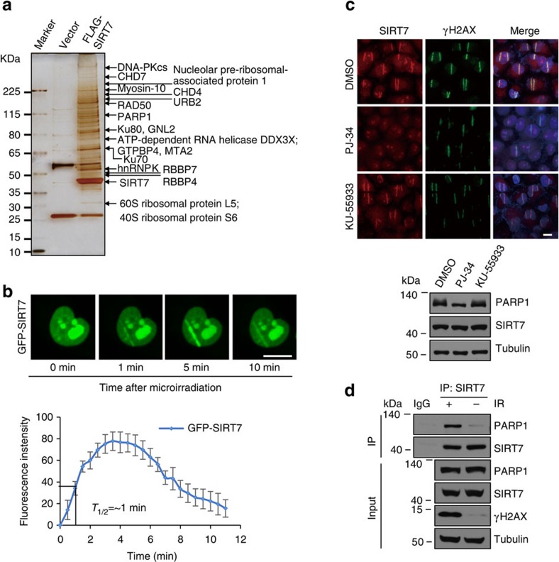
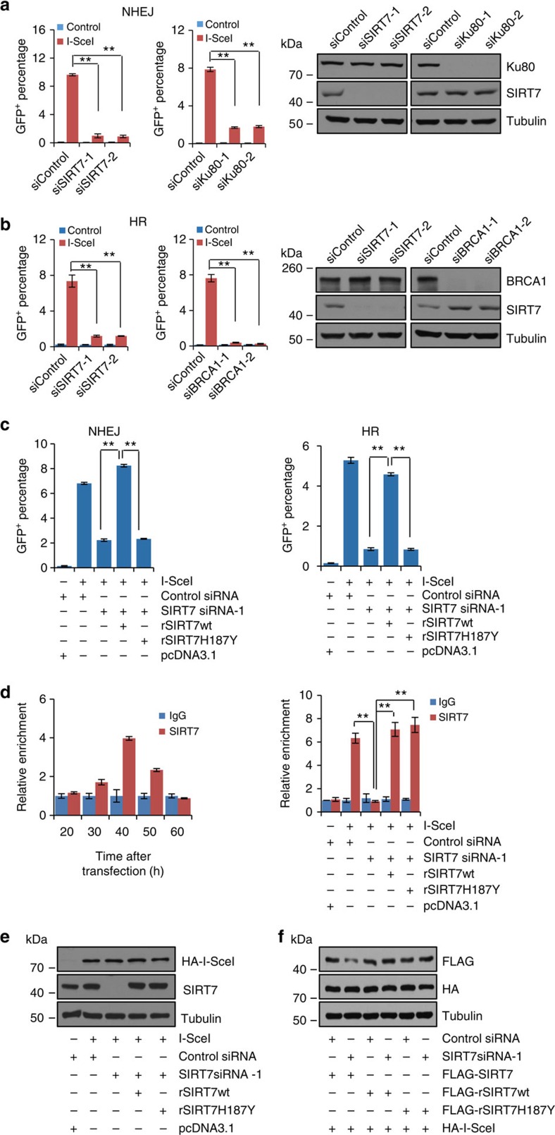
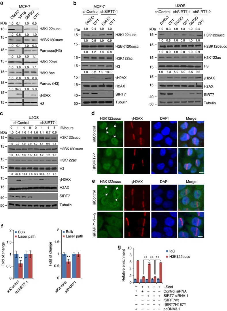
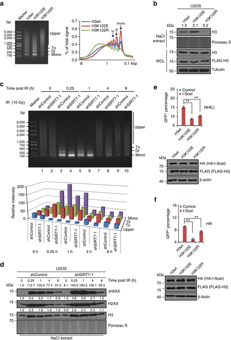
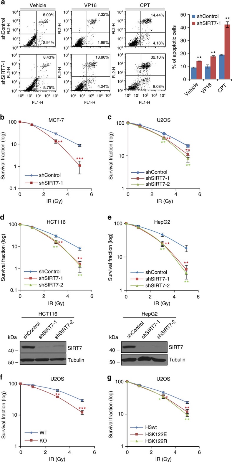
Although SIRT7 is a member of sirtuin family proteins that are described as NAD(+)-dependent class III histone deacetylases, the intrinsic enzymatic activity of this sirtuin protein remains to be investigated and the cellular function of SIRT7 remains to be explored. Here we report that SIRT7 is an NAD(+)-dependent histone desuccinylase. We show that SIRT7 is recruited to DNA double-strand breaks (DSBs) in a PARP1-dependent manner and catalyses desuccinylation of H3K122 therein, thereby promoting chromatin condensation and DSB repair. We demonstrate that depletion of SIRT7 impairs chromatin compaction during DNA-damage response and sensitizes cells to genotoxic stresses. Our study indicates SIRT7 is a histone desuccinylase, providing a molecular basis for the understanding of epigenetic regulation by this sirtuin protein. Our experiments reveal that SIRT7-catalysed H3K122 desuccinylation is critically implemented in DNA-damage response and cell survival, providing a mechanistic insight into the cellular function of SIRT7.


Figures

Figure 1
Figure 1. SIRT7 regulates histone succinylation.
(a) Western blotting analysis of SIRT7 expression in different cell lines. (b) The workflow of the integrated SILAC labelling, affinity enrichment and mass spectrometry-based quantitative proteomics to quantify dynamic changes of histone lysine crotonylation (Kcro), succinylation (Ksucc) and malonylation (Kmal) in control or SIRT7-depleted MCF-7 cells. (c) Heatmap of the changes in the levels of Kcro, Ksucc and Kmal of histones detected by SILAC labelling and mass spectrometry-based quantitative proteomics. All experiments were carried out in triplicate and the results are presented as base 2 logarithmic value of the ratio of control/KD. Results of detectable succinylation in one, two or three replicates are also presented as histogram. The efficiency of SIRT7 KD in MCF-7 cells was monitored by western blotting. (d) HEK293T cells were transfected with wild-type SIRT7, SIRT7 mutants or SIRT6. Histones were extracted and pan-succinylation of H3 and H3K18ac were analysed by western blotting. The bands were quantified with ImageJ software. The numbers indicate the relative levels of the indicated modifications. Whole-cell lysate was prepared for monitoring the efficiency of overexpression of SIRT7 and SIRT6 by western blotting. (e) High-throughput microscopic analysis of the mean relative fluorescence intensity of pan-succinylation in control or SIRT7-depleted MCF-7 cells. Scale bar, 10 μm. Each bar represents the mean±s.d. for triplicate experiments. **P<0.01 (two-tailed unpaired Student's t-test).
Figure 2
Figure 2. SIRT7 regulates histone H3K122 succinylation in vivo.
(a) Confirmation of the specificity of H3K122succ (upper) or H2BK120succ (lower) antibodies by peptide competition experiments. Different amounts of soluble histones extracted from U2OS cells were resolved on SDS–polyacrylamide gel electrophoresis (SDS–PAGE) gels, probed with anti-H3K122succ or anti-H2BK120succ with or without excessive H3K122succ or H2BK120succ peptides. The extracted histones were also resolved on SDS–PAGE and stained with Coomassie brilliant blue (CBB). FLAG-H3wt or FLAG-H3K122R was purified from U2OS cells, resolved on SDS–PAGE gels and probed with the anti-H3K122succ antibody alone or anti-H3K122succ antibody pre-adsorbed with H3K122succ peptides or H3K122 control peptides, with H3 analysis as an internal control (middle). (b) SIRT7 or SIRT6 was overexpressed in HEK293T cells or knocked down in MCF-7 cells. Histones were extracted and the succinylation and acetylation were analysed by western blotting with the indicated antibodies. The efficiency of overexpression or KD of SIRT7 and SIRT6 was monitored by western blotting of whole-cell lysate, with corresponding antibodies. (c) MCF-7 cells were treated with 10 μM trichostatin A (TSA), 4 mM of sodium butyrate or 10 mM NAM. Twenty four hours later, cells were collected and soluble histones were prepared and subjected to western blotting with antibodies as indicated.
Figure 3
Figure 3. SIRT7 catalyses histone H3K122 desuccinylation in vitro.
(a) FLAG-SIRT5, FLAG-SIRT6, FLAG-SIRT7wt or FLAG-SIRT7H187Y was expressed in and purified with anti-FLAG M2 affinity gel from HEK293T cells and stained with Coomassie brilliant blue. (b) In vitro desuccinylation assays with synthesized H3K122succ peptides. Two micrograms of purified FLAG-SIRT7wt, FLAG-SIRT7H187Y, FLAG-SIRT5 or FLAG-SIRT6 were incubated with 500 ng H3K122succ peptides in the presence or absence of 1.0 mM NAD+. The reaction mixtures containing 8, 16 or 25 ng peptides were boiled and subjected to dot blot analysis with anti-H3K122succ or anti-H3. The dots were quantified by densitometry with ImageJ software. The numbers indicate the relative levels of the indicated modifications. (c) In vitro desuccinylation assays with calf thymus histones. Different amounts of purified FLAG-SIRT7wt, FLAG-SIRT7H187Y, FLAG-SIRT5wt or FLAG-SIRT6wt were incubated with 1 μg calf thymus histones in the presence of 1.0 mM NAD+ and/or 10 mM NAM. The reaction mixtures were boiled and analysed by western blotting with the indicated antibodies. (d) The base peaks of H3K122succ in control and SIRT7-treated calf thymus histones. The peak areas were used for the quantification of H3K122succ in the two samples. (e) The MS/MS spectra for the identification of H3K122succ. b and y ions indicate peptide backbone fragment ions containing the N and C terminal, respectively. ++ indicates doubly charged ions. (f) The quantification ratios of several succinylation and acetylation sites in histones by comparing the peak areas in SIRT7-treated and control samples. (g) In vitro desuccinylation assays with mononucleosomes. Different amounts of purified FLAG-SIRT7wt or FLAG-SIRT7H187Y were incubated with 1 μg HeLa cell-derived mononucleosomes in the presence or absence of 1.0 mM NAD+ and/or 10 mM NAM. The reaction mixture was analysed by western blotting with the indicated antibodies.
Figure 4
Figure 4. SIRT7 is transiently recruited to DNA-damage sites in a PARP1-dependent manner.
(a) Whole-cell extracts from HEK293T cells expressing vector or FLAG-SIRT7wt were subjected to affinity purification with anti-FLAG M2 affinity gel. The purified protein complex was resolved on SDS–polyacrylamide gel electrophoresis and silver stained. The bands were retrieved and analysed by mass spectrometry. (b) U2OS cells stably expressing GFP-SIRT7 were subjected to laser microirradiation using micropoint system and analysed for the accumulation of GFP-SIRT7 in DSBs by fluorescent microscopy (upper). Scale bar, 10 μm. The real-time recruitment of GFP-SIRT7 was also analysed in 30 independent cells (lower). Error bars indicate mean±s.e.m. (c) U2OS cells were pre-treated with PARP1 inhibitor PJ-34 or ATM inhibitor KU-55933 for 1 h and subjected to laser microirradiation and immunofluorescent analysis of SIRT7 and γH2AX at 5 min after microirradiation (upper). Scale bar, 10 μm. The expression of SIRT7 was analysed by western blotting (lower). (d) U2OS cells were exposed to 6 Gy of IR. Whole-cell lysate was immunoprecipitated with antibodies against SIRT7 followed by immunoblotting with the indicated antibodies.
Figure 5
Figure 5. The catalytic activity of SIRT7 is required for efficient DSB repair.
(a) NHEJ efficiency was determined by FACS in SIRT7- or Ku80-deficient EJ5-HEK293 cells. Each bar represents the mean±s.d. for triplicate experiments. KD efficiency of SIRT7 and Ku80 was examined by western blotting. (b) HR efficiency was determined by FACS in SIRT7- or BRCA1-deficient DR-GFP-U2OS cells. Each bar represents the mean±s.d. for triplicate experiments. KD efficiency of SIRT7 and BRCA1 was examined by western blotting. (c) Rescue experiments for NHEJ or HR deficiency induced by SIRT7 depletion. EJ5-GFP-HEK293 cells (left) or DR-GFP-U2OS cells (right) stably expressing SIRT7 siRNA-1-resistant SIRT7wt (rSIRT7wt) or siRNA-1-resistant SIRT7H187Y (rSIRT7H187Y) were transfected with control siRNA or siSIRT7-1 as indicated. Twenty four hours later, the cells were transfected with pcDNA3.1 vector or I-SceI for 48 h, and collected and analysed by FACS. Each bar represents the mean±s.d. for triplicate experiments. (d) SIRT7 occupancy at chromatin flanking DSB generated by endonuclease I-SceI. DR-GFP-U2OS cells transfected with I-SceI were collected at different time points and subjected to ChIP assay, with antibodies against SIRT7. The final DNA extractions were amplified by quantitative real-time PCR using primer that covers the DNA sequences flanking the I-SceI site. Each bar represents the mean±s.d. for triplicate experiments (left). Control or DR-GFP-U2OS cells stably expressing rSIRT7wt or rSIRT7H187Y were transfected with control siRNA or siSIRT7-1 as indicated. Twenty four hours later, the cells were transfected with pcDNA3.1 vector or I-SceI for 40 h and subjected to qChIP analysis with antibodies against SIRT7. Each bar represents the mean±s.d. for triplicate experiments (right). (e) The efficiency of SIRT7 KD and overexpression of I-SceI, rSIRT7wt or rSIRT7H187Y in DR-GFP-U2OS cells. Tubulin was analysed as an internal control. (f) The KD specificity of SIRT7 siRNA-1 for FLAG-tagged wild-type SIRT7, siRNA-1-resistant rSIRT7wt or rSIRT7H187Y in DR-GFP-U2OS cells. The haemagglutinin (HA)-tagged I-SceI and tubulin were used as loading controls. **P<0.01 (two-tailed unpaired Student's t-test).
Figure 6
Figure 6. SIRT7 desuccinylates H3K122succ at DSB sites.
(a) MCF-7 cells were treated with 40 nM VP16 or 1 μM CPT for 8 h. Histones were extracted for western blotting analysis with the indicated antibodies. (b) Control or SIRT7-depleted MCF-7 cells (left) or U2OS cells (right) were treated with 1 μM CPT for 8 h followed by histone extraction and western blotting analysis with the indicated antibodies. The efficiency of SIRT7 KD and DNA-damage effect induced by CPT were monitored by western blotting of whole-cell lysate using antibodies against SIRT7 and γH2AX, respectively. (c) Control or SIRT7-depleted U2OS cells were exposed to 10 Gy of IR and collected at different time points for histone extraction and western blotting analysis with the indicated antibodies. The efficiency of SIRT7 KD and IR treatment was monitored by western blotting of whole-cell lysate using antibodies against SIRT7 and γH2AX, respectively. (d) Control or SIRT7-depleted U2OS cells were subjected to laser microirradiation and immunofluorescent analysis of H3K122succ and γH2AX at 5 min after microirradiation. Scale bar, 10 μm. (e) U2OS cells transfected with control siRNA or siPARP1 were subjected to laser microirradiation and immunofluorescent analysis of H3K122succ and γH2AX at 5 min after microirradiation. Scale bar, 10 μm. (f) H3K122succ levels in e,f were measured at and beside damage sites using ImageJ. At least 30 independent cells were scored. Data are represented as mean±s.e.m. **P<0.01 (two-tailed Student's t-test). (g) Control or DR-GFP-U2OS cells stably expressing rSIRT7wt or rSIRT7H187Y were transfected with control siRNA or siSIRT7-1 as indicated. Twenty four hours later, the cells were transfected with pcDNA3.1 vector or I-SceI for 40 h and subjected to qChIP analysis with antibodies against H3K122succ. Each bar represents the mean±s.d. for triplicate experiments. **P<0.01 (two-tailed unpaired Student's t-test).
Figure 7
Figure 7. SIRT7-catalysed H3K122succ desuccinylation is linked to chromatin condensation during DSB repair.
(a) Nuclei from U2OS cells stably expressing FLAG-H3wt, FLAG-H3K122E or FLAG-H3K122R were incubated with 40 gel units of MNase for 5 min followed by DNA extraction and ethidium bromide staining (left). The band densities were quantified using ImageJ software and expressed as percentage of signal minus background of the entire line from top to the bottom. Calibrated kilobase pair (kbp) sizes are indicated (right). (b) U2OS cells stably expressing FLAG-H3wt, FLAG-H3K122E or FLAG-H3K122R were extracted in lysate buffer containing 1.5 M NaCl, salt soluble proteins were separated by SDS–polyacrylamide gel electrophoresis (SDS–PAGE), and H3 was detected by western bloting. Ponceau S staining indicates loading. The efficiency of overexpression of FLAG-H3, H3 mutants or total H3 was monitored by western blotting of whole-cell lysate, with corresponding antibodies. (c) Control or SIRT7-depleted U2OS cells were exposed to 10 Gy of IR and collected at different time points. Nuclei were prepared and subjected to MNase assays. Mononucleosome, dinucleosome and trinucleosome are indicated (upper). The band densities were quantified using ImageJ software and the intensity values were background subtracted (lower). (d) Control or SIRT7-depleted U2OS cells were exposed to 10 Gy of IR and collected at different time points. Cells were extracted in lysate buffer containing 1.0 M NaCl, salt-soluble proteins were separated by SDS–PAGE, and γH2AX, H2AX and H3 were detected by western bloting. Ponceau S staining indicated loading. (e,f) Overexpression of H3K122 mutants affected the repair efficiency of NHEJ and HR. EJ5-GFP-HEK293 (e) or DR-GFP-U2OS (f) cells stably expressing FLAG-H3wt, FLAG-H3K122E or FLAG-H3K122R were transfected with I-SceI for 48 h and analysed by FACS. Each bar represents the mean±s.d. for triplicate experiments. **P<0.01 (two-tailed unpaired Student's t-test). The efficiency of overexpression of FLAG-H3, H3 mutants or HA-I-SceI was monitored by western blotting.
Figure 8
Figure 8. SIRT7-mediated H3K122succ desuccinylation is implemented in cellular response to DNA damage.
(a) Control or SIRT7-depleted MCF-7 cells were treated with 40 nM VP-16 or 1 μM CPT, and collected for annexin V and propidium iodide double staining. Cell apoptosis was determined by flow cytometry. Data were represented as mean±s.d. (be) Control or SIRT7-depleted MCF-7 (b), U2OS (c), HCT116 (d) or HepG2 (e) cells were treated with or without IR at the indicated doses and then subjected to clonogenic survival assays. The efficiency of SIRT7 KD in HCT116 (d) or HepG2 (e) cells was monitored by western blotting of whole-cell lysate, with corresponding antibodies. (f) Control or SIRT7-knockout U2OS cells were treated with or without IR at the indicated doses and then subjected to clonogenic survival assays. (g) U2OS cells stably expressing FLAG-H3wt, FLAG-H3K122E or FLAG-H3K122R were treated with or without IR at the indicated doses and then subjected to clonogenic survival assays. Data were represented as mean±s.d. for triplicate experiments. *P<0.05, **P<0.01 and ***P<0.001 (two-tailed unpaired Student's t-test).

Similar articles

Cited by

References

    1. Nasmyth K. A. The regulation of yeast mating-type chromatin structure by SIR: an action at a distance affecting both transcription and transposition. Cell 30, 567–578 (1982). - PubMed
    1. Frye R. A. Phylogenetic classification of prokaryotic and eukaryotic Sir2-like proteins. Biochem. Biophys. Res. Commun. 273, 793–798 (2000). - PubMed
    1. Blander G. & Guarente L. The Sir2 family of protein deacetylases. Annu. Rev. Biochem. 73, 417–435 (2004). - PubMed
    1. Bao X. et al. Identification of ‘erasers' for lysine crotonylated histone marks using a chemical proteomics approach. eLife 3,, e02999 (2014). - PMC - PubMed
    1. Jiang H. et al. SIRT6 regulates TNF-alpha secretion through hydrolysis of long-chain fatty acyl lysine. Nature 496, 110–113 (2013). - PMC - PubMed

Publication types