

Copy number variations in 375 patients with oesophageal atresia and/or tracheoesophageal fistula
- PMID: 27436264
- PMCID: PMC5117935
- DOI: 10.1038/ejhg.2016.86
Copy number variations in 375 patients with oesophageal atresia and/or tracheoesophageal fistula
Abstract
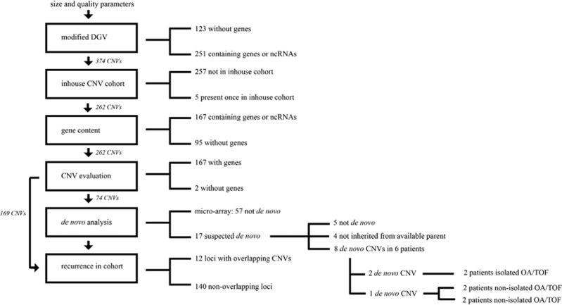
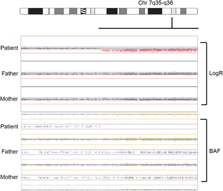
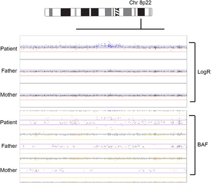
Oesophageal atresia (OA) with or without tracheoesophageal fistula (TOF) are rare anatomical congenital malformations whose cause is unknown in over 90% of patients. A genetic background is suggested, and among the reported genetic defects are copy number variations (CNVs). We hypothesized that CNVs contribute to OA/TOF development. Quantifying their prevalence could aid in genetic diagnosis and clinical care strategies. Therefore, we profiled 375 patients in a combined Dutch, American and German cohort via genomic microarray and compared the CNV profiles with their unaffected parents and published control cohorts. We identified 167 rare CNVs containing genes (frequency<0.0005 in our in-house cohort). Eight rare CNVs - in six patients - were de novo, including one CNV previously associated with oesophageal disease. (hg19 chr7:g.(143820444_143839360)_(159119486_159138663)del) 1.55% of isolated OA/TOF patients and 1.62% of patients with additional congenital anomalies had de novo CNVs. Furthermore, three (15q13.3, 16p13.3 and 22q11.2) susceptibility loci were identified based on their overlap with known OA/TOF-associated CNV syndromes and overlap with loci in published CNV association case-control studies in developmental delay. Our study suggests that CNVs contribute to OA/TOF development. In addition to the identified likely deleterious de novo CNVs, we detected 167 rare CNVs. Although not directly disease-causing, these CNVs might be of interest, as they can act as a modifier in a multiple hit model, or as the second hit in a recessive condition.
Figures
References
-
- Felix JF, de Jong EM, Torfs CP, de Klein A, Rottier RJ, Tibboel D: Genetic and environmental factors in the etiology of esophageal atresia and/or tracheoesophageal fistula: an overview of the current concepts. Birth Defects Res 2009; 85: 747–754. - PubMed
-
- Pedersen RN, Calzolari E, Husby S, Garne E: Oesophageal atresia: prevalence, prenatal diagnosis and associated anomalies in 23 European regions. Arch Dis Child 2012; 97: 227–232. - PubMed
-
- Genevieve D, de Pontual L, Amiel J, Sarnacki S, Lyonnet S: An overview of isolated and syndromic oesophageal atresia. Clin Genet 2007; 71: 392–399. - PubMed
-
- Quan L, Smith DW: The VATER association. Vertebral defects, anal atresia, T-E fistula with esophageal atresia, radial and renal dysplasia: a spectrum of associated defects. J Pediatr 1973; 82: 104–107. - PubMed
-
- Temtamy SA, Miller JD: Extending the scope of the VATER association: definition of the VATER syndrome. J Pediatr 1974; 85: 345–349. - PubMed
Publication types
MeSH terms
Supplementary concepts
Grants and funding
LinkOut - more resources
Full Text Sources
Other Literature Sources
