Review
. 2016 Sep 6;44(15):7007-78.
doi: 10.1093/nar/gkw530. Epub 2016 Jul 19.

Ribosomal frameshifting and transcriptional slippage: From genetic steganography and cryptography to adventitious use

Affiliations
Review

Ribosomal frameshifting and transcriptional slippage: From genetic steganography and cryptography to adventitious use

John F Atkins et al. Nucleic Acids Res. .

Abstract

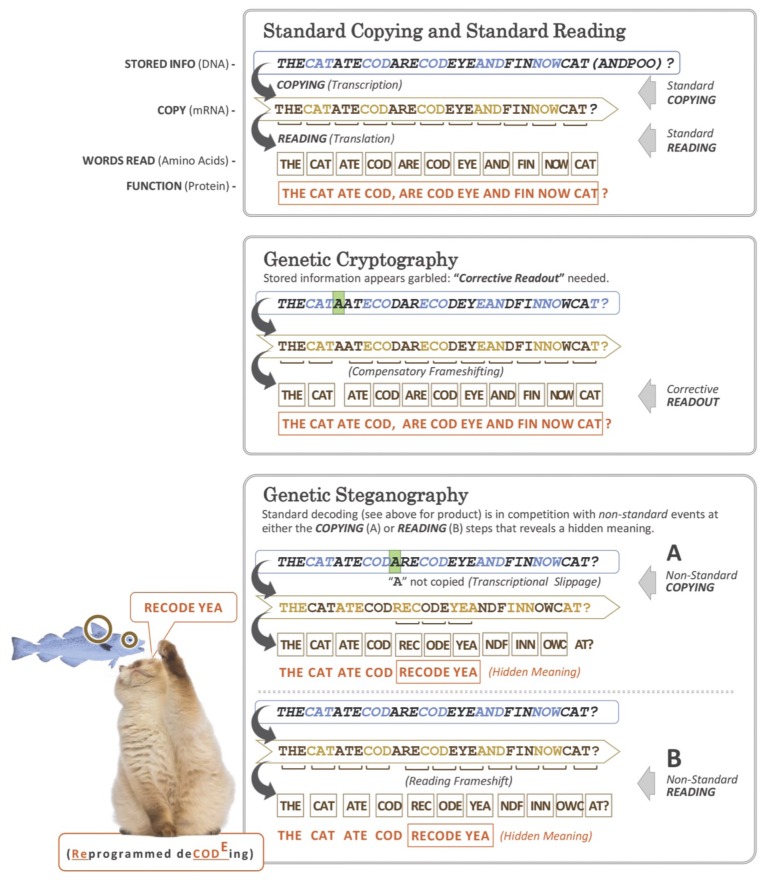
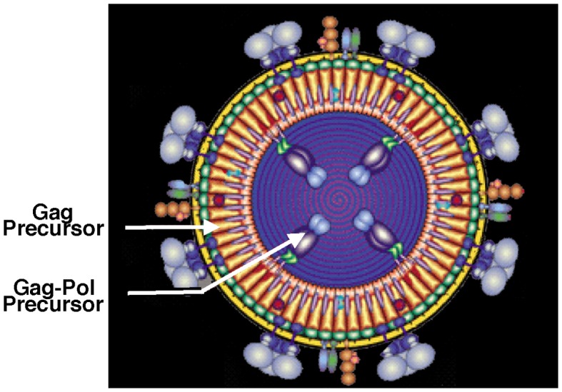
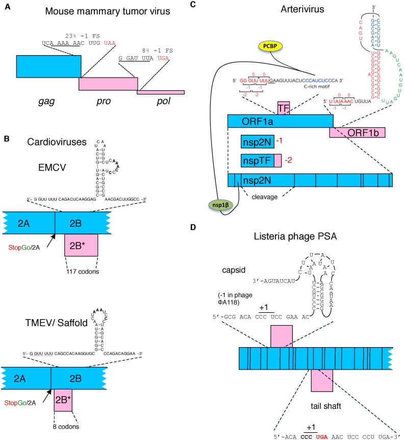
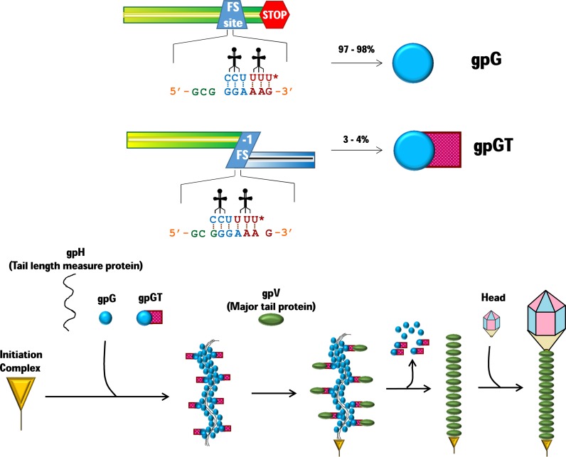
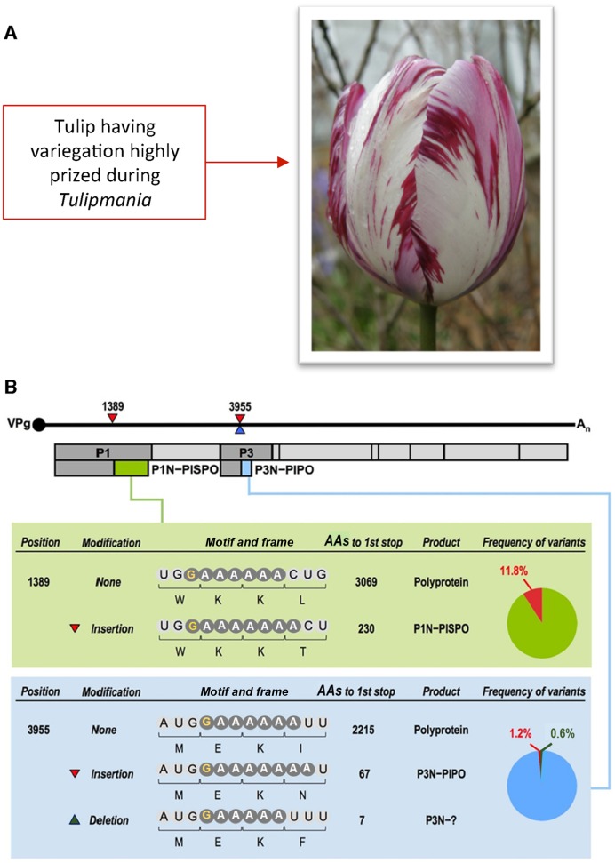
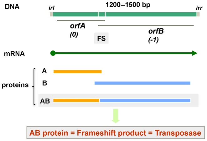
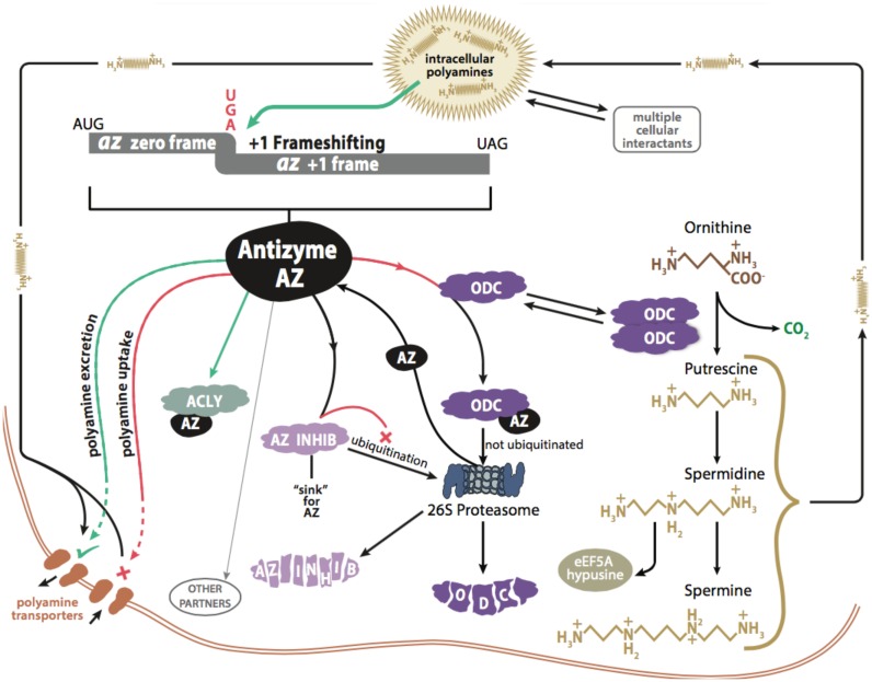
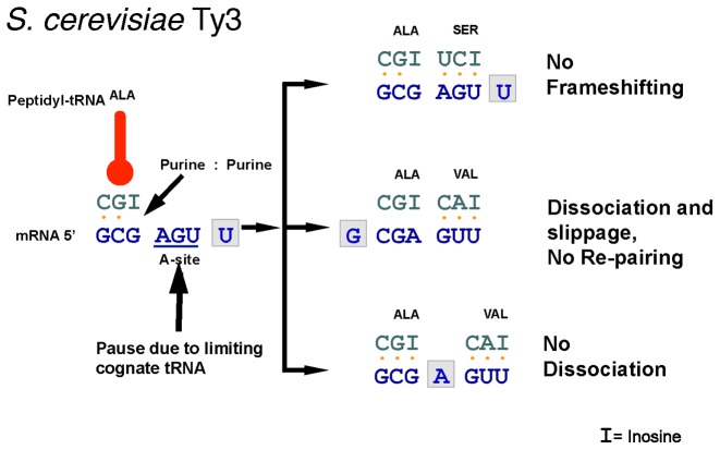
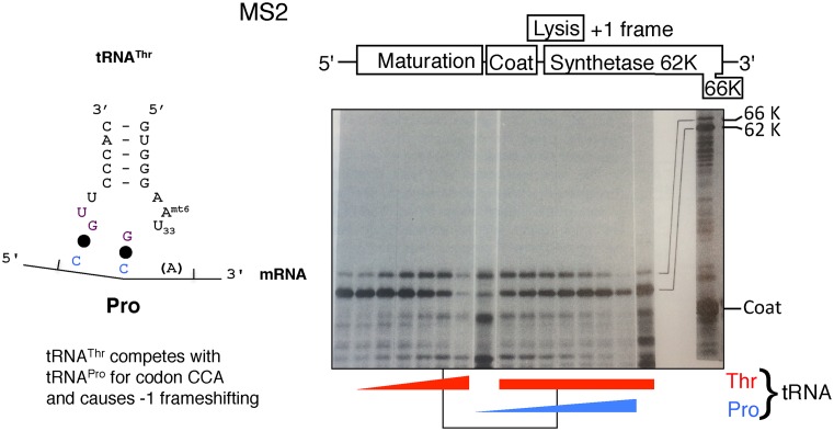
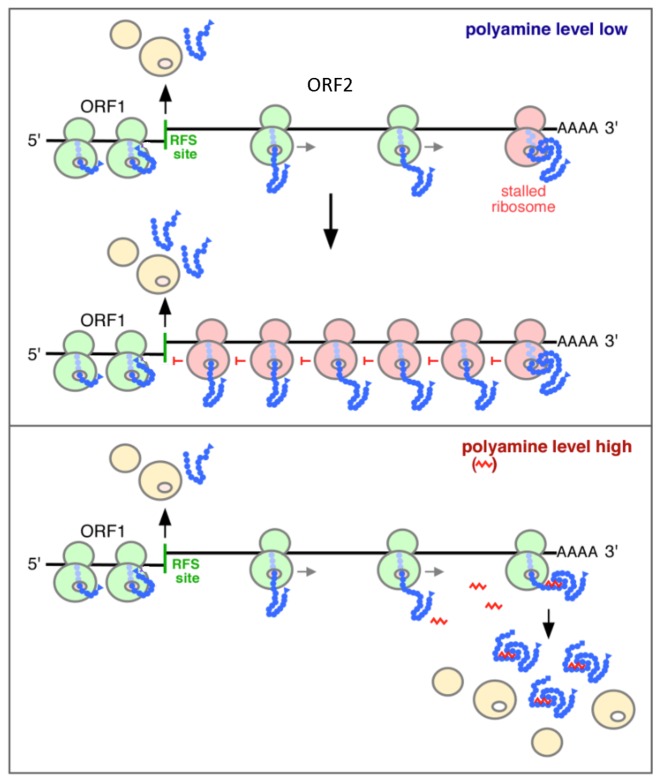
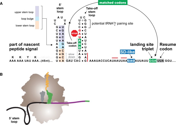
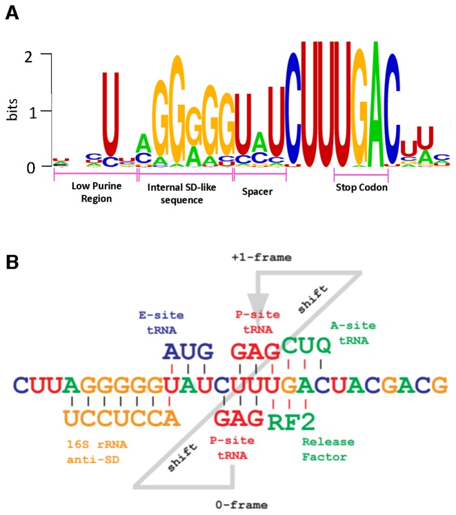
Genetic decoding is not 'frozen' as was earlier thought, but dynamic. One facet of this is frameshifting that often results in synthesis of a C-terminal region encoded by a new frame. Ribosomal frameshifting is utilized for the synthesis of additional products, for regulatory purposes and for translational 'correction' of problem or 'savior' indels. Utilization for synthesis of additional products occurs prominently in the decoding of mobile chromosomal element and viral genomes. One class of regulatory frameshifting of stable chromosomal genes governs cellular polyamine levels from yeasts to humans. In many cases of productively utilized frameshifting, the proportion of ribosomes that frameshift at a shift-prone site is enhanced by specific nascent peptide or mRNA context features. Such mRNA signals, which can be 5' or 3' of the shift site or both, can act by pairing with ribosomal RNA or as stem loops or pseudoknots even with one component being 4 kb 3' from the shift site. Transcriptional realignment at slippage-prone sequences also generates productively utilized products encoded trans-frame with respect to the genomic sequence. This too can be enhanced by nucleic acid structure. Together with dynamic codon redefinition, frameshifting is one of the forms of recoding that enriches gene expression.


Figures

Figure 1.
Figure 1.
Genetic ‘Bletchley-ism’: As illustrated with three letter words, the framing of genetic informational readout can be modified to convey meaning from genetic ‘hieroglyphs’ (cryptography) or additional and hidden meaning (steganography). Embellishing the old adage ‘From tapes to shapes’ (proteins), in several cases this involves ‘shapes-in-the-tapes’ unlike counterparts in many human languages. The process is dynamic, and the competition yields products from both standard reading and frameshifted reading. The relative proportions of the products from each are case dependent. Examples of genetic cryptography involving translational bypassing are in the decoding of phage T4 gene 60 and the mitochondrial genome of the yeast Magnusiomyces capitatus (56,65,158) and another type is in decoding the mitochondrial genome of glass sponges (252). The latter is a WT translation component counterpart of the suppression of frameshift mutants by suppressor mutants of translational components. Examples of genetic steganography involving transcriptional realignment are in the gene expression of paramyxoviruses, potyviruses and the bacterial insertion sequence Roseiflexus IS630 (42,99,617); examples of genetic steganography involving ribosomal frameshifting are in the decoding of influenza A virus expression (125,270) and D. melanogaster APC (46). While standard expression of most bacterial release factor 2 genes, and also probably eukaryotic antizyme genes except for antizyme 3, yields a product that is non-functional on its own, the +1 frameshifting required for productive expression has been positively selected. The representation was inspired in part by a genetic framing garden ‘rebus’ (812), a slide by V.N. Gladyshev and a recent publication (1).
Figure 2.
Figure 2.
Illustration of the Gag component of GagPol serving to incorporate and localize Pol within forming virions.
Figure 3.
Figure 3.
Schematic representation of several viral frameshifting cassettes. (A) Retroviral frameshifting with the thickness of the pro and pol ORFs (pink) reflecting frameshift efficiency and the proportion of ribosomes that decode them with respect to zero frame gag (blue). (B) The −1 ribosomal frameshift site in two cardioviruses that yields a transframe encoded protein (pink). The proximity of the Theiler's murine encephalomyelitis virus (TMEV) shift site to the StopGo is evident and the relevant amino acid sequence is shown in Table 2. (C) In arteriviruses, −1 ribosomal frameshifting near the end of a long 5′ gene, ORF1a, leads a proportion of ribosomes to continue synthesis by decoding a second and also long gene (ORF1b), with the products of both ORFs specifying non-structural polyproteins. At least eight shorter 3′ ORFs encode structural proteins. −2 ribosomal frameshifting in a central region of ORF1a causes some ribosomes to access the wholly overlapping +1 frame TF ORF to yield a C-terminal extension to nsp2 (the product liberated by proteolytic cleavage from that region of the polyprotein encoded by ORF1a). This frameshifting is stimulated in trans by virus-encoded nsp1β in complex with poly(C) binding protein (PCBP). (D) Listeria phage PSA. Capsid and tail shaft encoding genes utilize +1 ribosomal frameshifting on a proline codon just 5′ of either a stimulatory pseudoknot (capsid) or stop codon (tail shaft).
Figure 4.
Figure 4.
Frameshifting and phage tail assembly. Phage lambda and many other dsDNA phages gene g encodes gpG, a tail assembly chaperone, that binds to multiple regions of the tape measure protein gpH. Near the 3′ end of the g coding sequence, a small proportion of translating ribosomes shift –1 and continue to synthesize the fusion protein gpGT. The ‘G’ portion of gpGT binds to the tape measure protein gpH, while the ‘T’ region binds the major tail protein gpV, linking them and mediating the initiation of gpV on gpH. However, gpG and gpGT both dissociate from the assembly, aiding the fast polymerization of gpV on the initiator complex to form the mature tail. Finally, the head binds to the fully mature tail to form phage lambda. The tRNALys wobble anticodon nucleotide shown as U*, represents uridine with a 5-methylaminomethyl-2-thiouridine (mnm5s2U) modification.
Figure 5.
Figure 5.
Potyvirus transcriptional frameshifting. This permits expression of the overlapping ORF pipo whose product (light blue) is part of a protein, P3N-PIPO involved in viral movement between cells, and therefore essential to virus viability. Potyviruses are important as major pathogens of agricultural crops and for their effects on other plants. The effect of tulip mosaic potyvirus on flower pattern caused one bulb at the height of tulip-mania in the 1625 to cost ‘Four tons of wheat; eight tons of rye; four fat oxen; eight fat pigs; twelve fat sheep; two hogsheads of wine; four barrels of beer; two barrels of butter; one thousand pounds of cheese; one bed, with accessories; one full-dress suit; and one silver goblet.’ A second instance of transcriptional frameshifting in a small subset of potyviruses yields the product P1N-PISPO (PISPO in light green) [RNA Polymerase Slippage as a Mechanism for the Production of Frameshift Gene Products in Plant Viruses of the Potyviridae Family. Rodamilans, B., Valli, A., Mingot, A., San León, D., Baulcombe, D, López-Moya, J.J., and García, J.A. J. Virol. (2015) 89(13) 6965-6967, doi:10.1128/JVI.00337-15, reproduced with permission from American Society for Microbiology.].
Figure 6.
Figure 6.
Bacterial Insertion Sequence (IS) with two partially overlapping ORFs. Many IS elements have this type of gene organization. Utilization of ribosomal frameshifting to allow a proportion of ribosomes translating orfA to access orfB has been studied in depth in a variety of IS elements, but transcriptional slippage is now known to be used as an alternative by a substantial number of IS elements.
Figure 7.
Figure 7.
Schematic representation of synthesis of mammalian antizyme-1 (AZ) protein and the interactions between it, ornithine decarboxylase (ODC), antizyme inhibitor (AZ Inhib), ATP citrate lyase (ACYL) and polyamine transporters. Increasing polyamines enhances the ribosomal frameshifting required for synthesis of antizyme, which then acts to inhibit synthesis and uptake of polyamines. Antizyme binding to an ODC monomer prevents the ODC dimer formation required for catalyzing synthesis of putrescine from which the polyamines spermidine and spermine are derived.
Figure 8.
Figure 8.
Absence (or minimal) potential for re-pairing to mRNA at an overlapping +1 frame codon in S. cerevisiae Ty3 frameshifting. Biophysical studies are needed to determine whether the tRNAAla anticodon 3′GCI5′ (where I is inosine) dissociates at the time of frameshifting. Only a small number of codons can substitute for GCG, including CGA (422). See text and (453, 814) for discussion of this and other candidate occurrences.
Figure 9.
Figure 9.
Non-cognate frameshifting. In extracts of E. coli with unperturbed tRNA balance, tRNAThr decodes a proline codon to cause −1 frameshifting that results in synthesis of a 66 K form of the viral encoded component, synthetase, of replicase. Increasing the relative amount of tRNAThr (red wedge) increases the proportion of 66 K with respect to the product of standard decoding (62 K). With elevated levels of tRNAThr (uniform thickness red line) increasing the amount of cognate tRNAPro (blue wedge) decreases the relative amount of 66 K. Anticodon bases 35 and 34 are complementary to the first and second proline codon bases. The normal anticodon of tRNAThr is in purple. A model with 1:6 anticodon loop stacking is discussed in (453). Reprinted in part from Atkins, J.F., Gesteland, R.F., Reid, B.R. and Anderson, C.W. (1979) Normal tRNAs promote ribosomal frameshifting. Cell, 18, 1119–1131 with permission from Elsevier.
Figure 10.
Figure 10.
Scheme for S. cerevisiae antizyme mRNA +1 frameshifting. The top box represents low cellular polyamine concentrations, where ribosomes pause at the frameshifting site (RFS) with the ORF1 stop codon in the ribosomal A-site. Some terminate, while others continue translation in the +1 frame. An inhibitory element near the N-terminus of the nascent peptide, encoded near the start of ORF1, keeps frameshifting rates low. When a ribosome approaches the 3′ end of ORF2, a newly synthesized part of the nascent peptide, causes this ribosome (pink) to stall. Following translating ribosomes (green) encounter the stalled ribosome and their movement is also blocked (line 2 with multiple pink ribosomes). When polyamine levels are high (lower panel) the nascent peptide encoded near the 3′ end of ORF2, binds polyamines and is then unable to cause stalling. This allows the ribosome containing it to proceed to termination and release of functional antizyme. Reprinted by permission from Macmillan Publishers Ltd: Kurian,L., Palanimurugan,R., Godderz,D. and Dohmen,R.J. (2011) Polyamine sensing by nascent ornithine decarboxylase antizyme stimulates decoding of its mRNA. Nature, 477, 490–494.
Figure 11.
Figure 11.
(A) Features important for translational bypassing in decoding T4 gene 60. The matched take-off and landing codons, GGA, are shown in white letters in dark green boxes; the UAG stop codon immediately 3′ of the take-off site is in red letters next to the stop sign; stop codons within the coding gap are overlined in red; and sequences that slow ribosomes on approach to the second stem loop from the left, the take-off stem loop, are overlined in black. A Shine–Dalgarno-like sequence is shown in the blue box, and has a modest influence on landing site selection. The translational resume codon is indicated by the gray box. (B) Model depicting the predicted positioning of the 5′ stem loop and nascent peptide interaction when the GGA take-off codon is in the ribosomal P site. Part B Reproduced in part from Chen, J., Coakley, A., O'Connor, M., Petrov, A., O'Leary, S.E., Atkins, J.F. and Puglisi, J.D. (2015) Coupling of mRNA Structure Rearrangement to Ribosome Movement during Bypassing of Non-coding Regions. Cell, 163, 1267–1280 with permission from Elsevier.
Figure 12.
Figure 12.
The features of release factor 2 autoregulatory frameshifting are highly conserved in diverse bacteria. The level of the release factor for the underlined ORF1 stop codon UGA(C) governs the efficiency with which the anticodon paired to the 5′ adjacent codon, CUU, shifts +1 to pair with UUU permitting continued translation to synthesize release factor 2. The frameshift stimulatory internal Shine Dalgarno (SD) sequence is preceeded by sequence unable to pair with the anti-Shine Dalgarno sequence of translating ribosomes, permitting availability of the anti-Shine Dalgarno to form the correctly positioned frameshift stimulatory hybrid with mRNA. The figure was generated with Weblogo3 (815) using RF2 frameshift sites obtained from ARFA (348).
Figure 13.
Figure 13.
2D and 3D models of RNA structures that promote access to alternative reading frames in viral genes. (A) The intergenic Internal Ribosome Entry Site (IRES) of the dicistrovirus, Israeli acute paralysis virus, causes most ribosomes to initiate 1 base 5′ of where the others initiate. Stimulators for ribosomal frameshifting are shown for (B) beet western yellows virus (BWYV), (C) infectious bronchitis virus (IBV) and (D) mouse mammary tumor virus gag/pro (MMTV). Slippery sequences are underlined. Reproduced in part from Brierley, I., Gilbert, R.J.C. and Pennell, S., Pseudoknot-Dependent Programmed -1 Ribosomal Frameshifting: Structures, Mechanisms and Models (2010) In: Atkins, J.F. and Gesteland, R.F. (eds.), Recoding: Expansion of Decoding Rules Enriches Gene Expression. Springer New York, New York, pp. 149–174, with permission from Springer.
Figure 14.
Figure 14.
Sequence variability of antizyme frameshift stimulatory pseudoknot single-stranded regions contrasts with that involved in stem formation. The illustration is from pseudoknots from invertebrates (an oyster and an aphid) that differ from their mammalian counterparts. (A) Crassostrea gigas, (B) An aphid. The frameshift site is indicated with orange letters. Black arrowheads represent substitutions deduced from phylogenetic comparison to orthologous genes. Non-compensatory changes in the stems are shown in black letters; compensatory changes are shown in blue letters.
Figure 15.
Figure 15.
Alphavirus ribosomal frameshift stimulators are diverse. A single stem loop is the frameshifting stimulator for (A) Sindbis virus, (B) eastern equine encephalitis virus, (C) sleeping disease virus, and (D) Middelburg virus. Experimental support has been obtained for the second stem of the pseudoknot shown. (E) The stimulator for Semliki Forest virus frameshifting does not appear to involve intra-mRNA pairing, and is a candidate for exerting its effect via pairing with rRNA in the mRNA entrance channel. A similarly acting 3′ sequence may be acting as well as the pseudoknot, to stimulate the efficiency of frameshifting in Middelburg virus genome decoding to the high level of 49%. Reproduced from Chung B.Y.W., Firth A.E., Atkins J.F. (2010) Frameshifting in Alphaviruses: A Diversity of 3′ Stimulatory Structures, Journal of Molecular Biology, 397, 448–456 with permission from Elsevier.
Figure 16.
Figure 16.
Model suggesting −1 slippage during uncoupled translocation. smFRET experiments on a dnaX-derived motif show that slippage occurs during a state of single tRNA occupancy, where the P-site tRNAAla remains after translocation is uncoupled from E-site tRNA exit in an elongated pause state. This leads to a non-canonical structure of the ribosome where EF-G samples the ribosome >5 times per codon, in parallel with A-site sampling by the incoming tRNALys. tRNALys sampling defines the reading frame, and the non-canonical state is resolved by EF-G mediated translocation, resuming elongation in the −1 frame.
Figure 17.
Figure 17.
The mobile bacterial ribosome protein L9 that restrains forward mRNA slippage. Its N-terminal domain binds to the large subunit L1 stalk relevant to E-site tRNA egress. (A) Cryo-EM visualization of the L9 carboxy-terminal domain contacting ribosomal proteins (L9 is purple) versus the elongated form of L9 (in pink) detected in crystals. In crystals, instead of the L9 carboxy-terminal domain interacting with the 30S subunit of the same ribosome, it interacts with the 16S rRNA of a neighbouring ribosome. Whether this is a crystal artifact or indicative of a ‘strut’ function in polysomes relevant to frameshifting is unknown. (B) Model of L9 in the context of polysomes. This shows the arrangement of neighboring ribosomes (i-1 and i) in the major t-t form of E. coli polysomes, with the conformation of L9 (blue) revealed by cryo-EM. L9 is located close to protein S4 of the neighboring 30S subunit (30S i) according to the polysome model. The purple arrow indicates the rearrangement of L9 from the cryo-EM conformation to that seen in crystals. The black arrow denotes the location of the mRNA entry channel in the 30S subunit i. Images A and B are from (590). (C) Mass spectral analysis of protein products shows evidence for L9 functioning to restrain forward ribosome slippage on mRNA. Inactivation of L9 (lower panel) shows an additional product (2, in green) due to peptidyl-tRNA anticodon dissociation and re-pairing to mRNA at a 3′ position (indicated on sequence at right). The system used had additional mutants that exaggerate the effect. It involved polysomes, and the effect has not yet been tested with low ribosome loading (484) [Reprinted by permission from Macmillan Publishers Ltd: NATURE (Fischer, N., Neumann, P., Konevega, A.L., Bock, L.V., Ficner, R., Rodnina, M.V. and Stark, H., Structure of the E. coli ribosome-EF-Tu complex at <3 Å resolution by Cs-corrected cryo-EM, Nature, 2015, 520: 567-570), copyright 2015.].
Figure 18.
Figure 18.
Nascent RNA stem-loop formation stimulates DNA-dependent RNA polymerase slippage. In the top segment (adapted from (614)), the transcription bubble with the unpaired dsDNA and the 9–10 bp RNA–DNA are shown within the dotted ellipse. The position of the polymerase active site is marked in yellow and the incoming NTP substrate is in green. The RNA exit channel is between the Flap domain (blue) and the clamp domain (purple). The lower segment illustrates the nascent RNA stem loop dependent transcription slippage required for synthesis of the transposase of a Roseiflexus insertion sequence (IS). The DNA template strand slippage motif, 3′A5G5, hybrid with the growing RNA U5C4 in the post-translocated state with the catalytic center positioned at the 5th G. Formation of a nascent RNA stem loop adjacent to the hybrid and within the mRNA exit channel, has been proposed to melt the upstream part of the hybrid (99) and to open the polymerase clamp (614). In addition to stimulatory effects on forward realignment of the 3′ end of the RNA with respect to the template, the stem loop can also potentially stimulate the slippage by preventing RNA polymerase backtracking and favoring forward polymerase translocation.
Figure 19.
Figure 19.
Programmed transcriptional frameshifting in members of the Paramyxovirinae. In the P-gene mRNA of Sendai virus, the insertion of a G occurs over the minus strand template RNA slippery sequence UUUUUUCCC. In 30% of the population (middle panel), a pause in RdRp over the slippery sequence promotes slippage of a G-C bond to form G:U pairs (in red dotted lines). Polymerization resumes by addition of a G (in green) over the critical C (in pink), thus encoding mRNA for the V-protein in an overlapping translational −1 frame compared to genomic template. The panel to the right shows that the potential insertion of two G's has very poor likelihood due to multiple G-U pairs not being tolerated. Inset: Hexamer phasing in members of the Paramyxovirinae. The ribonucleoprotein (RNP) structure is a left-handed coil where each nucleocapsid protein binds precisely 6 nucleotides.
Figure 20.
Figure 20.
GWIPS-viz (816) screenshots of profiles of ribosome footprint densities. Expression of the selected genes involves ribosomal frameshifting from (A) bacteria, (B) yeast and (C) humans. Blue bars at the bottom represent annotation of protein coding genes according to the corresponding genomes (A and B) and RefSeq (C) with yellow boxes indicating positions of frameshifting sites. The density of ribosome footprints is shown as red columns corresponding to inferred positions of the A-sites. The top plots (A and B) show ORF organisation in genomes (green ticks are for ATG and red for stop codons). For more details on notations used see GWIPS-viz browser at http://gwips.ucc.ie

References

    1. Kwun H.J., Toptan T., Ramos da Silva S., Atkins J.F., Moore P.S., Chang Y. Human DNA tumor viruses generate alternative reading frame proteins through repeat sequence recoding. Proc. Natl. Acad. Sci. U.S.A. 2014;111:E4342–E4349. - PMC - PubMed
    1. Yanagitani K., Imagawa Y., Iwawaki T., Hosoda A., Saito M., Kimata Y., Kohno K. Cotranslational targeting of XBP1 protein to the membrane promotes cytoplasmic splicing of its own mRNA. Mol. Cell. 2009;34:191–200. - PubMed
    1. Keiler K.C. Mechanisms of ribosome rescue in bacteria. Nat. Rev. Microbiol. 2015;13:285–297. - PubMed
    1. Himeno H., Nameki N., Kurita D., Muto A., Abo T. Ribosome rescue systems in bacteria. Biochimie. 2015;114:102–112. - PubMed
    1. Hwang C.B., Horsburgh B., Pelosi E., Roberts S., Digard P., Coen D.M. A net +1 frameshift permits synthesis of thymidine kinase from a drug-resistant herpes simplex virus mutant. Proc. Natl. Acad. Sci. U.S.A. 1994;91:5461–5465. - PMC - PubMed

MeSH terms