. 2016 Jul 20:6:29667.
doi: 10.1038/srep29667.

Rescue of perfluorooctanesulfonate (PFOS)-mediated Sertoli cell injury by overexpression of gap junction protein connexin 43

Affiliations

Rescue of perfluorooctanesulfonate (PFOS)-mediated Sertoli cell injury by overexpression of gap junction protein connexin 43

Nan Li et al. Sci Rep. .

Abstract

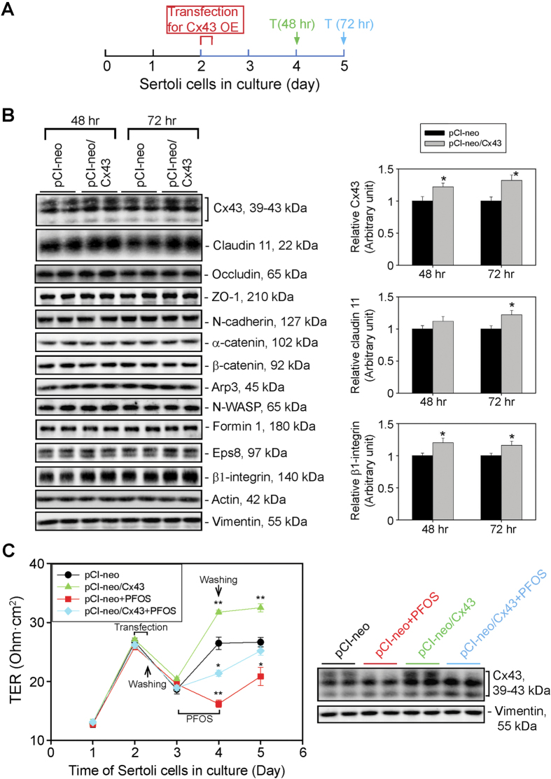
Perfluorooctanesulfonate (PFOS) is an environmental toxicant used in developing countries, including China, as a stain repellent for clothing, carpets and draperies, but it has been banned in the U.S. and Canada since the late 2000s. PFOS perturbed the Sertoli cell tight junction (TJ)-permeability barrier, causing disruption of actin microfilaments in cell cytosol, perturbing the localization of cell junction proteins (e.g., occluden-ZO-1, N-cadherin-ß-catenin). These changes destabilized Sertoli cell blood-testis barrier (BTB) integrity. These findings suggest that human exposure to PFOS might induce BTB dysfunction and infertility. Interestingly, PFOS-induced Sertoli cell injury associated with a down-regulation of the gap junction (GJ) protein connexin43 (Cx43). We next investigated if overexpression of Cx43 in Sertoli cells could rescue the PFOS-induced cell injury. Indeed, overexpression of Cx43 in Sertoli cells with an established TJ-barrier blocked the disruption in PFOS-induced GJ-intercellular communication, resulting in the re-organization of actin microfilaments, which rendered them similar to those in control cells. Furthermore, cell adhesion proteins that utilized F-actin for attachment became properly distributed at the cell-cell interface, resealing the disrupted TJ-barrier. In summary, Cx43 is a good target that might be used to manage PFOS-induced reproductive dysfunction.


Figures

Figure 1
Figure 1. Disruptive effects of PFOS on Sertoli cell F-actin organization and distribution of BTB-associated proteins at the cell-cell interface.
(A) Treatment regimen used for this experiment. Sertoli cells cultured alone at 0.4 × 106 cell/cm2 for 3 days following the establishment of a functional TJ-barrier were treated with 20 μM PFOS for 24 hr. Thereafter, cells were harvested for immunoblotting or for immunofluorescent microscopy. (B) Immunoblot analysis to assess changes in the steady-state level of Cx43 (a GJ protein), TJ- (e.g., claudin 11), basal ES (e.g., N-cadherin, ß-catenin), and actin binding/regulatory proteins (e.g., Arp3, N-WASP, formin 1 and Eps8) with ß-actin serving as the protein loading control. Cx43 appeared as three immunoreactive bands designated as p0, p1 and p2, corresponding to Mr of 39, 41, and 43 kDa due to differential phosphorylation, which is consistent with earlier reports. A study by qPCR (with GAPDH serving as the internal control for normalization) also confirmed a down-regulation of Cx43 expression. Immunoblot analysis also illustrated a down-regulation on the steady-state level of Cx43 protein when p0, p1 and p2 isoforms were analyzed as a whole, with a more considerable reduction for p2. Besides Cx43, claudin 11 and ß1-integrin were also down-regulated. Each bar in the two bar graphs is a mean ± SD of n = 5 experiments. **P < 0.01. (C) F-actin organization in Sertoli cells was assessed by phalloidin-FITC staining, illustrating PFOS induced truncation and defragmentation of actin microfilaments. PFOS treatment also considerably down-regulated the expression of Cx43 and claudin 11 at the Sertoli-Sertoli cell interface, consistent with data shown in (B). While the levels of N-cadherin and ß-catenin were not considerably affected based on immunoblotting results, these basal ES proteins localized diffusely at the Sertoli cell-cell interface, apparently rapidly internalized. Different cell densities were used to optimize the visualization of different target proteins including F-actin as noted in Materials and Methods. Micrographs shown herein are representative results of an experiment. All three independent experiments yielded similar results. Scale bar, 25 μm, which applies to all other micrographs. Each bar graph is a mean ± SD of n = 3 experiments. **P < 0.01.
Figure 2
Figure 2. Overexpression of Cx43 in Sertoli cells with an established TJ-permeability barrier promotes barrier function, rescuing PFOS-induced barrier disruption.
(A) Treatment regimen used to assess the effects of overexpression of Cx43 in Sertoli cells on the steady-state levels of BTB-associated proteins by immunoblot analysis. T, termination. (B) Immunoblot analysis using Sertoli cell lysates to assess changes in the steady-state levels of selected BTB-associated proteins, including Cx43 following its overexpression at 48- and 72-hr after transfection (i.e., on days 4 and 5, respectively). Among all the BTB-associated proteins examined, only the TJ protein claudin 11 and the hemidesmosome protein ß1-integrin displayed a moderate up-regulation following Cx43 overexpression as illustrated in the bar graphs on the right panel. Each bar in the bar graphs is a mean ± SD of n = 3 experiments. *P < 0.05. (C) Sertoli cells were cultured on Matrigel-coated bicameral units at 1.0 × 106 cells/cm2 for two days to establish a functional TJ-permeability barrier. Cells were then transfected with pCI-neo empty vector vs. pCI-neo vector containing Cx43 for overexpression of Cx43 (pCI-neo/Cx43) in these cells for 6 hr. Thereafter, cells were washed twice with F12/DMEM and cultured for another 18 hr, followed by an incubation with or without (i.e., equal amount of DMSO-containing medium) 20 μM PFOS for 24 hr. Cells were then rinsed twice to remove PFOS and cultured in F12/DMEM for another 24 hr. TER was recorded daily using quadruple bicameral units for each time-point. Overexpression of Cx43 in Sertoli cells was found to promote the TJ-permeability barrier and alleviate PFOS-induced TJ-disruption. Each data point is a mean ± SD of quadruple bicameral units of a representative experiment from n = 3 experiments, which yielded similar results. *P < 0.05; **P < 0.01. Sertoli cell lysates obtained on day 5 (see left panel, and 2A for the treatment regimen) were also used for immunoblotting, illustrating overexpression of Cx43 partially rescued the PFOS-induced Cx43 down-regulation. Vimentin served as the protein loading control.
Figure 3
Figure 3. Overexpression of Cx43 in Sertoli cells rescues PFOS-induced GJ communication dysfunction.
(A) Treatment regimen used to determine if overexpression (OE) of Cx43 can rescue PFOS-induced GJ communication function based on a dye-transfer FRAP (fluorescence recovery after photobleaching) functional assay. (B) GJ intercellular communication function between Sertoli cells cultured at 0.15 × 106 cells/cm2 was assessed by a dye-transfer FRAP assay based on the transfer of fluorescent dye (calcein AM) from neighboring cells via GJ following photobleaching of a single Sertoli cell. Sertoli cells were cultured at 0.15 × 106 cells/cm2 on Matrigel-coated glass-bottom dishes for 2 days. The cells were then transfected with pCI-neo/Cx43 vs. pCI-neo empty vector for 6 hr. Thereafter, cells were washed twice with F12-DMEM and cultured for another 18 hr, followed by treatment with 20 μM PFOS for 24 hr (with or without Cx43 overexpression vs. Cx43 overexpression alone or empty vector alone). Results from a typical FRAP assay are shown with n = 3 experiments, which yielded similar results. Scale bar, 40 μm, which applies to all other micrographs. (C) Typical results of a representative FRAP assay from n = 3 experiments that yielded similar results, illustrating GJ communication was impaired by PFOS treatment, but overexpression of Cx43 rescued the PFOS-induced GJ dysfunction. Each data point on the right panel is a mean ± SD of n = 4 independent experiments for the four experimental groups using different batches of Sertoli cells for the FRAP assay. **P < 0.01.
Figure 4
Figure 4. Overexpression of Cx43 in Sertoli cells rescues PFOS-induced Sertoli cell BTB disruption through proper localization of adhesion proteins at the cell-cell interface.
Sertoli cells were cultured at 0.1–0.15 × 106 cells/cm2 for two days. They were transfected with pCI-neo/Cx43 vs. pCI-neo empty vector (control, Ctrl) and then subjected to PFOS treatment, and cells were harvested for IF analysis as noted in the regimen shown in Fig. 3A. In cells overexpressed with Cx43 (pCI-neo/Cx43), the fluorescent intensity for Cx43 at cell-cell interface was moderately stronger than control (pCI-neo empty vector alone) cells. However, PFOS induced considerable down-regulation of Cx43 and claudin 11 at the cell-cell interface (pCI-neo+PFOS), PFOS also perturbed the localization of TJ protein ZO-1, and basal ES proteins N-cadherin and β-catenin. Importantly, overexpression of Cx43 in PFOS treated cells (pCI-neo/Cx43+PFOS) rescued the PFOS-mediated BTB disruption by maintaining the expression of Cx43 and claudin 11. This also induced proper localization of TJ protein ZO-1 and basal ES proteins N-cadherin and ß-catenin at the cell-cell interface. Plasmid DNA (pCI-neo vs. pCI-neo/Cx43) was labeled with Cy3 and appeared as red fluorescence to annotate successful transfection. Scale bar, 30 μm, in the first micrograph, which applies to the remaining micrographs in the same panel. These results are representative micrographs from an experiment, and n = 3 independent experiments were performed using different batches of Sertoli cells and yielded similar results. Each bar graph is a mean ± SD of n = 3 experiments. **P < 0.01. FI, fluorescent intensity; FS, fluorescent signal.
Figure 5
Figure 5. Overexpression of Cx43 in Sertoli cells restores actin microfilament organization via proper spatiotemporal expression of actin binding/regulatory proteins Arp3 and Eps8.
(A) We next examined the mechanism by which Cx43 overexpression rescues the PFOS-induced Sertoli cell TJ-barrier disruption. Exposure of Sertoli cells with an established functional TJ-permeability barrier to PFOS for 24 hr was found to induce mis-orgranization of actin microfilaments wherein they displayed extensive defragmentation across the Sertoli cell cytosol. However, overexpression of Cx43 restored organization of actin microfilaments in Sertoli cells, apparently via proper spatiotemporal expression of actin binding and regulatory proteins Arp3 and Eps8, such that these proteins localized to the Sertoli cell-cell interface, similar to the control cells expressed with the empty pCI-neo vector. Plasmid DNA (pCI-neo vs. pCI-neo/Cx43) was labeled with Cy3 and appeared as red fluorescence to annotate successful transfection. Scale bar, 30 μm, which applies to all other micrographs. (B) Relative fluorescent intensity of Arp3 and Eps8 at the Sertoli cell-cell interface was quantified in the PFOS treatment group (pCI-neo+PFOS) vs. control (Ctrl, transfected with pCI-neo vector alone), Cx43 overexpression (pCI-neo/Cx43) and Cx43 overexpression plus PFOS (pCI-neo/Cx43+PFOS) groups. Each bar is a mean ± SD by taking the average fluorescent intensity from two opposite ends between adjacent Sertoli cells (see corresponding colored boxed area for Arp3 vs. Eps8 from different groups) from 50 randomly selected cells in each experiment with n = 3 independent experiments. **P < 0.01.

References

    1. Bai D. Structural analysis of key gap junction domains - lessons from genome data and disease-linked mutants. Semin Cell Dev Biol, 10.1016/j.semcdb.2015.11.015 (2015). - DOI - PubMed
    1. Balse E. et al.. Dynamic of ion channel expression at the plasmam membrane of cardiomyocytes. Physiol Rev 92, 1317–1358 (2012). - PubMed
    1. Gerber J., Heinrich J. & Brehm R. Blood-testis barrier and Sertoli cell function: lessons from SCCx43KO mice. Reproduction 151, R15–27, 10.1530/REP-15-0366 (2016). - DOI - PubMed
    1. Li M. W. M., Mruk D. D. & Cheng C. Y. Gap junctions and blood-tissue barriers. Adv Exp Med Biol 763, 260–280 (2012). - PMC - PubMed
    1. Pointis G., Gilleron J., Carette D. & Segretain D. Physiological and physiopathological aspects of connexins and communicating gap junctions in spermatogenesis. Philos Trans R Soc Lond B Biol Sci 365, 1607–1620 (2010). - PMC - PubMed

Publication types