Review
. 2016 Dec:60:136-145.
doi: 10.1016/j.semcdb.2016.07.018. Epub 2016 Jul 18.

Auto-fusion and the shaping of neurons and tubes

Affiliations
Review

Auto-fusion and the shaping of neurons and tubes

Fabien Soulavie et al. Semin Cell Dev Biol. 2016 Dec.

Abstract

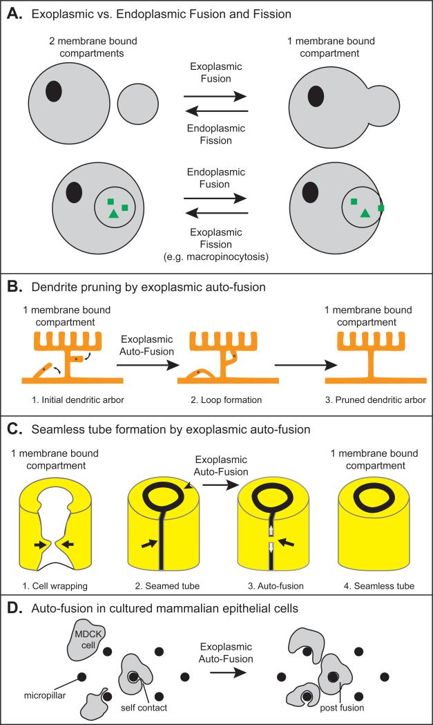
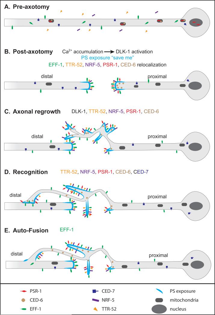
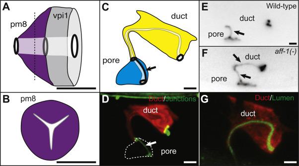
Cells adopt specific shapes that are necessary for specific functions. For example, some neurons extend elaborate arborized dendrites that can contact multiple targets. Epithelial and endothelial cells can form tiny seamless unicellular tubes with an intracellular lumen. Recent advances showed that cells can auto-fuse to acquire those specific shapes. During auto-fusion, a cell merges two parts of its own plasma membrane. In contrast to cell-cell fusion or macropinocytic fission, which result in the merging or formation of two separate membrane bound compartments, auto-fusion preserves one compartment, but changes its shape. The discovery of auto-fusion in C. elegans was enabled by identification of specific protein fusogens, EFF-1 and AFF-1, that mediate cell-cell fusion. Phenotypic characterization of eff-1 and aff-1 mutants revealed that fusogen-mediated fusion of two parts of the same cell can be used to sculpt dendritic arbors, reconnect two parts of an axon after injury, or form a hollow unicellular tube. Similar auto-fusion events recently were detected in vertebrate cells, suggesting that auto-fusion could be a widely used mechanism for shaping neurons and tubes.

Keywords: Auto-fusion; Dendrite morphogenesis; Fusogen; Neurons regeneration; Seamless tube.


Figures

Figure 1
Figure 1. Fission, Fusion and Auto-fusion
(A) Comparison of different types of fission and fusion, adapted from []. Cytoplasmic domains are marked with grey coloring. (B-D) Examples of exoplasmic auto-fusion maintaining one membrane bound compartment. (B) A C. elegans arborized dendrite (“menorah”) has two ectopic branches. Auto-fusion induces loop formation to prune the ectopic branches and maintain only the appropriate right-angled branches []. (C) The three steps of seamless tube formation [, , ]. (1) Cell wrapping. White, apical surface; yellow, basal surface. The arrows show the path of cell wrapping. (2) Formation of a seamed tube. An auto-cellular junction (arrow) maintains the cell in the shape of a tube. A ring junction (arrow-head) seals the cell with its neighbors (not shown) to continue the tube. (3) Proposed model of Auto-fusion. A fusion pore forms (black arrow) and extends (white arrows) to form a seamless tube (4). (D) Model of auto-fusion in cultured mammalian MDCK cells [].
Figure 2
Figure 2. Model of axon reconnection by auto-fusion
Model based on refs. [, , ]. (A) Before axotomy, PSR-1 (red) localizes in mitochondria and in the nucleus, and CED-6 (brown) localizes in mitochondria only. EFF-1 (green) is both intracellular and on the plasma membrane. (B) Immediately after axon injury, calcium is released and phosphatidylserine (PS, light blue) is exposed to the external lipid layer. Calcium release induces DLK-1 activation to promote axon regrowth. PSR-1 and CED-6 accumulate at the proximal tip. TTR-52 (orange), NRF-5 (purple) and EFF-1 accumulate on external membranes at both proximal and distal tips. (C) DLK-1 and the phagocytosis pathway promote axonal regrowth. (D) The phagocytosis pathway promotes recognition of distal axon by the proximal growth cone. (E) EFF-1 stimulates auto-fusion to reconnect the proximal part to the distal part of the axon.
Figure 3
Figure 3. Seamless tubes formed by auto-fusion can have complex shapes
(A) Lateral view of C. elegans pharyngeal-intestinal valve cells (pm8 and vpi1), which are both seamless tubes. The tubes are linked to neighbors by ring-shaped junctions (black ovals). (B) A transverse section of pm8 (dashed line in A) shows the tri-radiate shape of the lumen []. (C) The C. elegans excretory duct is a seamless tube, in contrast to the adjacent cell, the excretory pore, which maintains an auto-junction (arrow). Both tubes are linked to neighbors by ring-shaped junctions (black ovals). (D) The excretory duct has an elongated shape. Red, duct cytoplasmic marker lin-48pro::mRFP []. Green, junction marker AJM-1::GFP []. (E, F) AFF-1 is required for duct seamlessness. Cell junctions marked by AJM-1::GFP. (E) Wild-type excretory duct is a seamless tube. (F) In an aff-1 loss of function mutant, the excretory duct is a seamed tube with an auto-junction (arrows) []. (G) The excretory duct has an elongated lumen. Red, duct cytoplasmic marker lin-48pro::mRFP. Green, luminal marker let-653pro::LET-653::GFP []. Scale bars. 2 μm.

Similar articles

Cited by

References

    1. Lefebvre JL, Sanes JR, Kay JN. Development of dendritic form and function. Annu Rev Cell Dev Biol. 2015;31:741–77. - PubMed
    1. Andrew DJ, Ewald AJ. Morphogenesis of epithelial tubes: Insights into tube formation, elongation, and elaboration. Dev Biol. 2010;341(1):34–55. - PMC - PubMed
    1. Ghosh-Roy A, Wu Z, Goncharov A, Jin Y, Chisholm AD. Calcium and cyclic AMP promote axonal regeneration in Caenorhabditis elegans and require DLK-1 kinase. J Neurosci. 2010;30(9):3175–83. - PMC - PubMed
    1. Oren-Suissa M, Hall DH, Treinin M, Shemer G, Podbilewicz B. The fusogen EFF-1 controls sculpting of mechanosensory dendrites. Science. 2010;328(5983):1285–8. - PMC - PubMed
    1. Rasmussen JP, English K, Tenlen JR, Priess JR. Notch signaling and morphogenesis of single-cell tubes in the C. elegans digestive tract. Dev Cell. 2008;14(4):559–69. - PMC - PubMed

Publication types

LinkOut - more resources