. 2016 Aug 2;113(31):8843-8.
doi: 10.1073/pnas.1608264113. Epub 2016 Jul 19.

Transcriptional control of amino acid homeostasis is disrupted in Huntington's disease

Affiliations

Transcriptional control of amino acid homeostasis is disrupted in Huntington's disease

Juan I Sbodio et al. Proc Natl Acad Sci U S A. .

Abstract

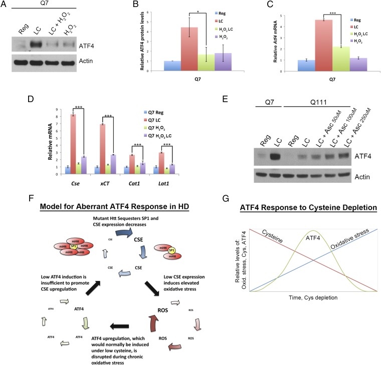
Disturbances in amino acid metabolism, which have been observed in Huntington's disease (HD), may account for the profound inanition of HD patients. HD is triggered by an expansion of polyglutamine repeats in the protein huntingtin (Htt), impacting diverse cellular processes, ranging from transcriptional regulation to cognitive and motor functions. We show here that the master regulator of amino acid homeostasis, activating transcription factor 4 (ATF4), is dysfunctional in HD because of oxidative stress contributed by aberrant cysteine biosynthesis and transport. Consistent with these observations, antioxidant supplementation reverses the disordered ATF4 response to nutrient stress. Our findings establish a molecular link between amino acid disposition and oxidative stress leading to cytotoxicity. This signaling cascade may be relevant to other diseases involving redox imbalance and deficits in amino acid metabolism.

Keywords: ATF4; CSE; Huntington’s disease; cysteine; oxidative stress.


Conflict of interest statement

The authors declare no conflict of interest.

Figures

Fig. 1.
Fig. 1.
CSE is induced in response to cysteine deprivation and is regulated by ATF4. (A) Schematic representation of the reverse transsulfuration pathway leading to cysteine and glutathione biosynthesis. CSE uses cystathionine to generate cysteine, which in turn is used to produce hydrogen sulfide (H2S). (B) Kinetics of CSE induction in MEFs. MEFs derived from wild-type (WT) and CSE−/− (CSE KO) mice were incubated in cysteine-free medium, and induction of CSE was monitored at various time points by Western blotting using actin as a loading control. (C) CSE levels are inversely proportional to the cysteine content in the growth medium. MEFs were incubated with media containing varying concentrations of cysteine for 24 h and analyzed by Western blotting. (D) CSE and ATF4 are induced in response to low cysteine. Wild-type and CSE KO MEFs were grown in low-cysteine (LC) medium for 24 h. The levels of CSE, ATF4, SP1, and Actin (loading control) were monitored by Western blotting. (E) Quantitation of D. n = 3 (means ± SEM). *P < 0.05; ***P < 0.001. (F) Schematic representation of SP1 sequestration by mHtt disrupting CSE expression in Huntington’s disease. (G) ATF4 is induced in response to cysteine depletion in Q7, but not in Q111, cells. Q7 and Q111 striatal cells were grown in medium containing different cysteine concentrations for 24 h, and ATF4 induction was monitored by Western blotting. (H) CSE is up-regulated under low-cysteine conditions in Q7, but not in Q111, striatal cells. Cell lysates used in G were used to monitor CSE induction. (I) Striatal Q7 and Q111 cells were grown in low cysteine (0.05 mM) for different time periods, and induction of CSE and ATF4 levels was monitored by Western blotting.
Fig. S1.
Fig. S1.
Cysteine becomes an essential amino acid in the absence of CSE. (A) CSE−/− (CSE KO) mice lose weight on a cysteine-free (Cys-free) diet. Wild-type (WT) and CSE KO mice were placed on a cysteine-free or regular (0.3% cysteine) diet starting 4 wk of age and weights were recorded. Cysteine was replenished at day 5 post deprivation and changes in weight measured. (B) CSE KO mice exhibited motor deficits on a cysteine-free diet. The motor functions of the wild-type (WT) and CSE KO mice used in A were assessed by the rotarod assay (Movie S1). (C) Cell proliferation of wild-type MEFs in media containing different concentrations of cysteine. Cells were incubated in medium containing varying concentrations of cysteine, and viability was assessed using the MTT assay. (D) ATF4 expression responds rapidly to cysteine concentrations. Wild-type MEFs were incubated in low-cysteine (LC) medium for 24 h. Cysteine was added back to the growth medium, and the levels of ATF4 and CSE were monitored by Western blotting at different time points. (E) CSE levels in striatal cells and MEFs. Q7 and Q111 striatal cells and wild-type (CSE WT) and CSE−/− (CSE KO) MEFs were harvested, and protein levels of CSE were assessed by Western blotting using actin as the loading control. (F) Overexpression of ATF4 rescues CSE expression. Q111 cells were transfected with a plasmid encoding ATF4. CSE levels were compared with that in Q7 cells by Western blotting using actin as a loading control. (G) Overexpression of ATF4 in striatal Q111 cells restores growth in cysteine-free medium. Striatal Q7 and Q111 cells were transfected with either the empty vector or that encoding ATF4, and growth in cysteine-free medium was monitored by MTT assay. Shown is a representative six-well plate showing enhanced growth of Q111 cells harboring ATF4 plasmid. (H) Striatal cell viability was not significantly affected by low cysteine medium growth. Striatal Q7 and Q111 cells were incubated in regular or low-cysteine medium for 24 h, and cell survival was assessed by MTT assay. Data are presented as means ± SE.
Fig. 2.
Fig. 2.
Lack of induction of ATF4 in striatal cells expressing mutant huntingtin is specific to cysteine deprivation. (A) Transcriptional targets of ATF4 are not induced in response to cysteine deprivation in Q111 cells. Striatal Q7 and Q111 cells were grown in regular or low-cysteine media, and transcript levels of Cse, xCT, Cat1, and Lat1, which encode CSE, cystine transporter, cationic amino acid transporter 1, and large amino acid transporter 1, respectively, were monitored by real-time qPCR. n = 4 (means ± SEM). ***P < 0.001. (B) Inhibition of cystine uptake also fails to induce ATF4 in Q111 cells. Striatal Q7 and Q111 cells were treated with 0.5 µM erastin (ERA), an inhibitor of xCT, the cystine transporter, to induce cysteine deprivation for 24 h. ATF4 was monitored by Western blotting. (C) Transcriptional targets of ATF4 were not induced in response to erastin treatment in Q111 cells. Striatal Q7 and Q111 cells were grown in regular medium and treated with 0.5 µM ERA. Transcript levels of Cse, xCT, Cat1, and Lat1 were monitored by real-time qPCR. n = 3 (means ± SEM). ***P < 0.001. (D) ATF4 is induced in response to endoplasmic reticulum (ER) stress in both Q7 and Q111 cells. Striatal cells were incubated in regular or low-cysteine (LC) medium or in regular medium (Reg) containing DMSO (vehicle) or 1 µM thapsigargin (TG) for 24 h. ATF4 and SP1, the markers of ER stress (not induced in LC) phosphorylated PERK, the ER chaperone binding Ig protein (BiP), and actin (loading control) levels were monitored by Western blotting. (E) Immunofluorescence analysis depicting the up-regulation and nuclear localization of ATF4 in response to low cysteine and ER stress. Q7 and Q111 cells were incubated as in D and the cells were fixed, permeabilized, and stained for ATF4 (green) and DNA (DAPI). (Magnification, 60×.) (F) The up-regulation of ATF4 and CSE occurs at the transcriptional level. Striatal Q7 and Q111 cells were incubated as in D. Cells were scraped and the RNA was isolated and analyzed by real-time qPCR. n = 4 (means ± SEM). ***P < 0.001; ns: not significant. (G) ATF4 is induced in response to deprivation of other amino acids. Striatal cells were grown in media individually deprived of the amino acids arginine (−R), lysine (−K), glutamine (−Q), or leucine (−L) for 24 h. ATF4 induction was assessed by Western blotting.
Fig. S2.
Fig. S2.
Amino acid sensing and H2S involvement in HD. (A) Schematic representation of the sensing of amino acid deprivation by the general control nonderepressible2 kinase (GCN2). When amino acid levels are sufficient, charging of tRNAs by the aminoacyl tRNA synthetase (aaRS) occurs. When cells are starved for any amino acid, its cognate tRNA remains uncharged and binds to GCN2, the amino acid sensor, inducing its autophosphorylation. Activated GCN2 phosphorylates the eukaryotic translation initiation factor 2α (eIF2α) and inhibits its activity, leading to a global translational arrest. Under these conditions, translation of a few select mRNAs such as the ATF4 mRNA occurs, which orchestrates the response of cell survival pathways. (B) Amino acid deprivation sensing is not affected in Q111 cells. Q7 and Q111 striatal cells were incubated in medium deprived of cysteine for different time periods. CSE, ATF4, phosphorylated GCN2, and total GCN2 were monitored by Western blotting with actin as a loading control. (C) H2S does not restore ATF4 induction. Q7 and Q111 cells were incubated with DMSO (vehicle) or the H2S donors GYY4137 (50 and 100 µM) and NaHS (100 µM) in low- or regular-cysteine media. ATF4 and CSE were monitored by Western blotting.
Fig. 3.
Fig. 3.
Elevated ROS is the causative factor for diminished ATF4 response in Q111 striatal cells. (A) CSE−/− (CSE KO) MEFs exhibit elevated oxidative stress. Wild-type (WT) and CSE KO MEFs were incubated in regular medium or in regular medium containing 100 µM H2O2 for 24 h. Oxidation was assessed by measuring protein carbonylation. n = 3 (means ± SEM). *P < 0.05; **P < 0.01. (B) Primary cortical neurons from CSE KO mice are more vulnerable to oxidative stress. Primary cortical neurons from wild-type or CSE KO mice were incubated in medium with or without H2O2 for 18 h. Cell viability was determined by MTT assay. n = 3 (means ± SEM). **P < 0.01; ***P < 0.001. (C) Primary cortical neurons from CSE KO mice are more susceptible to the oxidant homocysteic acid (HCA). Primary cortical neurons from wild-type or CSE KO mice were treated with or without HCA for 18 h. Cell viability was assessed by MTT assay. n = 3 (means ± SEM). ***P < 0.001. (D) Striatal Q111 cells have elevated reactive oxygen species. Striatal Q7 and Q111 cells were grown in regular or low-cysteine medium for 24 h. Cells were treated with the fluorescent indicator CellROX green to assay levels of ROS. n = 3 (means ± SEM). ***P < 0.001. (E) Striatal Q7 and Q111 cells were grown in low-cysteine (LC) medium for 24 h and incubated with the redox-sensitive fluorescent indicator CM-H2DCFDA and analyzed by fluorescence microscopy. (Magnification, 10×.) (F) Relative oxidative stress in striatal Q7 and Q111 cells. Q7 cells were incubated with different concentrations of H2O2 for 24 h and compared with Q111 cells incubated in regular or low-cysteine medium for 24 h. Oxidative stress was measured as in D. n = 3 (means ± SEM).
Fig. 4.
Fig. 4.
Diminished ATF4 induction caused by oxidative stress is reversed by antioxidant supplementation. (A) Oxidative stress suppresses ATF4 response to cysteine deprivation in Q7 cells. Q7 striatal cells were grown in regular and low-cysteine (LC) medium with or without 100 µM H2O2 for 24 h. ATF4 levels were monitored by Western blotting using actin as a control. (B) Quantitation of A. n = 4 (means ± SEM). *P < 0.05. (C) Q7 striatal cells were treated as in A and harvested, and Atf4 mRNA levels were analyzed by real-time qPCR. n = 3 (means ± SEM). ***P < 0.001. (D) Transcriptional targets of ATF4 are diminished in wild-type striatal Q7 cells under oxidative stress. Striatal Q7 cells were treated with 100 µM H2O2 to induce oxidative stress and grown in low-cysteine medium, and targets of ATF4, Cse, xCT, Cat1, and Lat1 were analyzed at the transcript level. n = 3 (means ± SEM). ***P < 0.001. The induction of these transcripts was decreased under oxidative stress in Q7 cells. (E) Reducing oxidative stress by antioxidant supplementation rescues ATF4 response to cysteine deprivation. Striatal Q7 and Q111 cells were incubated in regular or low-cysteine medium for 24 h. Q111 cells were also grown in low-cysteine medium containing different concentrations of ascorbate for 24 h, and ATF4 levels were monitored by Western blotting. (F) Model for aberrant ATF4 response in HD. Schematic representation of the model for lack of ATF4 response in HD. Early in the disease, levels of CSE, the biosynthetic enzyme for cysteine, an amino acid with major antioxidant properties, are normal. With disease progression, CSE levels decrease due to sequestration of SP1, the basal transcription factor for CSE, by mHtt. Low levels of CSE and low levels of cysteine transporters observed in HD result in decreased cysteine levels in cells and consequent oxidative stress. The progressive increase in oxidative stress elicited by cysteine deficit would normally be ameliorated by induction and activity of the transcription factor ATF4. Under these stress conditions, ATF4 regulates CSE and other target genes. However, chronic oxidative imbalance, such as that in HD, disrupts the up-regulation of ATF4 in response to stress. Poor induction of ATF4 thus results in further elevation of oxidative stress, which leads to a further inhibition of ATF4 up-regulation and disruption of amino acid homeostasis. This vicious cycle culminates in reduced viability of cells. (G) Schematic representation of diminished ATF4 response as a function of oxidative stress caused by cysteine deprivation. When cysteine is depleted, ATF4 is induced (depicted by green line). In addition, cysteine deprivation (red line) also results in increased oxidative stress (blue line). The induction of ATF4, which is optimal during low-grade oxidative stress, starts declining with greater accumulation of reactive oxygen species, suggesting a “threshold” beyond which ATF4 induction is compromised.

Similar articles

Cited by

References

    1. The Huntington’s Disease Collaborative Research Group A novel gene containing a trinucleotide repeat that is expanded and unstable on Huntington’s disease chromosomes. Cell. 1993;72(6):971–983. - PubMed
    1. Davies SW, et al. Formation of neuronal intranuclear inclusions underlies the neurological dysfunction in mice transgenic for the HD mutation. Cell. 1997;90(3):537–548. - PubMed
    1. Sugars KL, Rubinsztein DC. Transcriptional abnormalities in Huntington disease. Trends Genet. 2003;19(5):233–238. - PubMed
    1. Zhai W, Jeong H, Cui L, Krainc D, Tjian R. In vitro analysis of huntingtin-mediated transcriptional repression reveals multiple transcription factor targets. Cell. 2005;123(7):1241–1253. - PubMed
    1. Mochel F, Benaich S, Rabier D, Durr A. Validation of plasma branched chain amino acids as biomarkers in Huntington disease. Arch Neurol. 2011;68(2):265–267. - PubMed

Publication types

LinkOut - more resources