. 2016 Aug 2;113(31):E4467-75.
doi: 10.1073/pnas.1605497113. Epub 2016 Jul 19.

Outer membrane protein functions as integrator of protein import and DNA inheritance in mitochondria

Affiliations

Outer membrane protein functions as integrator of protein import and DNA inheritance in mitochondria

Sandro Käser et al. Proc Natl Acad Sci U S A. .

Abstract

Trypanosomatids are one of the earliest diverging eukaryotes that have fully functional mitochondria. pATOM36 is a trypanosomatid-specific essential mitochondrial outer membrane protein that has been implicated in protein import. Changes in the mitochondrial proteome induced by ablation of pATOM36 and in vitro assays show that pATOM36 is required for the assembly of the archaic translocase of the outer membrane (ATOM), the functional analog of the TOM complex in other organisms. Reciprocal pull-down experiments and immunofluorescence analyses demonstrate that a fraction of pATOM36 interacts and colocalizes with TAC65, a previously uncharacterized essential component of the tripartite attachment complex (TAC). The TAC links the single-unit mitochondrial genome to the basal body of the flagellum and mediates the segregation of the replicated mitochondrial genomes. RNAi experiments show that pATOM36, in line with its dual localization, is not only essential for ATOM complex assembly but also for segregation of the replicated mitochondrial genomes. However, the two functions are distinct, as a truncated version of pATOM36 lacking the 75 C-terminal amino acids can rescue kinetoplast DNA missegregation but not the lack of ATOM complex assembly. Thus, pATOM36 has a dual function and integrates mitochondrial protein import with mitochondrial DNA inheritance.

Keywords: evolution; mitochondria; mitochondrial genome; protein import; trypanosomes.


Conflict of interest statement

The authors declare no conflict of interest.

Figures

Fig. 1.
Fig. 1.
Ablation of pATOM36 primarily affects OM proteins. (A) Global changes in the proteome of procyclic pATOM36-RNAi cells were analyzed by SILAC-MS by comparing protein abundances in 2-d-induced versus uninduced cells. For proteins quantified in at least two of the three replicates, the mean log10 of normalized ratios (induced/uninduced) were plotted against the –log10 P value (two-sided t test). Proteins with a P value < 0.05 that were down-regulated more than 1.5-fold are depicted as black dots in the green square. (Right) Graph showing which proteins were down-regulated more than 1.5-fold. A selection of proteins that were not down-regulated is depicted on the right side of the graph. SDs are depicted. COXIV, cytochrome c oxidase subunit IV; CP450r, NADPH-cytochrome p450 reductase; CytC, cytochrome c; hyp. cons., hypothetical conserved; KREN3, RNA editing endoribonuclease; mASAT, mitochondrial aspartate aminotransferase; mtHSP70, mitochondrial heat shock protein 70; put., putative; Sam50, sorting and assembly machinery 50; TbMRPS6, mitochondrial ribosomal protein of small subunit 6; TXN1, tryparedoxin 1a. For a complete list of proteins, see Dataset S1. (B, Top) Immunoblots of whole-cell extracts analyzed by SDS/PAGE at the indicated time points after induction of pATOM36-RNAi. (B, Bottom) As in B, Top, but analysis was done with mitochondrial-enriched fractions by BN-PAGE. (C) Immunoblots of whole-cell extracts from the uninduced and 3-d-induced pATOM36-RNAi cell line separated by SDS/PAGE. The samples were either left untreated or incubated with 500 nM of the proteasome inhibitor MG132.
Fig. S1.
Fig. S1.
Ablation of pATOM36 in bloodstream form (NYsm) cells inhibits ATOM complex assembly. Immunoblots of mitochondria-enriched fractions analyzed by BN-PAGE at the indicated time points after induction of pATOM36-RNAi in bloodstream form cells.
Fig. 2.
Fig. 2.
pATOM36 is required for assembly but not for insertion of ATOM subunits. (A) In vitro insertion assay of the indicated 35S-Met–labeled OM proteins using mitochondria isolated from the uninduced (–Tet) and induced (+Tet, 2 d) procyclic pATOM36-RNAi cell line. Incubation time is indicated at the top. After alkaline carbonate extraction, the integral membrane proteins were separated by SDS/PAGE and analyzed by autoradiography. Sections of the Coomassie-stained gels serve as loading controls. (B) Assembly of 35S-Met–labeled ATOM46 into the ATOM complex is monitored by BN-PAGE followed by autoradiography. Incubation time is indicated at the top. A section of the Coomassie-stained gel serves as a loading control. (C) pATOM36 transiently interacts with ATOM46. 35S-labeled ATOM46 was incubated for 15 min with mitochondria isolated from wild-type cell (427 wt) and from a cell line that exclusively expresses C-terminally HA-tagged pATOM36. Subsequently mitochondria were solubilized and subjected to IP using anti-HA antiserum. Ten percent of the input (In), 100% of the eluate (IP), and 100% of the flow-through (FT) fractions were analyzed by SDS/PAGE. 35S-Met–ATOM46 and ATOM40, which serves as a control, were detected by autoradiography and immunoblots, respectively.
Fig. 3.
Fig. 3.
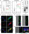
pATOM36 interacts with a previously uncharacterized TAC component. (A) Reciprocal SILAC-IPs of C-terminally HA-tagged pATOM36 and C-terminally Myc-tagged TAC65 from digitonin-solubilized mitochondrial extracts. Mean log10 ratios (induced/uninduced) of proteins detected by quantitative MS in three independent experiments are plotted against the corresponding –log10 P values (one-sided t test). Horizontal dashed lines indicate a P value of 0.05, whereas the vertical black dashed lines mark a threefold-enrichment. The bait proteins are indicated in green. For a complete list of proteins, see Datasets S2 and S3. (B) BN-PAGE of mitochondria-enriched fractions of the procyclic cells expressing either pATOM36-HA or TAC65-Myc. Immunoblots were probed with anti-HA or anti-Myc antibodies, respectively. (C) IF analysis of procyclic T. brucei cells expressing either TAC65-Myc or pATOM36-HA, respectively. DNA is stained with DAPI (blue). ATOM40 serves as a mitochondrial marker (green). HA- or Myc-tagged proteins are shown in red. (Scale bar, 2 µm.) (D) Isolated flagella from procyclic cells expressing TAC65-Myc or pATOM36-HA, respectively. DNA is stained with DAPI (blue). The anti-PFR antiserum stains the paraflagellar rod (green). HA- and Myc-tagged proteins are shown in red. Differential interference contrast (DIC). (Scale bar, 2 µm.) (E) Costaining of TAC65-Myc and pATOM36-HA in isolated flagella. TAC65-Myc is shown in green, pATOM36-HA is shown in red, and the DNA is stained with DAPI (blue). [Scale bar, (Left) 2 µm and (Right) 1 µm.]
Fig. S2.
Fig. S2.
IF analysis of procyclic T. brucei cells expressing pATOM36-HA. (A) Whole cells (Top) and isolated flagella (Bottom) were stained for HA-tagged pATOM36 (red) and ATOM40 (green), respectively. DNA is stained with DAPI (blue). [Scale bar, 5 µm and (Insets) 1 µm.] (B) As in A, but staining was done for HA-tagged pATOM36 (red) and VDAC (green), respectively.
Fig. 4.
Fig. 4.
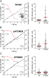
Ablation of TAC65 and pATOM36 causes missegregation of kDNA. (Left) Graphs showing growth curves and loss of kDNA of the indicated knockdown cell lines. Red lines depict percentage of cells still having the kDNA. (Right) Fluorescent intensities of kDNA networks were measured in the indicated knockdown cell lines. Red lines mark the median. Time of induction is indicated. (A) TAC65-RNAi cell line. (Inset) Northern blot showing ablation of TAC65 mRNA. EtBr-stained rRNAs serve as a loading control. (B) pATOM36-RNAi cell line targeting the ORF. (C) ATOM40-RNAi cell line. Northern blots confirming the ablation of ATOM40 and pATOM36 in the corresponding RNAi cell lines have been published previously (13, 14).
Fig. S3.
Fig. S3.
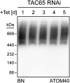
Ablation of TAC65 does not affect assembly of the ATOM complex. Immunoblots of mitochondria-enriched fractions analyzed by BN-PAGE at the indicated time points after induction in the procyclic TAC65-RNAi cell line.
Fig. S4.
Fig. S4.
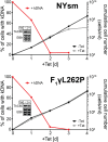
TAC65 is essential in bloodstream form cells (NYsm) but dispensable in an engineered bloodstream from a cell line that can grow in the absence of the kDNA (F1γL262P). Graphs show growth curves and loss of kDNA of the indicated bloodstream form cell lines. Red lines depict percentage of cells still having the kDNA. (Insets) Northern blots demonstrating ablation of endogenous TAC65 mRNA. The corresponding EtBr-stained rRNAs serve as loading controls.
Fig. S5.
Fig. S5.
IF analysis of a procyclic T. brucei TAC65-RNAi cell line in which one allele of TAC65 is tagged with the Myc epitope. Whole cells of uninduced and 40 h-induced cells were stained for Myc-tagged TAC65 using anti Myc antibodies (red) and for the basal bodies using the monoclonal anti–α-tubulin antibody YL1/2 (green). DNA is stained with DAPI (blue). In the merged pictures, the positions of the kDNA (K) and the basal bodies (*) are indicated. [Scale bar, 5 µm and (Insets) 1 µm.]
Fig. 5.
Fig. 5.
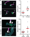
pATOM36 is necessary for kDNA segregation and connects the outer mitochondrial membrane to the basal body. (A) pATOM36 ablation leads to accumulation of unsegregated kDNAs that are connected to the old flagellum. kDNA/basal body regions of uninduced and induced pATOM36-RNAi cells were visualized by SBF-SEM. (Top Left) Uninduced cell in which a single kDNA network each is connected to the old and the new flagellum. (Bottom Left) Induced cell (3 d) in which a large kDNA network is connected only to the old flagellum, indicating a kDNA segregation defect. (Right) Graph depicts volume measurements of kDNAs from uninduced (n = 19) and induced (3 d, n = 19) pATOM36-RNAi cells. Red line, median. (B) Ablation of pATOM36 prevents the formation of the connection between the outer mitochondrial membrane and the basal body. kDNA/basal body regions of uninduced and induced pATOM36-RNAi cells were visualized by SBF-SEM and used to measure distances between the basal body and the mitochondrial membranes. (Top Left) Uninduced cell in which a single kDNA network is connected to a single flagellum. (Bottom Left) kDNA/basal body region of an induced cell (3 d) that lacks kDNA. (Right) Graph depicts the distances between the mitochondrial membrane (mtm) and the basal body (bb) from uninduced (–Tet, 22 bb analyzed) and from induced pATOM36-RNAi cells (+Tet, 12 bb analyzed). For induced cells, only basal bodies lacking an attached kDNA were included in the analysis. Dark green, Mitochondrion; light green, kDNA; purple, flagella; red line, median; white, basal and probasal body.
Fig. 6.
Fig. 6.
pATOM36 has distinct functions in the OM and the TAC. (A) pATOM36-3′UTR-RNAi cell line. (B) pATOM36-3′UTR-RNAi cell line complemented with an ectopically HA-tagged version of pATOM36 lacking the C-terminal 60 amino acids (ΔC60). (C) Same as in B, but complementation was done with a pATOM36 version lacking the last 75 amino acids (ΔC75). (Upper) Graphs showing growth curves and loss of kDNA of the indicated knockdown and complemented 3′UTR-RNAi cell lines. Red lines depict percentage of cells still having the kDNA. (Insets) Northern blots demonstrating ablation of endogenous pATOM36 mRNA. The corresponding EtBr-stained rRNAs serve as loading controls. For the complemented cell lines, immunoblots showing the expression of the inducible HA-tagged versions of pATOM36 are also shown. (Bottom Left) Graph of the fluorescent intensities of kDNA networks in the indicated knockdown cell lines. Time of induction is indicated. Red lines mark the median. (Bottom Right) BN-PAGE (BN) immunoblots of mitochondrial-enriched fractions at the indicated time points after induction of pATOM36-RNAi stained with the anti-ATOM40 antiserum.
Fig. S6.
Fig. S6.
C-terminally truncated HA-tagged versions of pATOM36 cofractionate with crude mitochondria and are integral membrane proteins. (Top) Immunoblots of total (Tot), digitonin-extracted cytosolic (Cyt), and crude mitochondrial (Mit) fractions from cell lines expressing C-terminally truncated HA-tagged versions (ΔC60, ΔC75) of pATOM36 probed with anti-HA antibodies (HA). ATOM40 and elongation factor 1a (EF-1a) serve as mitochondrial and cytosolic markers, respectively. (Bottom) Immunoblots of an alkaline carbonate extraction from mitochondria-enriched fractions of the indicated transgenic cell lines. Pe, pellet; Sup, supernatant; Tot, total. The integral membrane protein ATOM40 and the peripheral membrane protein CytC serve as markers.
Fig. S7.
Fig. S7.
pATOM36 has two predicted transmembrane domains that contain conserved GxxxG motifs. Shown is the protein sequence alignment of pATOM36 orthologs. Lbra, Leishmania braziliensis; linf, Leishmania infantum; lmaj, Leishmania major; lmex, Leishmania mexicana; tbrg, T. brucei gambiense; tbr4, T. brucei brucei 427; tbr9, T. brucei brucei 927; tcon, Trypanosoma congolense; tcru, Trypanosoma cruzi; tviv, Trypanosoma vivax; teva, Trypanosoma evansi. Predicted transmembrane domains (phobius.sbc.su.se) are highlighted in red rectangles (TMD1, TMD2). Conserved residues are marked with asterisks, and GxxxG motifs are highlighted with gray bars. Sites of deletion for truncated versions of pATOM36 are indicated (ΔC60, ΔC75).

Similar articles

Cited by

References

    1. Friedman JR, Nunnari J. Mitochondrial form and function. Nature. 2014;505(7483):335–343. - PMC - PubMed
    1. Gray MW. Mitochondrial evolution. Cold Spring Harb Perspect Biol. 2012;4(9):a011403. - PMC - PubMed
    1. Mishra P, Chan DC. Mitochondrial dynamics and inheritance during cell division, development and disease. Nat Rev Mol Cell Biol. 2014;15(10):634–646. - PMC - PubMed
    1. Westermann B. Mitochondrial inheritance in yeast. Biochim Biophys Acta. 2014;1837(7):1039–1046. - PubMed
    1. Chacinska A, Koehler CM, Milenkovic D, Lithgow T, Pfanner N. Importing mitochondrial proteins: Machineries and mechanisms. Cell. 2009;138(4):628–644. - PMC - PubMed

Publication types

MeSH terms

Substances

LinkOut - more resources