

The Carbofix™ "Piccolo Proximal femur nail": A new perspective for treating proximal femur lesion. A technique report
- PMID: 27436924
- PMCID: PMC4939471
- DOI: 10.1016/j.jor.2016.07.001
The Carbofix™ "Piccolo Proximal femur nail": A new perspective for treating proximal femur lesion. A technique report
Abstract
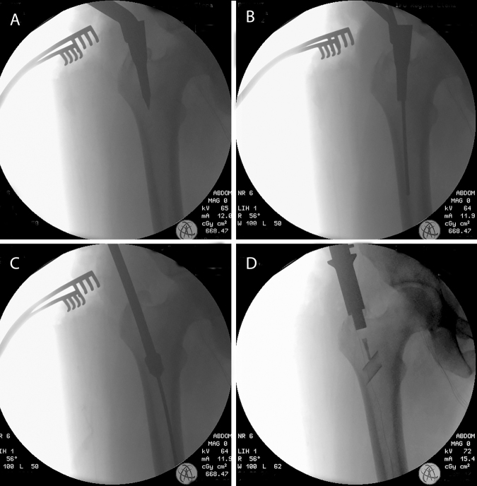
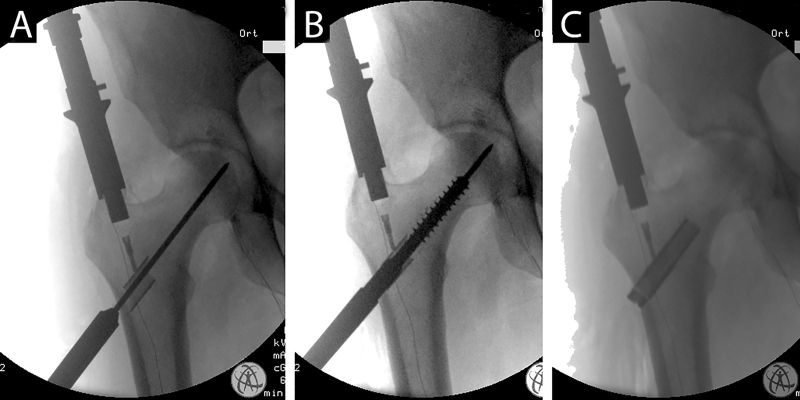
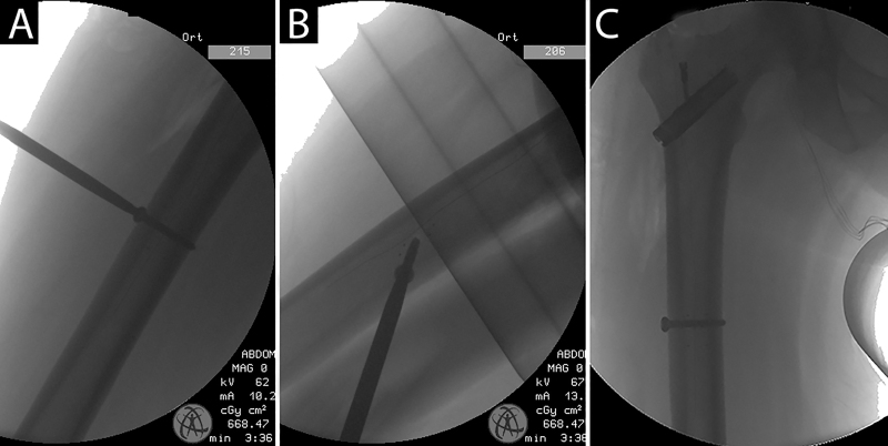
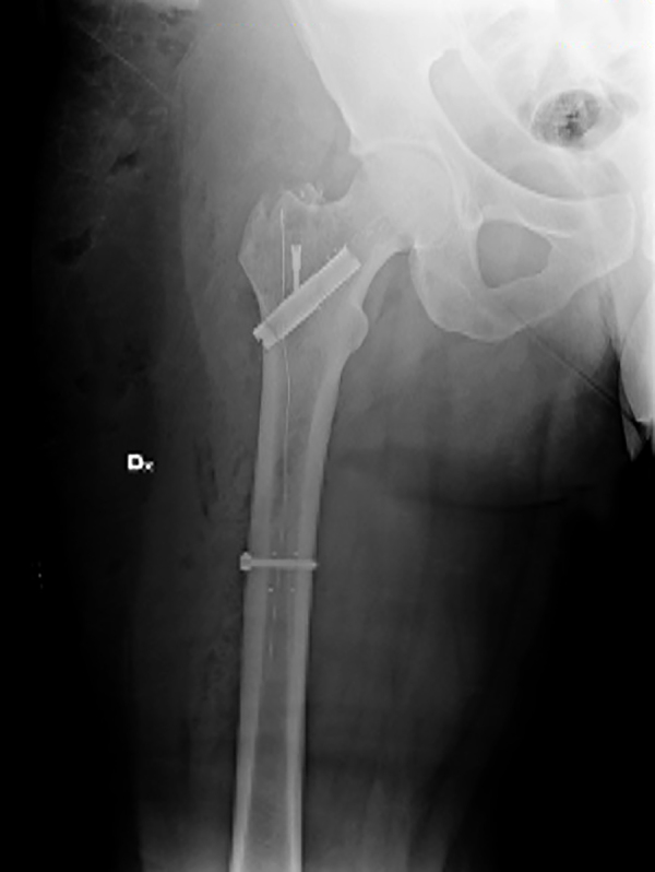
Metastases to proximal femur are common and surgery is often suggested to prevent fractures; otherwise it is necessary in cases where this has already occurred. Adjuvant radiotherapy is necessary to reduce the risk of local progression. Nevertheless, the success or failure of radiation therapy treatments depends upon the accuracy in which target identification is correct and dose prescription is fulfilled. Unfortunately, the use of titanium nails consistently limits radiation dose; indeed, the presence of ferromagnetic artifacts interferes with target identification. We present the technique for implant a new carbon fiber nail useful to reduce the ferromagnetic artifacts which allows a better adjuvant radiotherapy.
Keywords: Artifacts; Osteolysis; Pathologic fracture; Proximal femur metastases; Radiotherapy; Tumor.
Figures
References
-
- Hage W.D., Aboulafia A.J., Aboulafia D.M. Incidence, location, and diagnostic evaluation of metastatic bone disease. Orthop Clin N Am. 2000;31(4):515–528. - PubMed
-
- Sim F.H. Metastatic bone disease of the pelvis and femur. Instr Course Lect. 1992;41:317–327. - PubMed
-
- Jacofsky D.J., Haidukewych G.J. Management of pathologic fractures of the proximal femur: state of the art. J Orthop Trauma. 2004;18(7):459–469. - PubMed
-
- Camnasio F., Scotti C., Peretti G.M., Fontana F., Fraschini G. Prosthetic joint replacement for long bone metastases: analysis of 154 cases. Arch Orthop Trauma Surg. 2008;128(8):787–793. - PubMed
-
- Townsend P.W., Smalley S.R., Cozad S.C., Rosenthal H.G., Hassanein R.E. Role of postoperative radiation therapy after stabilization of fractures caused by metastatic disease. Int J Radiat Oncol Biol Phys. 1995;31(1):43–49. - PubMed
LinkOut - more resources
Full Text Sources
Other Literature Sources
