. 2016 Aug 4;536(7614):86-90.
doi: 10.1038/nature18935. Epub 2016 Jul 20.

CD47-blocking antibodies restore phagocytosis and prevent atherosclerosis

CD47-blocking antibodies restore phagocytosis and prevent atherosclerosis

Yoko Kojima et al. Nature. .

Abstract

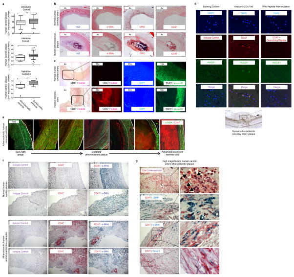
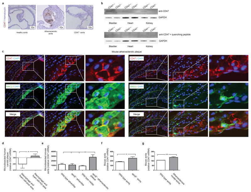
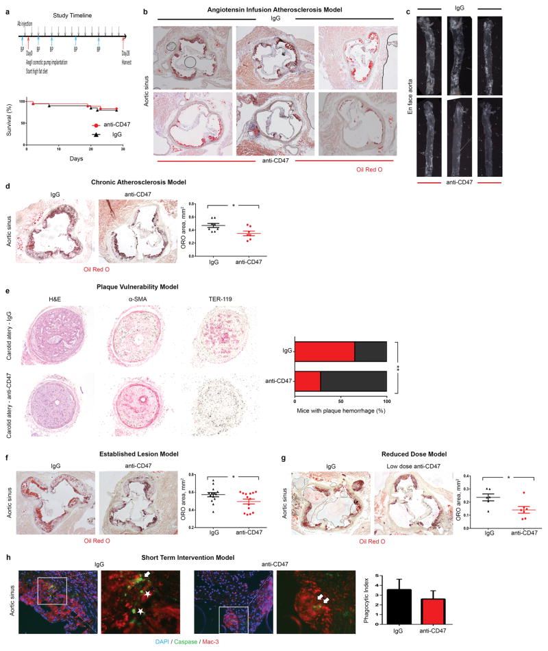
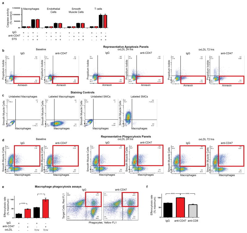
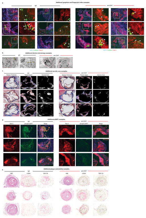
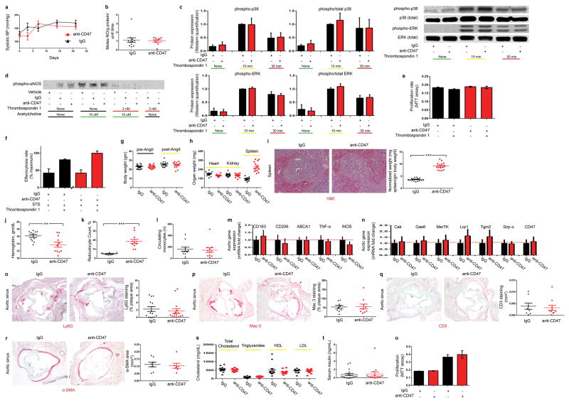
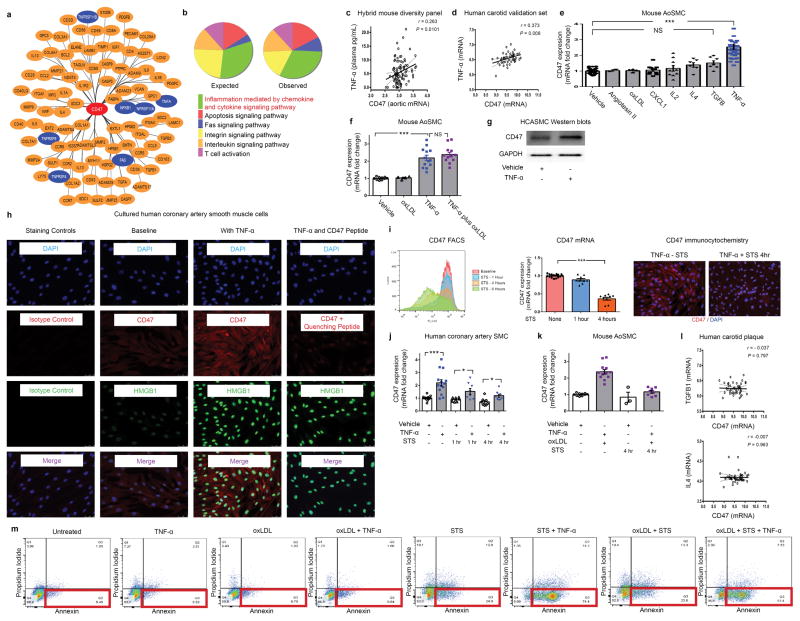
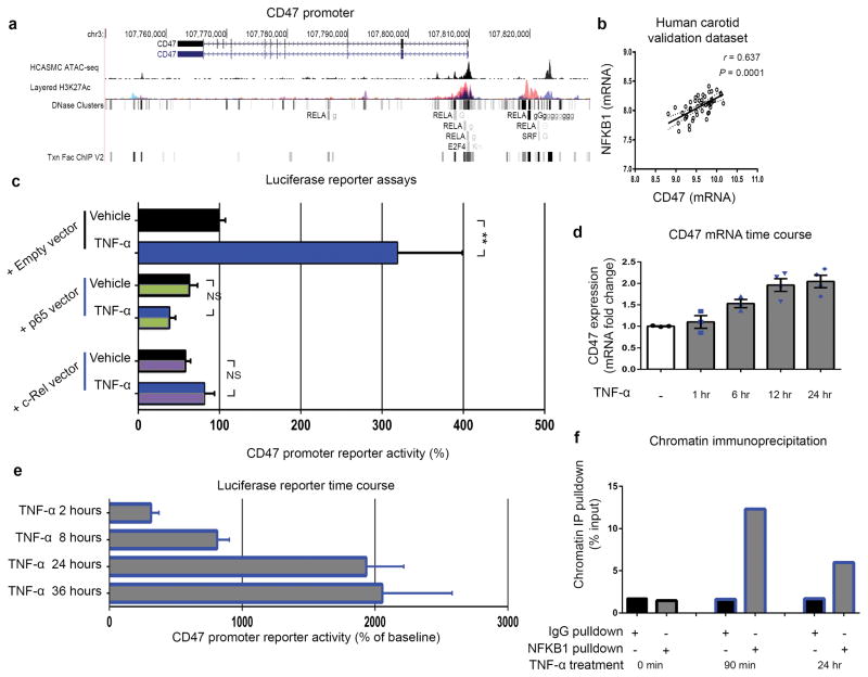
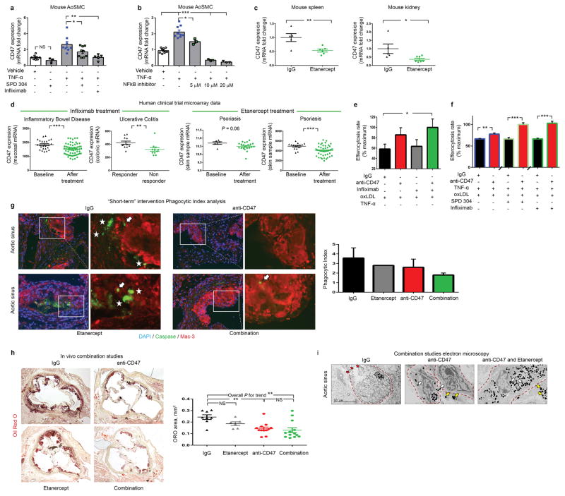
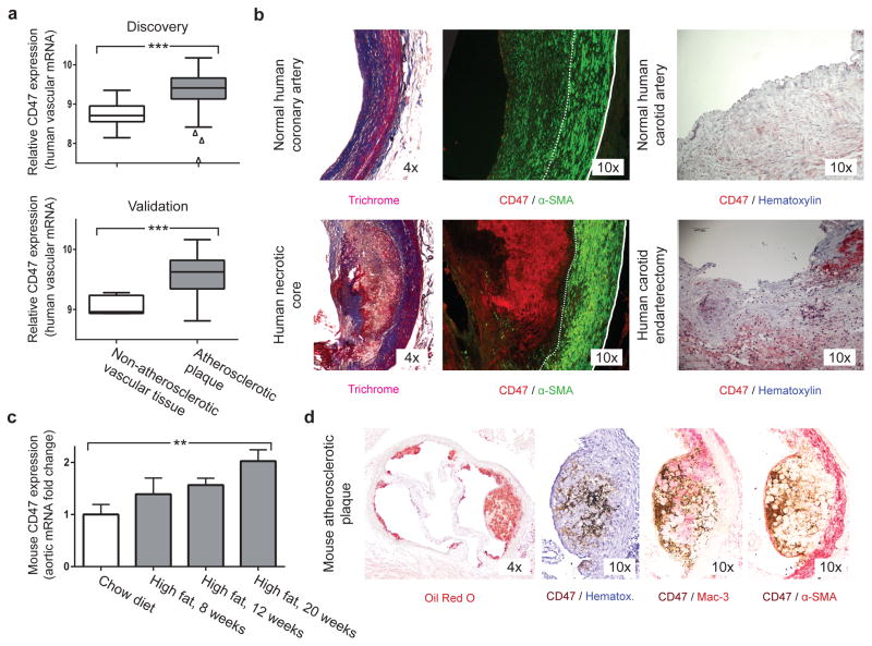
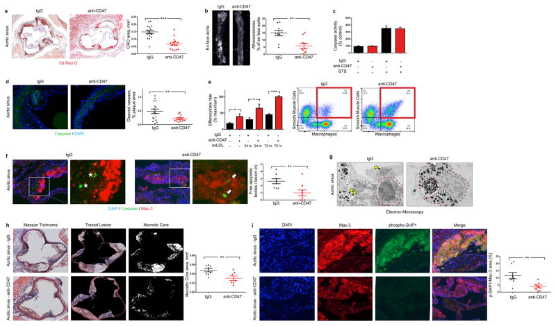
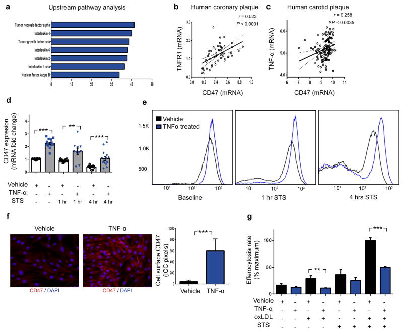
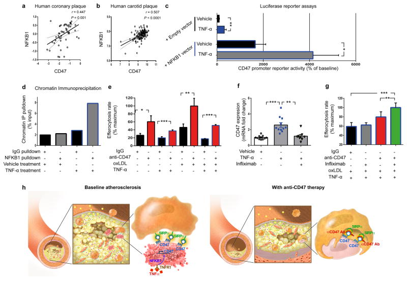
Atherosclerosis is the disease process that underlies heart attack and stroke. Advanced lesions at risk of rupture are characterized by the pathological accumulation of diseased vascular cells and apoptotic cellular debris. Why these cells are not cleared remains unknown. Here we show that atherogenesis is associated with upregulation of CD47, a key anti-phagocytic molecule that is known to render malignant cells resistant to programmed cell removal, or 'efferocytosis'. We find that administration of CD47-blocking antibodies reverses this defect in efferocytosis, normalizes the clearance of diseased vascular tissue, and ameliorates atherosclerosis in multiple mouse models. Mechanistic studies implicate the pro-atherosclerotic factor TNF-α as a fundamental driver of impaired programmed cell removal, explaining why this process is compromised in vascular disease. Similar to recent observations in cancer, impaired efferocytosis appears to play a pathogenic role in cardiovascular disease, but is not a fixed defect and may represent a novel therapeutic target.


Conflict of interest statement

The authors declare competing financial interests: N.J.L. and I.L.W. have filed a patent describing inhibition of CD47 as a method to prevent atherosclerosis.

Figures

Extended Data Figure 1
Extended Data Figure 1. CD47 expression correlates with risk for clinical cardiovascular events and is progressively upregulated in the necrotic core of human blood vessels during atherogenesis
(a). cDNA microarray expression profiling in the BiKE carotid endarterectomy biobank reveals that the relative expression of CD47 is increased in vascular homogenates taken from subjects with symptomatic disease (stroke or TIA, n=85) compared to those with stable, asymptomatic lesions (n=40). Similar findings were observed in the non-overlapping discovery and validation cohorts from BiKE (n=55), and a second validation cohort from the Helsinki Carotid Endarterectomy Study (HeCES, n=21). Data presented as Tukey boxplots. (b). Immunohistochemical staining reveals that CD47 co-localizes with lipidated plaque within human coronary lesions, as measured by Oil-Red-O (ORO) staining. (c). Immunofluorescent staining of coronary samples confirms that CD47 is upregulated within the necrotic core. (d). High magnification (40x) imaging of atherosclerotic coronary plaque confirms that CD47 expression is present on the surface of nucleated cells undergoing cell death, as indicated by HMGB1 staining. Specificity of the anti-CD47 Ab is confirmed in assays where the signal was quenched by preincubating the sections with recombinant CD47 peptide prior to primary antibody exposure. (e). Additional representative coronary artery segments spanning the spectrum of progressive coronary artery disease (non-atherosclerotic coronary, early ‘fatty streak’, inwardly remodeled plaque, and advanced ulcerated lesion with necrotic core) confirm that CD47 is progressively upregulated during the development of coronary artery disease. The tunica media is indicated by dotted lines. (f). Additional staining in human carotid artery sections confirms that CD47 expression is upregulated in atherosclerosis relative to healthy tissue, and appears most pronounced within the necrotic core. (g). High magnification (100x) imaging confirms that the CD47 expression is specific to lesional cells, including SMCs (αSMA), macrophages (CD68) and cells undergoing programmed cell death (Casp3). Comparisons made by two-tailed t tests. ** = P < 0.01, * = P < 0.05.
Extended Data Figure 2
Extended Data Figure 2. CD47 expression is increased in mouse models of atherosclerosis
(a). Mice injected with biotin labeled anti-CD47 Ab reveal that this Ab accumulates in the vasculature of atherosclerotic mice (middle), relative to non-atherosclerotic control mice (left). No staining is detected in CD47−/− mice (right), indicating specificity of the Ab. (b). Western blotting of tissue homogenates obtained from WT and CD47−/− mice (with and without quenching CD47 peptide) further confirms the specificity of the antibody. For gel source data, see Supplementary Figure 1. (c). High resolution immunofluorescent staining of murine atherosclerotic plaques indicate that CD47 is specifically expressed on the surface of lesional cells, rather than extracellular debris. (d). Publically available microarray data from laser capture microdissected (LCM) vascular tissue reveals that CD47 expression is increased within the macrophage and foam cell-rich area of human plaque, relative to macrophage and foam cell-poor areas (GSE23303). (e). Similar results were observed in LCM tissue from mouse atherosclerotic plaque tissue, relative to non-atherosclerotic medial and adventitial tissue (GSE21419). (f) and (g). Additional results from the Gene Expression Omnibus (GEO) database reveal that aortic CD47 expression is upregulated in murine models of atherosclerosis, as observed in the current study (GSE2372 and GSE19286). ** = P < 0.03, * = P < 0.05.
Extended Data Figure 3
Extended Data Figure 3. Anti-CD47 Ab reduces atherosclerotic burden in several orthogonal in vivo models
(a). Study timeline detailing osmotic minimpump implantation and high fat feeding to induce atherosclerosis in the apoE−/−-“Angiotensin infusion” model used herein. Kaplan Meier curves indicate no change in mortality with anti-CD47 treatment during 28 days of follow up. Additional representative examples confirm that anti-CD47 Ab: (b). Reduces atherosclerosis content in the aortic sinus; and (c). Reduces the percent of the en-face aorta covered by atherosclerotic plaque. Several additional atherosclerosis models were also used in this study to confirm the beneficial effects of anti-CD47 Ab therapy, and to model additional aspects of human cardiovascular disease. These include: (d). A “chronic atherosclerosis” model, where Ab therapy was given for 12 weeks (with no angiotensin infusion); (e). A “plaque vulnerability” model, where the impact of Ab therapy on plaque rupture and intraplaque hemorrhage was quantified; (f). An “established disease” model, where therapy was given for 7 weeks after mice had already developed advanced plaques of equivalent size; and (g). A “reduced dose” model, where the dose of anti-CD47 Ab was reduced by 75%, relative to the preceding studies. (h). Additionally, a “short term” study was performed where mice with established lesions of equivalent size and identical apoptosis rates were pulsed with only five days of anti-CD47 Ab therapy prior to harvest, to quantify the impact of therapy on efferocytosis rates, independent of lesion size (Phagocytic Index indicated by the ratio of ‘free’ (white stars) to ‘associated’ (white arrows) apoptotic bodies). Additional methodological details are provided in the Methods. Comparisons made by two-tailed t tests. ** = P < 0.03, * = P < 0.05. Error bars represent the SEM.
Extended Data Figure 4
Extended Data Figure 4. Anti-CD47 Ab promotes the phagocytosis of diseased SMCs and macrophages, without altering apoptosis
(a). In vitro caspase activity assays reveal that anti-CD47 Ab does not alter rates of programmed cell death in any vascular cell type (b). Flow cytometry assays confirm that anti-CD47 Ab has no effect on apoptosis at baseline, or in vascular SMCs exposed to 24 or 72 hours of oxLDL. (c). Staining controls for the in vitro phagocytosis assays. (d). Representative FACS plots for the in vitro efferocytosis conditions displayed in Figure 2E. The right upper quadrant (highlighted in red) includes double positive cells which are taken to represent a macrophage that has ingested a target cell. (e). In vitro efferocytosis assays using lipid-loaded macrophages as the target cell confirm that anti-CD47 Ab also stimulates the clearance of this vascular cell type, similar to the findings observed with SMCs. (f). Additional in vitro efferocytosis assays confirm that anti-CD47 Ab stimulates phagocytosis of vascular cells in a specific manner. Error bars represent the SEM.
Extended Data Figure 5
Extended Data Figure 5. Additional examples confirm the pro-efferocytic properties of anti-CD47 Ab in vivo
(a). Additional representative images detail that mice treated with anti-CD47 Ab have a lower overall burden of apoptotic debris (Caspase in green), as well as fewer examples of ‘free’ apoptotic bodies (white stars). Those apoptotic bodies that are present in these lesions are more often found in close proximity to macrophages (Mac-3 in red) and are considered ‘associated’ with a phagocyte if physically co-localized (white arrows). (b). Additional electron microscopy examples provide further qualitative evidence that phagocytes present in the lesions of mice treated with anti-CD47 Ab are more likely to have ingested several apoptotic bodies (white arrows) compared to lesions from IgG treated mice which are more likely to have a high burden of ‘free’ apoptotic bodies (yellow arrows). Additional representative examples of the necrotic core analysis, the phospho-SHP1 staining, and the plaque hemorrhage analysis are provided in (c), (d) and (e), respectively, as described in the Methods.
Extended Data Figure 6
Extended Data Figure 6. Full dose anti-CD47 Ab induces anemia, but does not appear to alter nitric oxide (NO) elaboration, thrombospondin-1 dependent signaling, or any other processes relevant to vascular biology
(a). No significant change in blood pressure is observed between mice treated with IgG or anti-CD47 Ab, arguing against a systemic difference in NO production due to Ab therapy. (b). Direct measurement of pulmonary NO release via the Griess reaction indicates that anti-CD47 Ab does not increase NO elaboration in vivo. (c). Western blot analysis of cultured murine vascular cells reveals that anti-CD47 Ab has no effect on the expected induction of p38 and ERK phosphorylation secondary to Tsp-1 treatment. (d). Similarly, anti-CD47 Ab has no effect on Tsp-1-dependent inhibition of eNOS phosphorylation, nor Acetylcholine-dependent induction of eNOS phosphorylation. (e). MTT assays show that anti-CD47 Ab does not affect cellular proliferation rates in the presence of Tsp-1. (f). In vitro efferocytosis assays show that the expected basal increase in phagocytosis observed after apoptotic cells are exposed to Tsp-1 (black bars) is not altered in the presence of anti-CD47 Ab (red bars). (g). Compared to mice receiving control IgG, mice receiving anti-CD47 Ab treatment have similar body weights at baseline and at sacrifice. (h). No difference is observed for the weight of any organ between groups, with the exception of splenomegaly observed in the anti-CD47 Ab treated animals. (i). Histological analysis of the explanted splenic tissue reveals an increase in the red pulp of anti-CD47 treated mice without any change in fibrosis or white pulp content, suggestive of increased erythrophagocytosis in this reticuloendothelial organ. Dot plots detail the hemoglobin count (j), reticulocyte count (k) and circulating monocyte count (l) for each animal in the acute 4 week angiotensin-infusion atherosclerosis model. Note that this anemia appears to be self-limited, and no anemia was observed in the chronic atherosclerosis model or the reduced dose model (P = 0.54 and 0.57, respectively). (m). mRNA analysis of aortic tissue reveals that anti-CD47 Ab has no significant impact on the expression of macrophage polarization factors in vivo. (n). anti-CD47 Ab also has no effect on the aortic expression of any other candidate efferocytosis gene. Additional quantitative analyses reveals that anti-CD47 Ab has no effect on in vivo: (o). Neutrophil content (as assessed by Ly6G positive area normalized to lesion size); (p) Macrophage content (as assessed by Mac-3 positive area normalized to lesion size); (q) T cell content (as assessed by CD3 positive area across the lesion and adventitia); or (r). Smooth muscle cell content (as quantified by α-SMA positive area in the aortic sinus from the external elastic lamina to the lumen). (s). anti-CD47 Ab also had no effect on lipid level or serum insulin (t). (u). MTT assays reveal that anti-CD47 Ab has no effect on the proliferation of primary aortic SMCs obtained from apoE−/− deficient mice either at baseline (left) or in the presence of 10% serum (right). Comparisons made by two-tailed t tests, unless otherwise specified. *** = P < 0.001, ** = P < 0.01, * = P < 0.05. Error bars represent the SEM. For gel source data, see Supplementary Figure 1; for detailed serological data, see Extended Data Table 1.
Extended Data Figure 7
Extended Data Figure 7. Additional bioinformatic and experimental analyses further implicate a central role for the pro-inflammatory cytokine, TNF-α, in vascular CD47 signaling
(a). Cytoscape network visualization of the genes which are significantly correlated with CD47 expression in both human and murine atherosclerotic plaque reveals a high number of TNF-α-related factors (indicated in blue), including ligands, receptors, and downstream signaling factors. (b). PANTHER pathway analysis of those genes which were (a) significantly associated with CD47 expression in mouse and human vascular tissue and (b) have been previously associated with atherosclerosis through the STAGE study, identifies “inflammation mediated by chemokine and cytokine signaling pathway” as the most over-abundant pathway associated with CD47 expression in vascular tissue. (c). Using the Hybrid Mouse Diversity Panel (HMDP), which correlates aortic gene expression with Luminex cytokine array data of plasma samples from over 100 inbred strains of mice, we found that vascular CD47 expression is positively correlated with three inflammatory cytokines in vivo, including TNF-α, IL-2 and CXCL1. Correlation data shown for CD47 and TNF-α. (d). Co-expression studies confirm that TNF-α and CD47 expression are positively correlated in human carotid endarterectomy samples from the BiKE validation study. The Pearson correlation coefficient was determined assuming a Gaussian distribution and P values were determined using a two-tailed test. (e). Experiments with primarily cultured mouse aortic SMCs indicate that TNF-α reproducibly induces CD47 mRNA upregulation, while a number of other classical pro-atherosclerotic stimuli have no significant effect. Notably, CXCL1, IL4, TGFβ and IL-2 fail to induce CD47 expression in vitro, as assessed by ANOVA. (f). Additional studies suggest that the effect of TNF-α on CD47 expression persists in the presence of oxidized LDL, as occurs in the atherosclerotic plaque. (g). Western blotting confirms that TNF-α induces CD47 expression in vascular cells at the protein level. For gel source data, see Supplementary Figure 1. (h). Immunocytochemistry studies of HCASMCs confirm that CD47 expression is induced on the cell surface of TNF-α treated cells. TNF-α effect is assessed by co-staining for HMGB1, and antibody specificity is confirmed with isotype control and recombinant CD47 peptide quenching assays. (i). Multiple assays (including FACS, Taqman and immunocytochemistry studies) reveal that CD47 expression is downregulated on vascular SMCs during programmed cell death, as has previously been observed with inflammatory cells. (j). Confirmatory assays in cultured human coronary artery SMC reveal that TNF-α induces changes similar to those observed in murine cells (Fig 3D), including an induction of CD47 under physiological conditions and a blunting of its expected downregulation during apoptosis. (k). TNF-α’s capacity to impair CD47 downregulation during programmed cell death is also observed in mouse SMCs simultaneously exposed to pro-apoptotic stimuli and oxidized LDL. (l). No correlation between CD47 and other candidate cytokines was observed in the BiKE biobank, further supporting a specific relationship between CD47 and TNF-α. (m). Representative FACS-based apoptosis panels from cells exposed to the conditions used in Fig 3G confirm that TNF-α suppresses efferocytosis (Fig 3G) despite increasing programmed cell death. Comparisons made by two-tailed t tests, unless otherwise specified. *** = P < 0.001, * = P < 0.05. Error bars represent the SEM.
Extended Data Figure 8
Extended Data Figure 8. The CD47 promoter contains predicted binding sites for the TNF-α-related transcription factor, NFKB1
(a). UCSC genome browser screenshot showing overlay of human CD47 transcript with ENCODE transcription factor binding sites (including RELA, E2F4, and SRF), along with the active H3K27ac histone modification ChIP-seq track, and a custom track for chromatin accessibility in human coronary artery smooth muscle cells (HCASMC) using the Assay for Transposase Accessible Chromatin followed by sequencing (ATAC-seq). These chromatin, DNase hypersensitivity sites, and published ChIP-seq data suggest that members of the NFKB family of transcription factors could regulate CD47 expression in vascular tissue. (b). Additional co-expression studies in the BiKE validation study confirm that NFKB1 and CD47 expression are positively correlated in human carotid endarterectomy samples. The Pearson correlation coefficient was determined assuming a Gaussian distribution and P values were determined using a two-tailed test. (c). Additional luciferase promoter reporter assays reveal that induction of CD47 expression requires the presence of NFKB1 and cannot be induced by other NFKB co-factors such as RELA or c-REL. Time course studies confirm that CD47 expression is induced by TNF-α within 24 hours, suggesting a direct transcriptional relationship ((d) Taqman mRNA expression assays; (e) luciferase reporter assays). (f). Additional chromatin immunoprecipitation studies confirm that NFKB1 protein binds the CD47 promoter within 90 minutes of TNF-α-treatment in human coronary artery SMCs. ** = P < 0.01, * = P < 0.05. Error bars represent the SEM.
Extended Data Figure 9
Extended Data Figure 9. Dual inhibition of CD47 and TNF-α provides a combinatorial effect
(a). Pretreatment of mouse vascular SMCs with a chemical inhibitor (SPD 304) or a monoclonal Ab (infliximab) directed against TNF-α prevents the increase in CD47 expression normally seen after TNF-α exposure. (b). Similar effects were observed with the NFkB inhibitor, BAY 11–7085, confirming the molecular mechanism outlined in Fig 4. (c). Mice injected with four weeks of the decoy TNF-α receptor, etanercept, display a significant reduction in their in vivo expression of CD47 in splenic (left) and renal (right) tissue. (d). Publically available microarray data from human clinical trials of commercially available TNF-α inhibitors reveal that subjects treated with these agents also express lower levels of CD47 in vivo (as assessed by two-tailed t tests), confirming the mouse findings above (GSE accession #s from left to right: 16879 n=85, 12251 n=22, 47751 n=28 and 41663 n=66). Additional in vitro efferocytosis assays confirm a synergistic effect of anti-CD47 Ab with a variety of TNF-α inhibitors in both the absence (d) and presence (f) of exogenous TNF-α. (g). Mice with established plaques of identical size and with equivalent rates of apoptosis were treated with a short course (5 days) of IgG, anti-CD47 Ab, Etanercept, or combination therapy prior to harvest. As shown the phagocytic index (indicated by the ratio of ‘free’ (white stars) to ‘associated’ (white arrows) apoptotic bodies) displayed a non-significant trend toward improvement for combination therapy (P > 0.05). (h). When treated for a full 28 days in the Angiotensin-infusion model, individual comparisons showed that etanercept alone had no effect on atherosclerosis, and combination therapy was not significantly different from anti-CD47 alone, likely due to the potent effect of anti-CD47 monotherapy. ANOVA post-test analysis did identify a significant linear trend across all four groups (P for trend < 0.01). (i). Electron microscopy provides additional qualitative evidence that combination therapy may provide an incremental effect on efferocytosis, as suggested by an increased prevalence of macrophages within the plaque which had ingested a large number of apoptotic bodies (white arrows), a reduced prevalence of free apoptotic bodies (yellow arrows), and a reduced prevalence of uncleared cells undergoing secondary necrosis (red arrows). *** = P < 0.001, ** = P < 0.01, * = P < 0.05. Error bars represent the SEM.
Figure 1
Figure 1. The ‘don’t eat me’ ligand, CD47, is upregulated in atherosclerosis
(a). Microarray expression profiling in two carotid endarterectomy (CEA) cohorts reveals that CD47 expression is significantly increased in human atherosclerotic plaque, relative to non-diseased vascular tissue (data displayed as Tukey boxplots, n=182 subjects). (b). Immunostaining identifies intense CD47 upregulation within the necrotic core of human atherosclerotic coronary artery lesions (left) and carotid plaques (right). (c). Taqman mRNA analysis confirms that vascular CD47 expression progressively increases in a mouse model of atherosclerosis (apoE−/− mice fed hi fat diet, grey), relative to control animals (C57BL/6 mice fed chow, white, n=4 mice/timepoint). (d). Immunohistochemistry staining with a biotin labeled antibody (brown) reveals that CD47 expression co-localizes with apoptotic tissue within murine atherosclerotic plaque. *** = P < 0.001, ** = P for trend < 0.03. Error bars represent the standard error of the mean (SEM).
Figure 2
Figure 2. Inhibition of CD47 stimulates efferocytosis and prevents atherosclerosis
(a). Compared to mice treated with a control Ab (IgG, n=16), mice treated with an inhibitory anti-CD47 Ab (n=15) develop significantly smaller atherosclerotic plaques, as measured by Oil-Red-O (ORO) content in the aortic sinus. (b). Total aortic atherosclerosis content is also reduced. Inhibition of CD47 signaling does not alter the rate of programmed cell death in vitro (c) but does reduce the accumulation of apoptotic bodies in vivo (d). (e). Anti-CD47 Ab promotes efferocytosis of vascular cells at baseline and after exposure to pro-atherosclerotic lipids. Representative FACS phagocytosis plots for lipid loaded (72hr) SMCs shown at right (all assays repeated in triplicate). (f). In vivo, anti-CD47 Ab reduces the number of ‘free’ apoptotic bodies not associated with phagocytic macrophages, potentially indicative of increased efferocytosis (stars indicate ‘free’ apoptotic bodies, arrows indicate ‘not-free’ apoptotic bodies). (g). Electron microscopy confirms that mice treated with anti-CD47 Ab display features of enhanced intraplaque efferocytosis, including an increased prevalence of macrophages which had ingested multiple apoptotic bodies (white arrows) and a reduced burden of ‘free’ apoptotic bodies (yellow arrows). (h). Mice treated with anti-CD47 Ab develop smaller necrotic cores than mice treated with IgG. (i). Anti-CD47 Ab inhibits phosphorylation of lesional SHP1, a key anti-phagocytic effector molecule known to signal downstream of CD47. STS = staurosporine. Comparisons made by two-tailed t tests. *** = P < 0.001, ** = P < 0.01, * = P < 0.05. Error bars represent the SEM.
Figure 3
Figure 3. The pro-atherosclerotic cytokine, TNF-α, induces CD47 expression and renders vascular cells resistant to phagocytic clearance
(a). Ingenuity Pathway Analysis identifies “Tumor necrosis factor alpha” as the regulator most likely to be upstream of genes which are co-expressed with CD47 in vascular tissue ex vivo. Co-expression studies confirm that CD47 is positively correlated with the canonical TNF-α receptor, TNFR1, in human coronary plaque (b) and TNF-α levels in human carotid plaque (c). The Pearson correlation coefficient was determined assuming a Gaussian distribution and P values were determined using a two-tailed test shown with the 95% confidence band of the best fit line. (d). In vitro, TNF-α treatment significantly increases the basal expression of CD47 in vascular SMCs, and blunts the decrease expected to occur during apoptosis. Flow cytometry (e) and fluorescent microscopy (f) confirm that TNF-α increases the cell-surface expression of CD47 on vascular cells at baseline and during programmed cell death. (g). In vitro efferocytosis assays indicate that TNF-α treatment renders vascular SMCs resistant to programmed cell clearance under a variety of pro-atherosclerotic conditions. Comparisons made by two-tailed t tests. *** = P < 0.001, ** = P < 0.01. Error bars represent the SEM.
Figure 4
Figure 4. TNF-α promotes CD47 expression via NFKB1 and is a translational cardiovascular target
Co-expression analyses confirm that NFKB1 is significantly correlated with CD47 expression in both human coronary (a) and carotid (b) atherosclerotic plaque. Pearson correlation coefficients were determined assuming a Gaussian distribution and P values were determined using a two-tailed test. (c). Dual luciferase reporter assays reveal that CD47 promoter activity is stimulated in cells treated with TNF-α (top) but that this effect is significantly enhanced in cells co-transfected with an NFKB1 expression vector. (d). Chromatin immunoprecipitation studies confirm significant enrichment of NFKB1 protein on the CD47 promoter in TNF-α-treated human coronary artery SMCs. (e). In vitro efferocytosis assays reveal that anti-CD47 Ab enhances the clearance of cells exposed to TNF-α, and that its pro-efferocytic capacity is enhanced under pro-atherosclerotic conditions. (f). Pretreatment with the anti-TNF-α monoclonal Ab, Infliximab, prevents the upregulation in CD47 mRNA that normally occurs in SMCs exposed to TNF-α. (g). Concomitant inhibition of CD47 and TNF-α using anti-CD47 Ab and Infliximab, respectively, produces synergistic benefit in the clearance of diseased vascular cells as assessed by ANOVA. (h). Putative mechanism explaining why efferocytosis is impaired in cardiovascular disease, and how inhibition of CD47-SIRPα signaling could represent a new therapeutic target. Comparisons made by two-tailed t tests, unless otherwise specified. *** = P < 0.001, ** = P < 0.01, * = P < 0.05. Error bars represent the SEM.

Comment in

References

    1. Libby P, Ridker PM, Hansson GK. Progress and challenges in translating the biology of atherosclerosis. Nature. 2011;473:317–25. - PubMed
    1. Schrijvers DM, De Meyer GR, Kockx MM, Herman AG, Martinet W. Phagocytosis of apoptotic cells by macrophages is impaired in atherosclerosis. Arterioscler Thromb Vasc Biol. 2005;25:1256–61. - PubMed
    1. Thorp E, Tabas I. Mechanisms and consequences of efferocytosis in advanced atherosclerosis. J Leukoc Biol. 2009;86:1089–95. - PMC - PubMed
    1. Oldenborg PA, Zheleznyak A, Fang YF, Lagenaur CF, Gresham HD, Lindberg FP. Role of CD47 as a marker of self on red blood cells. Science. 2000;288:2051–4. - PubMed
    1. Chao MP, Majeti R, Weissman IL. Programmed cell removal: a new obstacle in the road to developing cancer. Nat Rev Cancer. 2012;12:58–67. - PubMed

Publication types

MeSH terms