Review
. 2016 Oct 10;591(1):279-291.
doi: 10.1016/j.gene.2016.07.033. Epub 2016 Jul 18.

FBN1: The disease-causing gene for Marfan syndrome and other genetic disorders

Affiliations
Review

FBN1: The disease-causing gene for Marfan syndrome and other genetic disorders

Lynn Y Sakai et al. Gene. .

Abstract

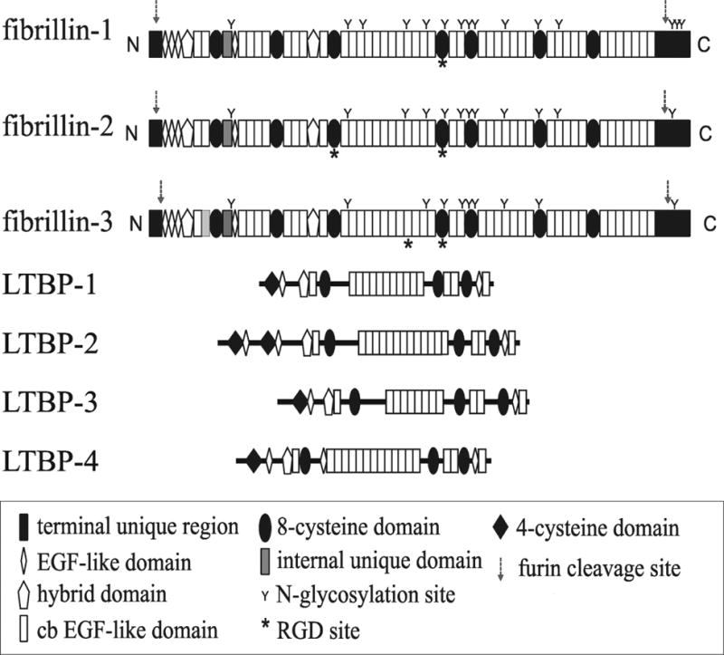
FBN1 encodes the gene for fibrillin-1, a structural macromolecule that polymerizes into microfibrils. Fibrillin microfibrils are morphologically distinctive fibrils, present in all connective tissues and assembled into tissue-specific architectural frameworks. FBN1 is the causative gene for Marfan syndrome, an inherited disorder of connective tissue whose major features include tall stature and arachnodactyly, ectopia lentis, and thoracic aortic aneurysm and dissection. More than one thousand individual mutations in FBN1 are associated with Marfan syndrome, making genotype-phenotype correlations difficult. Moreover, mutations in specific regions of FBN1 can result in the opposite features of short stature and brachydactyly characteristic of Weill-Marchesani syndrome and other acromelic dysplasias. How can mutations in one molecule result in disparate clinical syndromes? Current concepts of the fibrillinopathies require an appreciation of tissue-specific fibrillin microfibril microenvironments and the collaborative relationship between the structures of fibrillin microfibril networks and biological functions such as regulation of growth factor signaling.

Keywords: FBN1; Fibrillin; Fibrillinopathies; Marfan syndrome; Microenvironment; Thoracic aortic aneurysm; Weill–Marchesani syndrome.


Figures

Figure 1
Figure 1. Ultrastructure of Fibrillin Microfibrils
A. High magnification images of fibrillin microfibrils in human amnion show fibrils of uniform diameter with alternating hollow and filled (light and dark) regions. B. Fibrillin microfibrils exist in bundles (*), especially in close proximity to basement membranes. Here are two bundles of microfibrils intersecting the lamina densa at the dermal-epidermal junction in human skin. C. Fibrillin microfibrils surround amorphous elastin (e) in all elastic fibers. Shown here is an elastic fiber in human skin. D. In the aorta, elastic fibers are organized circumferentially in lamellae around the lumen of the vessel. Using high pressure freezing techniques, cell processes are seen directly adjacent to and even within the elastic fiber. In the mouse aorta, microfibrils are barely visible around the amorphous elastin. Scale bars = 200 nm (A,C,D); 500 nm (B).
Figure 2
Figure 2. Rotary-shadowed Images of Fibrillin and Extracted Microfibrils
Fibrillin microfibrils were extracted from human fetal membranes with guanidine (A) or with collagenase (B). A fibrillin monomer isolated from the medium of fibroblast cultures is shown as an inset (A). Scale bars = 200 nm.
Figure 3
Figure 3. Model of Fibrillin Molecules Within the Microfibril
A. In this model (taken from reference 20), single fibrillin molecules span two bead lengths and are staggered. The N- and C- termini are marked for 4 molecules with colored domains (yellow=calcium-binding EGF-like domains; red=8-cysteine domains; blue=hybrid domains; green=generic EGF-like domains; purple=proline-rich domain). The dark green ovals represent the beads seen in beaded string microfibrils (Figure 2). B. Domains in which mutations cause Weill-Marchesani syndrome (WMS), geleophysic dysplasia (GD), acromicric dysplasia (AD), and Stiff Skin Syndrome (SSkS) are boxed. These domains are predicted to be close together within the microfibril and may form a special microenvironment. The region containing mutations leading to neonatal Marfan syndrome is also boxed. C. Pro-BMP complexes (BMP), large latent TGFβ complexes (LTBP), and perlecan with an associated pro-GDF-8 complex (myostatin) are shown bound to fibrillin.
Figure 4
Figure 4. Domain Structures of the Fibrillin-LTBP Family of Proteins
Fibrillins and LTBPs are composed of the same types of domains. The 8-cysteine domain is present only in this family of proteins. The fibrillins are the same size, whereas the LTBPs vary in size.

Similar articles

Cited by

References

    1. Ross R, Bornstein P. The elastic fiber. I. The separation and partial characterization of its macromolecular components. J Cell Biol. 1969;40:366–381. - PMC - PubMed
    1. Gibson MA, Hughes JL, Fanning JC, Cleary EG. The major antigen of elastin-associated microfibrils is a 31-kDa glycoprotein. J Biol Chem. 1986;261:11429–11436. - PubMed
    1. Reinhardt DP, Sasaki T, Dzamba BJ, Keene DR, Chu ML, Gӧhring W, Timpl R, Sakai LY. Fibrillin-1 and fibulin-2 interact and are colocalized in some tissues. J Biol Chem. 1996;271:19489–19496. - PubMed
    1. El-Hallous E, Sasaki T, Hubmacher D, Getie M, Tiedemann K, Brinckmann J, Bӓtge B, Davis EC, Reinhardt DP. Fibrillin-1 interactions with fibulins depend on the first hybrid domain and provide an adaptor function to tropoelastin. J Biol Chem. 2007;282:8935–8946. - PubMed
    1. Dallas SL, Miyazono K, Skerry TM, Mundy GR, Bonewald LF. Dual role for the latent transforming growth factor-beta binding protein in storage of latent TGF-beta in the extracellular matrix and as a structural matrix protein. J Cell Biol. 1995;131:539–549. - PMC - PubMed