. 2016 Jul 20;11(7):e0159677.
doi: 10.1371/journal.pone.0159677. eCollection 2016.

ASPP2 Is a Novel Pan-Ras Nanocluster Scaffold

Affiliations

ASPP2 Is a Novel Pan-Ras Nanocluster Scaffold

Itziar M D Posada et al. PLoS One. .

Abstract

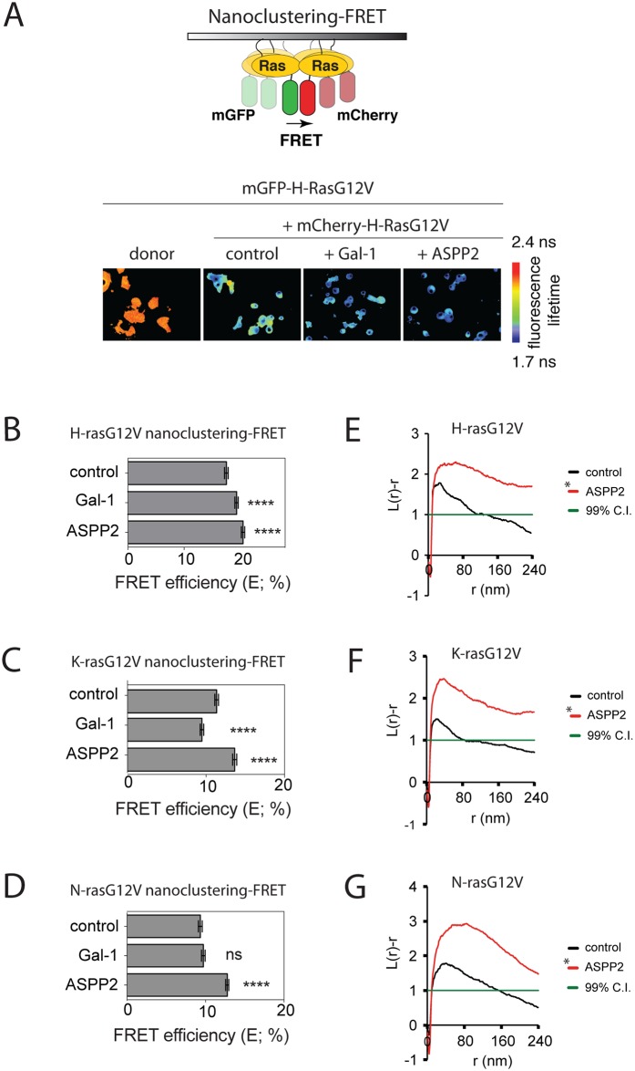
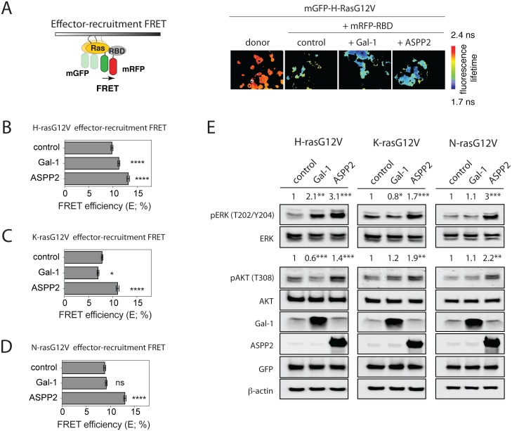
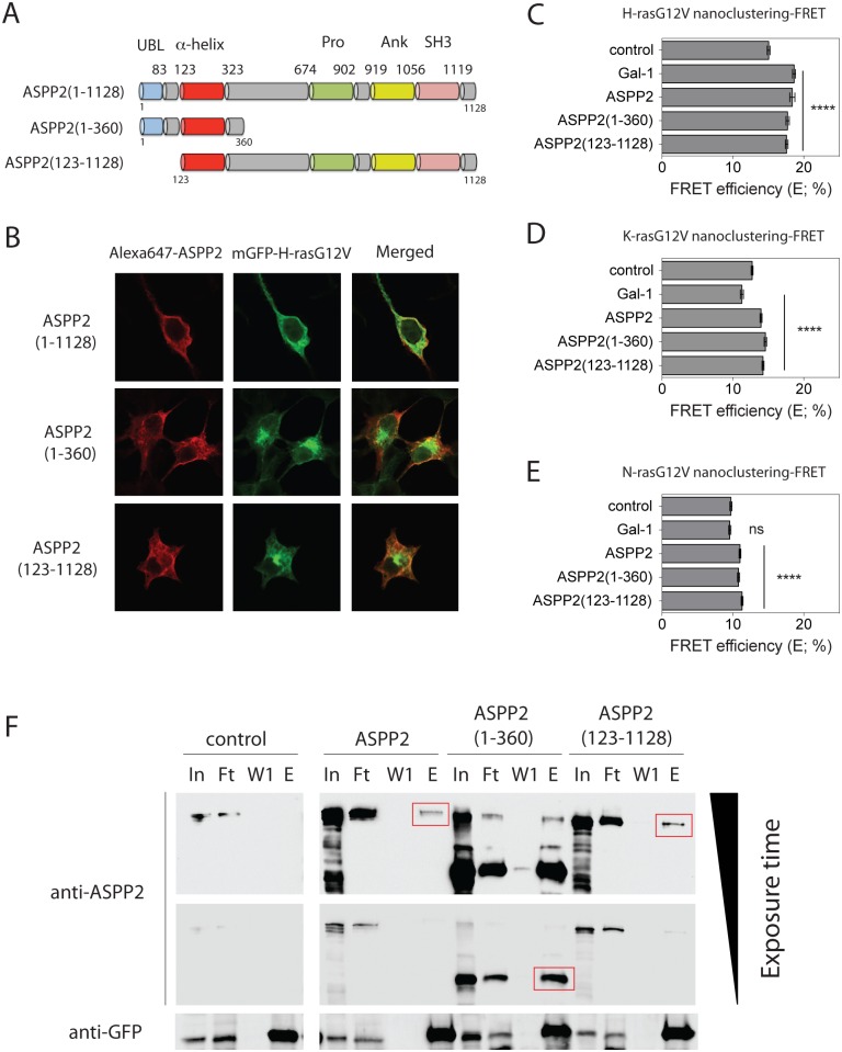
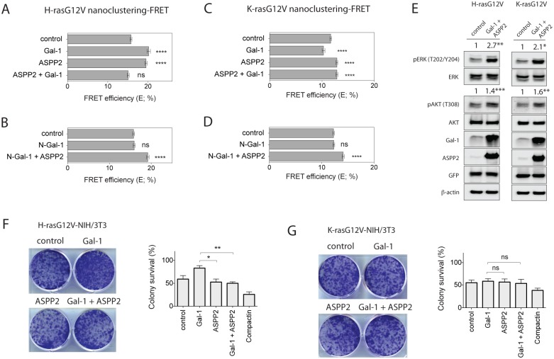
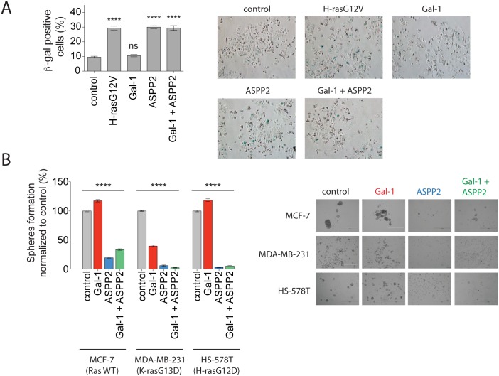
Ras-induced senescence mediated through ASPP2 represents a barrier to tumour formation. It is initiated by ASPP2's interaction with Ras at the plasma membrane, which stimulates the Raf/MEK/ERK signaling cascade. Ras to Raf signalling requires Ras to be organized in nanoscale signalling complexes, called nanocluster. We therefore wanted to investigate whether ASPP2 affects Ras nanoclustering. Here we show that ASPP2 increases the nanoscale clustering of all oncogenic Ras isoforms, H-ras, K-ras and N-ras. Structure-function analysis with ASPP2 truncation mutants suggests that the nanocluster scaffolding activity of ASPP2 converges on its α-helical domain. While ASPP2 increased effector recruitment and stimulated ERK and AKT phosphorylation, it did not increase colony formation of RasG12V transformed NIH/3T3 cells. By contrast, ASPP2 was able to suppress the transformation enhancing ability of the nanocluster scaffold Gal-1, by competing with the specific effect of Gal-1 on H-rasG12V- and K-rasG12V-nanoclustering, thus imposing ASPP2's ERK and AKT signalling signature. Similarly, ASPP2 robustly induced senescence and strongly abrogated mammosphere formation irrespective of whether it was expressed alone or together with Gal-1, which by itself showed the opposite effect in Ras wt or H-ras mutant breast cancer cells. Our results suggest that Gal-1 and ASPP2 functionally compete in nanocluster for active Ras on the plasma membrane. ASPP2 dominates the biological outcome, thus switching from a Gal-1 supported growth-promoting setting to a senescence inducing and stemness suppressive program in cancer cells. Our results support Ras nanocluster as major integrators of tumour fate decision events.


Conflict of interest statement

Competing Interests: The authors have declared that no competing interests exist.

Figures

Fig 1
Fig 1. ASPP2 increases oncogenic H-ras, K-ras and N-ras nanoclustering.
(A) Top, scheme explaining nanoclustering-FRET analysis in HEK cells. Green and red ovals represent mGFP- and mCherry-tags, respectively. Bottom, examples of FLIM-FRET images of HEK cells from the different FRET samples as indicated. (B-D) Nanoclustering-FRET analysis in HEK cells coexpressing mGFP- and mCherry-tagged (B) H-rasG12V, (C) K-rasG12V or (D) N-rasG12V. The effect of Gal-1 or ASPP2 expression on nanoclustering-FRET was compared to control samples. Statistical significance of differences between controls and treated samples was examined using one-way ANOVA (mean ± SEM, n = 3; ns, not significant; *, p<0.05, ****, p< 0.0001). (E-G) Electron microscopic nanoclustering analysis of BHK cells expressing mGFP-tagged (E) H-rasG12V, (F) K-rasG12V or (G) N-rasG12V alone or together with ASPP2. mGFP was immunolabeled with 4.5 nm gold nanoparticles coupled to anti-GFP antibody. The spatial distribution of gold particles was evaluated using univariate K-function, where L(r)–r values indicate the extent of nanoclustering as a function of length scale, r, in nm. At least 15 images were analysed for each condition. Statistical significance between different conditions was evaluated using bootstrap tests. Averaged curves are shown for each condition.
Fig 2
Fig 2. ASPP2 increases oncogenic H-ras, K-ras and N-ras-effector-recruitment, as well as ERK- and AKT-signalling.
(A) Left, scheme explaining effector-recruitment FRET analysis in HEK cells. Right, examples of FLIM-FRET images of HEK cells from the different FRET samples as indicated. (B-D) Effector-recruitment FRET analysis in HEK cells coexpressing (B) mGFP-H-rasG12V, (C) mGFP-K-rasG12V or (D) mGFP-NrasG12V and mRFP-RBD from C-Raf. The effect of Gal-1 or ASPP2 expression on effector-recruitment FRET was compared to control samples. (E) Representative Western blots from HEK cells expressing mGFP-H-rasG12V (left), K-rasG12V (middle) or N-rasG12V (right) without or with Gal-1 or ASPP2. Statistical significance of differences between controls and treated samples was examined using one-way ANOVA (mean ± SEM, n = 3; ns, not significant; *, p<0.05, **, p< 0.01, ***, p<0.001, ****, p< 0.0001).
Fig 3
Fig 3. N- and C-terminal truncation mutants of ASPP2 can still promote Ras nanoclustering.
(A) Schematic of full-length ASPP2, as well as ASPP2(1–360) and ASPP2(123–1128) truncation mutants. ASPP2 domains from left to right: Ubl, ubiquitin-like domain; α-helical domain; Pro, proline-rich domain; Ank, Ankyrin repeats; SH3, SRC homology 3 domain. (B) Confocal microscopic images of HEK cells cotransfected with mGFP-H-rasG12V (green) and full-length or truncated ASPP2 (red). (C-E) Nanoclustering-FRET analysis of HEK cells coexpressing mGFP- and mCherry-tagged (C) H-rasG12V, (D) K-rasG12V or (E) N-rasG12V. Cells were analysed after overexpression of Gal-1, full-length ASPP2 or its truncation mutants. (C-E) Statistical significance of differences between controls and treated samples was examined using one-way ANOVA (mean ± SEM, n = 3; ns, not significant; ****, p< 0.0001). (F) Western blot of anti-GFP immunoprecipitation samples probed with anti-ASPP2- (top) or anti-GFP- (bottom) antibodies. Samples were lysates prepared from mGFP-H-rasG12V transfected HEK cells that were cotransfected with full-length ASPP2 or its truncation mutants or an empty plasmid (control), as indicated. In, input; Ft, flow-through; W1, wash; E, elution. Red boxes indicate the immunoprecipitated ASPP2 fragments.
Fig 4
Fig 4. ASPP2 blocks Gal-1 dependent nanoclustering and halts oncogenic H-ras induced transformation.
Nanoclustering-FRET analysis in HEK cells coexpressing mGFP- and mCherry-tagged (A, B) H-rasG12V or (C, D) K-rasG12V. Cells were analysed after overexpression of either Gal-1 or ASPP2 plasmids, or both (1:1 ratio). Plotted are the means ± SEM, n = 3. (E) Representative Western blots from HEK cells expressing mGFP-H-rasG12V (left) or K-rasG12V (right) alone or together with Gal-1 and ASPP2. Statistical significance of differences was examined using t-test (n = 3; *, p<0.05, **, p< 0.01, ***, p<0.001). (F, G) Colony survival assay of NIH/3T3 cells stably expressing (F) H-rasG12V or (G) K-rasG12V and transiently expressing indicated constructs. Colony survival was graphed based on mean foci areas calculated from at least 4 independent biological repeats. (A-D, F-G) Statistical significance of differences between controls and treated samples was examined using one-way ANOVA (ns, not significant; *, p<0.05; **, p<0.01; ****, p<0.0001).
Fig 5
Fig 5. ASPP2 dominates over Gal-1 thus robustly inducing senescence and abrogating mammosphere formation.
(A) SA-β-gal assay of MCF-7 cells transfected with plasmids encoding H-rasG12V, ASPP2, Gal-1 or the combination of the latter two, as indicated. Cells were stained 7 days after transfection. On the left, percentages of SA-β-gal positive cells are shown in the graph (mean ± SEM, n = 3). On the right, representative images from the assay. (B) Mammosphere formation assay with MCF-7, MDA-MB-231 or HS-578T breast cancer cell lines. Mammospheres were transfected with Gal-1, ASPP2, or both (1:1 ratio) and cells were then grown under non-adherent conditions for 9 days. On the right, representative images of mammospheres are shown as indicated. (A, B) Statistical significance of differences between controls and treated samples was examined using one-way ANOVA (mean ± SEM n≥3; ns, not significant; ****, p<0.0001).

References

    1. Serrano M, Lin AW, McCurrach ME, Beach D, Lowe SW. Oncogenic ras provokes premature cell senescence associated with accumulation of p53 and p16INK4a. Cell. 1997;88: 593–602. - PubMed
    1. Downward J. Targeting RAS signalling pathways in cancer therapy. Nat Rev Cancer. 2003;3: 11–22. 10.1038/nrc969 - DOI - PubMed
    1. Godin-Heymann N, Wang Y, Slee E, Lu X. Phosphorylation of ASPP2 by RAS/MAPK pathway is critical for its full pro-apoptotic function. PLoS ONE. 2013;8: e82022 10.1371/journal.pone.0082022 - DOI - PMC - PubMed
    1. Ahearn IM, Haigis K, Bar-Sagi D, Philips MR. Regulating the regulator: post-translational modification of RAS. Nat Rev Mol Cell Biol. 2012;13: 39–51. 10.1038/nrm3255 - DOI - PMC - PubMed
    1. Yan J, Roy S, Apolloni A, Lane A, Hancock JF. Ras isoforms vary in their ability to activate Raf-1 and phosphoinositide 3-kinase. J Biol Chem. 1998;273: 24052–24056. - PubMed

MeSH terms