. 2016 Aug 9;7(32):51626-51639.
doi: 10.18632/oncotarget.10539.

Establishment and antitumor effects of dasatinib and PKI-587 in BD-138T, a patient-derived muscle invasive bladder cancer preclinical platform with concomitant EGFR amplification and PTEN deletion

Affiliations

Establishment and antitumor effects of dasatinib and PKI-587 in BD-138T, a patient-derived muscle invasive bladder cancer preclinical platform with concomitant EGFR amplification and PTEN deletion

Nakho Chang et al. Oncotarget. .

Abstract

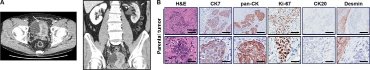
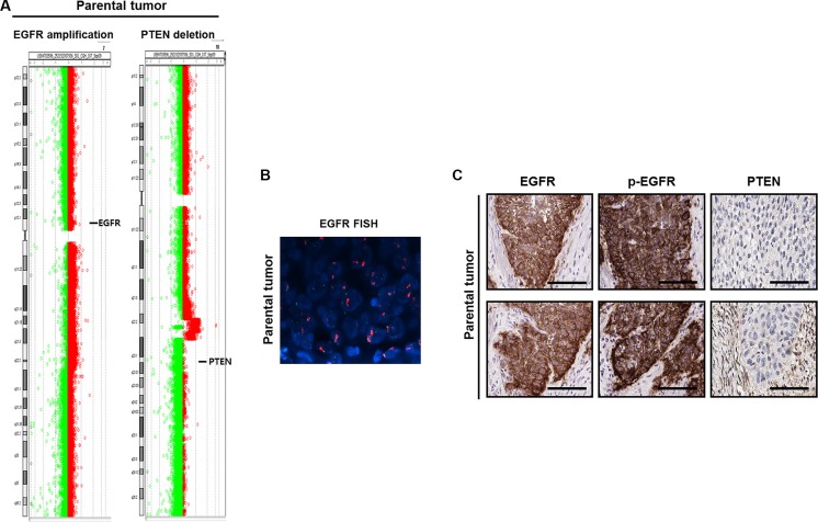
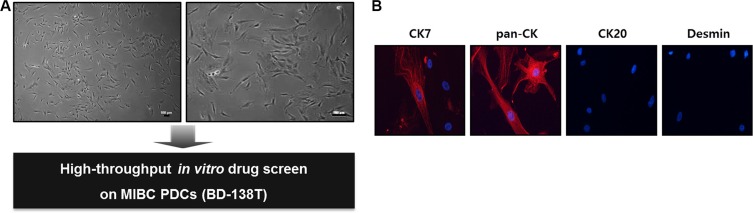
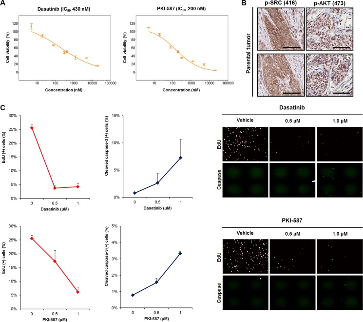
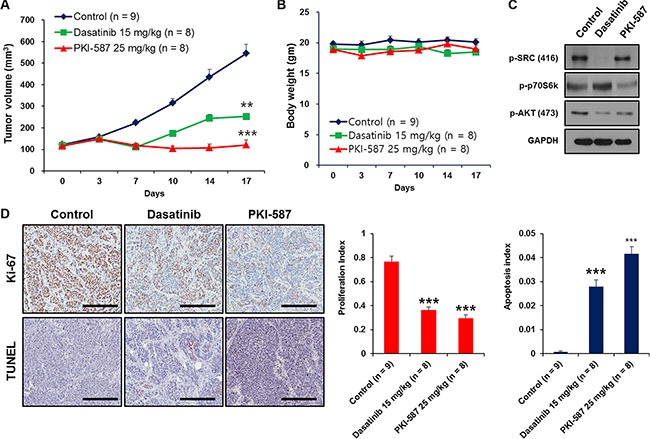
Muscle-invasive bladder cancer (MIBC) consists of a heterogeneous group of tumors with a high rate of metastasis and mortality. To facilitate the in-depth investigation and validation of tailored strategies for MIBC treatment, we have developed an integrated approach using advanced high-throughput drug screening and a clinically relevant patient-derived preclinical platform. We isolated patient-derived tumor cells (PDCs) from a rare MIBC case (BD-138T) that harbors concomitant epidermal growth factor receptor (EGFR) amplification and phosphatase and tensin homolog (PTEN) deletion. High-throughput in vitro drug screening demonstrated that dasatinib, a SRC inhibitor, and PKI-587, a dual PI3K/mTOR inhibitor, exhibited targeted anti-proliferative and pro-apoptotic effects against BD-138T PDCs. Using established patient-derived xenograft models that successfully retain the genomic and molecular characteristics of the parental tumor, we confirmed that these anti-tumor responses occurred through the inhibition of SRC and PI3K/AKT/mTOR signaling pathways. Taken together, these experimental results demonstrate that dasatinib and PKI-587 might serve as promising anticancer drug candidates for treating MIBC with combined EGFR gene amplification and PTEN deletion.

Keywords: EGFR; PTEN; drug screening; muscle invasive bladder cancer; patient-derived xenograft.


Conflict of interest statement

The authors declare that the research was conducted in the absence of any commercial or financial relationships that could be construed as a potential conflicts of interest.

Figures

Figure 1
Figure 1. Case presentation of patient 138T with muscle-invasive bladder cancer and validation of patient-derived tumor tissue
(A) Computed tomography (CT) scan of the pelvis indicates a large bladder tumor invading the base of the bladder of patient 138T. The bladder is visible at the bottom center of the scan. The arrow indicates the tumor region. The tumor tissue appears as cloudy material within the bladder, whereas the remaining area in the bladder is eclipsed. (B) Representative images of hematoxylin/eosin (H&E) and immunohistochemistry staining of BD-138T tumor tissue with antibodies for the indicated proteins. CK7, cytokeratin 7; pan-CK, pan-cytokeratin; CK20, cytokeratin 20. Scale bars: 100 μm (Top), 20 μm (Bottom).
Figure 2
Figure 2. Identification of gene alterations in 138T muscle-invasive bladder cancer
(A) Comparative genomic hybridization array analysis of the tumor from patient 138T, indicating the mutual EGFR amplification and PTEN deletion. Individual chromosome ratio plots are shown with red representing amplified regions and green representing deleted regions. High-level EGFR amplification in chromosome 7 (left). PTEN deletion in chromosome 10 (right). (B) Epidermal growth factor receptor (EGFR) amplification as assessed by FISH in 138T patient-derived cells (PDCs). Orange signal: EGFR; green signal: CEP7; blue signal: DAPI counterstaining. (C) Representative images of immunohistochemical staining of 138T PDC tumor tissue showing EGFR, p-EGFR (Tyr1068), and PTEN. Scale bars, 100 μm.
Figure 3
Figure 3. Establishment and validation of patient-derived cells (PDCs) from 138T muscle-invasive bladder cancer (MIBC)
(A) The establishment of PDCs from 138T MIBC tissues. The tissues were dissociated into single cells for in-vitro drug screening using a high-throughput screening (HTS) system. Scale bars: 100 μm. (B) Representative confocal microscopy images of immunofluorescence staining of cytokeratin (CK) 7, pan-CK, CK 20, and desmin in 138T PDCs. Scale bars, 100 μm. Red: indicated protein. Blue: DAPI.
Figure 4
Figure 4. Anti-tumor efficacy of dasatinib (a SRC inhibitor) and PKI-587 (a PI3K/mTOR inhibitor) in 138T muscle-invasive bladder cancer harboring mutual EGFR amplification and PTEN deletion
(A) Dose-response relationship curves of dasatinib and PKI-587 in 138T patient derived cell (PDC) tumors. Error bar represent the means ± SEM (triplicate). (B) Representative images of immunohistochemical staining of 138T PDC tumor tissue with antibodies against p-SRC (Tyr416) and p-AKT (Ser473). Scale bars, 100 μm. (C) 5′-ethynyl-2′-deoxyuridine (EdU) and cleaved caspase-3 analysis of 138T PDC tumors treated with dasatinib (Dose = 0, 0.5, or 1 μM) or PKI-587 (Dose = 0, 0.5, or 1 μM). Representative graphical analysis of EdU (+) and cleaved caspase-3 (+) cells following dasatinib or PKI-587 treatment; error bars represent the means ± SD. Representative images of EdU and cleaved caspase-3 staining following treatment (right).
Figure 5
Figure 5. Establishment of patient-derived xenograft (PDX) models in 138T muscle-invasive bladder cancer (MIBC) and validation of the genetic, molecular, and histologic similarity between parental and PDX tumors
(A) Schematic of the establishment of PDX tumors in 138T MIBC tissues. A PDX tumor was established by subcutaneous implantation in a BALB/c-nu mouse to validate the histologic, genomic, and molecular similarity of parental and PDX tumors. (B) Representative images of hematoxylin & eosin (H&E) and immunohistochemical (IHC) staining of 138T PDX tumor tissues with the indicated antibodies. CK7, cytokeratin 7; pan-CK, pan-cytokeratin; CK20, cytokeratin 20. Scale bars, 100 μm (Top), 20 μm (Bottom). (C) Array comparative genomic hybridization (CGH) analysis of 138T parental and PDX tumors. Gene amplifications and deletions were analyzed and compared between tumors to validate genetic similarity. (D) Array CGH analysis of 138T PDX tumors, illustrating the mutual EGFR amplification and PTEN deletion in the PDX and parental tumors. Individual chromosome ratio plots are shown with red representing the amplified region and green representing the deleted region. High-level EGFR amplification in chromosome 7 (left). PTEN deletion in chromosome 10 (right). (E) EGFR amplification assessed by FISH analysis in the 138T PDX tumor. Orange signal: EGFR; green signal: CEP7; blue signal: DAPI counterstaining. (F) Representative images of IHC staining of 138T PDX tumor tissue with EGFR, p-EGFR (Tyr1068), PTEN, p-SRC (Tyr416), and p-AKT (Ser473) antibodies. Scale bars, 100 μm.
Figure 6
Figure 6. Dasatinib and PKI-587 inhibit tumor growth of 138T muscle-invasive bladder cancer with EGFR amplification and PTEN deletion in the patient-derived xenograft (PDX) tumor mouse model
(A) Measurement of subcutaneous xenograft tumor size after treatment with dasatinib or PKI-587. **P < 0.01. ***P < 0.001. PDX tumors were treated with dasatinib (15 mg/kg, n = 8) and PKI-587 (25 mg/kg, n = 8) for 17 days, harvested, and processed for immunoblots. (B) Measurement of mouse body weight after treatment with dasatinib or PKI-587 showing no toxicity from the drug therapy in the PDX mouse model. Body weights were measured after treatment with dasatinib or PKI-587 for 17 days. (C) Immunoblots of p-SRC (Tyr416), p-p70S6K (Thr389) and p-AKT (Ser473) in xenograft tumors from (A). (D) Cell proliferation and apoptosis were measured immunohistochemically (IHC) by performing Ki-67 and TUNEL staining using PDX tissues treated with dasatinib or PKI-587 (left). Scale bar, 100 μm. Representative bar graphs of the IHC images (right), **P < 0.01, ***P< 0.001 (n = 5 fields; means ± SD, analyzed by two-tailed paired t-test).

References

    1. Jemal A, Siegel R, Ward E, Murray T, Xu J, Thun MJ. Cancer statistics, 2007. CA Cancer J Clin. 2007;57:43–66. - PubMed
    1. Vishnu P, Mathew J, Tan WW. Current therapeutic strategies for invasive and metastatic bladder cancer. Onco Targets Ther. 2011;4:97–113. - PMC - PubMed
    1. Zachos I, Konstantinopoulos PA, Tzortzis V, Gravas S, Karatzas A, Karamouzis MV, Melekos M, Papavassiliou AG. Systemic therapy of metastatic bladder cancer in the molecular era: current status and future promise. Expert Opin Investig Drugs. 2010;19:875–887. - PubMed
    1. Wu XR. Urothelial tumorigenesis: a tale of divergent pathways. Nat Rev Cancer. 2005;5:713–725. - PubMed
    1. Cordon-Cardo C. Molecular alterations associated with bladder cancer initiation and progression. Scand J Urol Nephrol Suppl. 2008:154–165. - PubMed

MeSH terms