

Review
doi: 10.1590/abd1806-4841.20164446.
High frequency ultrasound with color Doppler in dermatology
Affiliations
- PMID: 27438191
- PMCID: PMC4938268
- DOI: 10.1590/abd1806-4841.20164446
Item in Clipboard
Review
High frequency ultrasound with color Doppler in dermatology
An Bras Dermatol.
2016 May-Jun.
Abstract
Ultrasonography is a method of imaging that classically is used in dermatology to study changes in the hypoderma, as nodules and infectious and inflammatory processes. The introduction of high frequency and resolution equipments enabled the observation of superficial structures, allowing differentiation between skin layers and providing details for the analysis of the skin and its appendages. This paper aims to review the basic principles of high frequency ultrasound and its applications in different areas of dermatology.
Conflict of interest statement
Conflict of interest: none.
Figures
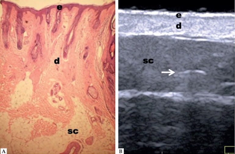
Non-glabrous skin anatomy. (A) Normal skin histology.
(B) HFUS, transverse view. (e) Epidermis.
(d) Dermis. (sc) Subcutaneous tissue with
the presence of fibrous septa (⇒ )
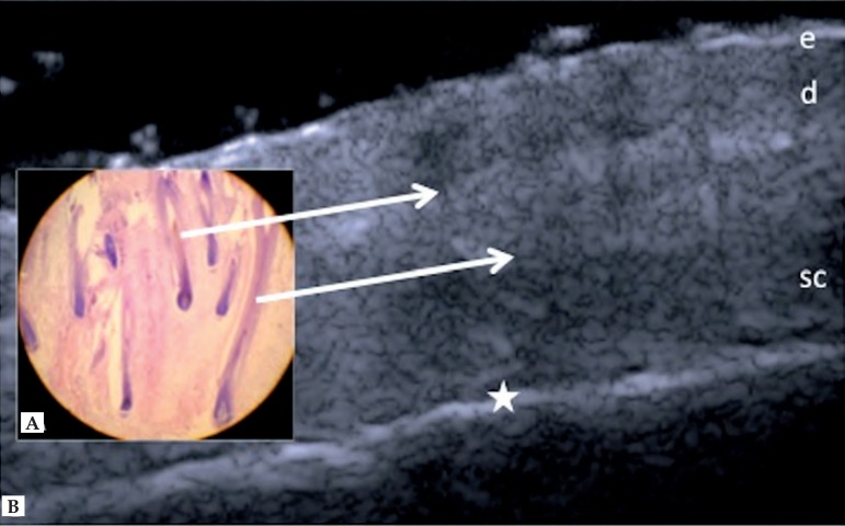
Scalp. (A) Histology, longitudinal section. (B)
HFUS, longitudinal view. (e) Epidermis. (d)
Dermis. (sc) Subcutaneous tissue. (⇒) Hypoechoic
oblique bands corresponding to hair follicles. (★) Margin of cranial
bone
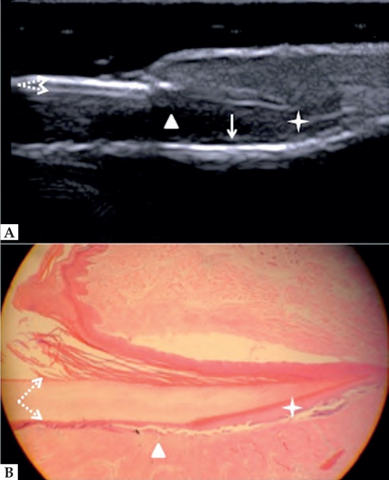
Normal nail unit, longitudinal section. (A) HFUS.
(B) Histological section. (➚)Floor plate. (➘) Dorsal
plate. (★) Nail matrix. (Δ) Nail bed and (↓) distal
phalanx bone
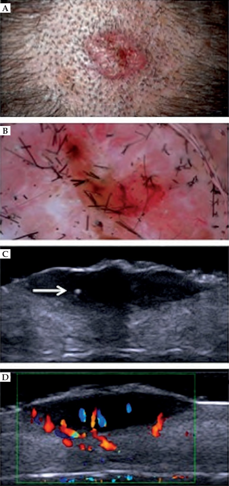
Basal cell carcinoma in the scalp. (A) Clinical aspect. (B)
Dermatoscopy presenting arboriform vessels. (C) HFUS, transversal
view. Hyperechoic point within the lesion (⇒). (D) Color Doppler.
Blood vessels inside and at the base of the lesion (blue and
red)
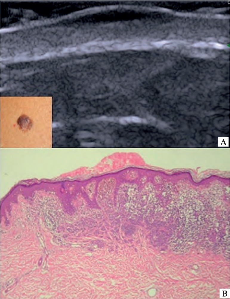
Melanoma. (A) HFUS, longitudinal view. Fusiform
hypoechoic lesions affecting the epidermis and dermis. (B)
Occupation of the central area of the epidermis and superior
dermis by proliferation of anaplastic melanocytic cells, with
irregular distribution of melanin pigment. Melanoma, superficial
spreading type, Breslow 0.62 mm and Clark level III. Hematoxylin
& Eosin 10X
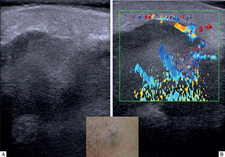
Metastatic melanoma. (A) HFUS, longitudinal view. Epidermis and
dermis with normal appearance. In the subcutaneous tissue, irregular
lesion with variable echogenicity. (B) Color Doppler. Intralesional
vascularity
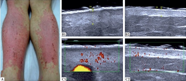
Psoriasis. (A) Posterior region of the legs. (B)
HFUS, transversal view. (B1) Psoriasis plaque.
Epidermis with 0.50 mm thickness and dermis with 2.56 mm thickness.
(B2) Healthy contralateral region. Epidermis with
0.34 mm thickness and dermis with 1.67 mm thickness.
(C) Color Doppler. (C1) Increased
blood flow of the affected area, setting up disease activity.
(C2) Healthy contralateral region
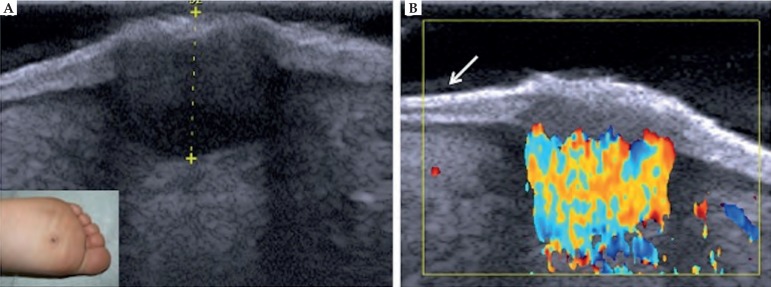
Plantar wart. (A) HFUS, transversal view. Fusiform
hypoechoic lesions localized in the epidermis and in the dermis.
(B) Color Doppler. Intense vascularization within
the lesion with predominance of arterial vessels. (⬋)
Ultrasonographic bilaminar aspect of the plantar region skin
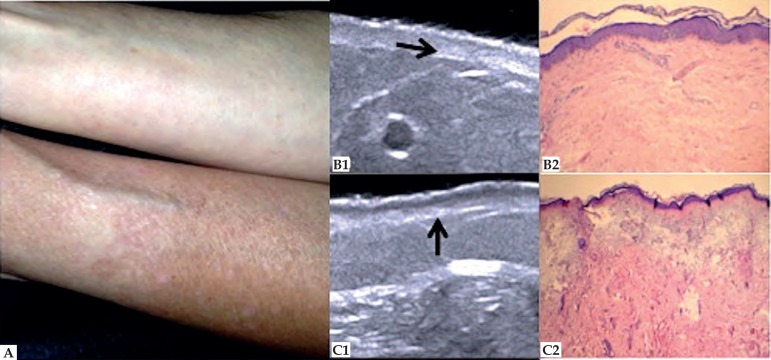
Photoaging. (A) Ventral and dorsal region of the forearm. (B1) HFUS,
longitudinal view. Decreased dermis echogenicity (➛). (B2) Discrete
degeneration of collagen fibers. Hematoxylin & Eosin 10X. (C1)
HFUS, longitudinal view. Subepidermal low echogenicity band
(↑). (C2) Solar elastosis. Hematoxylin & Eosin 10X
References
-
- Mogensen M, Morsy HA, Thrane L, Jemec GB. Morphology and epidermal thickness of normal skin imaged by optical coherence tomography. Dermatology. 2008;217:14–20. - PubMed
-
- Lallas A, Giacomel J, Argenziano G, García-García B, González-Fernández D, Zalaudek I, et al. Dermoscopy in general dermatology:practical tips for the clinician. Br J Dermatol. 2014;170:514–526. - PubMed
-
- Giacomel J, Lallas A, Argenziano G, Reggiani C, Piana S, Apalla Z, et al. Dermoscopy of basosquamous carcinoma. Br J Dermatol. 2013;169:358–364. - PubMed
-
- Longo C, Farnetani F, Ciardo S, Cesinaro AM, Moscarella E, Ponti G, et al. Is confocal microscopy a valuable tool in diagnosing nodular lesions? A study of 140 cases. Br J Dermatol. 2013;169:58–67. - PubMed
-
- Soyer HP, Prow TW. Reflectance confocal microscopy in the diagnosis of nodular skin lesions. Br J Dermatol. 2013;169:4–4. - PubMed
Publication types
MeSH terms
LinkOut - more resources
Full Text Sources
Other Literature Sources