. 2016 Jul 20;11(7):e0158212.
doi: 10.1371/journal.pone.0158212. eCollection 2016.

Immunological Cross-Reactivity between Malaria Vaccine Target Antigen P48/45 in Plasmodium vivax and P. falciparum and Cross-Boosting of Immune Responses

Affiliations

Immunological Cross-Reactivity between Malaria Vaccine Target Antigen P48/45 in Plasmodium vivax and P. falciparum and Cross-Boosting of Immune Responses

Yi Cao et al. PLoS One. .

Abstract

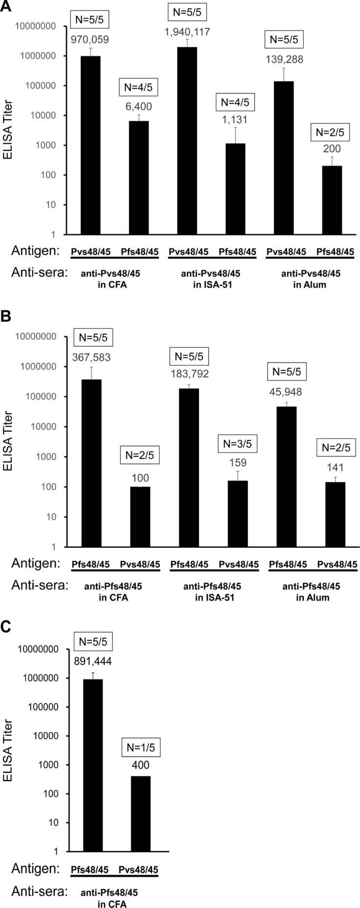
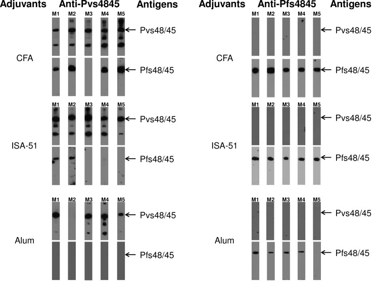
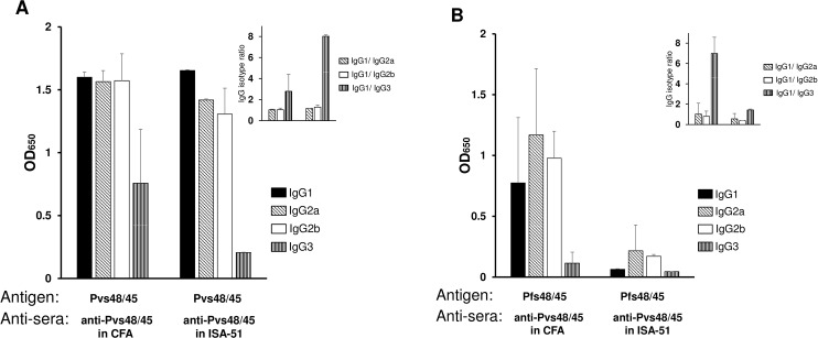
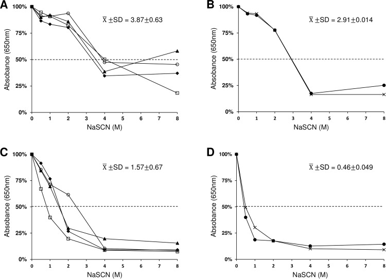
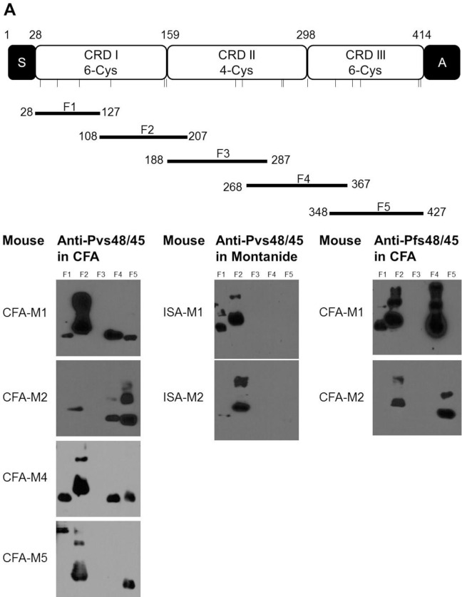
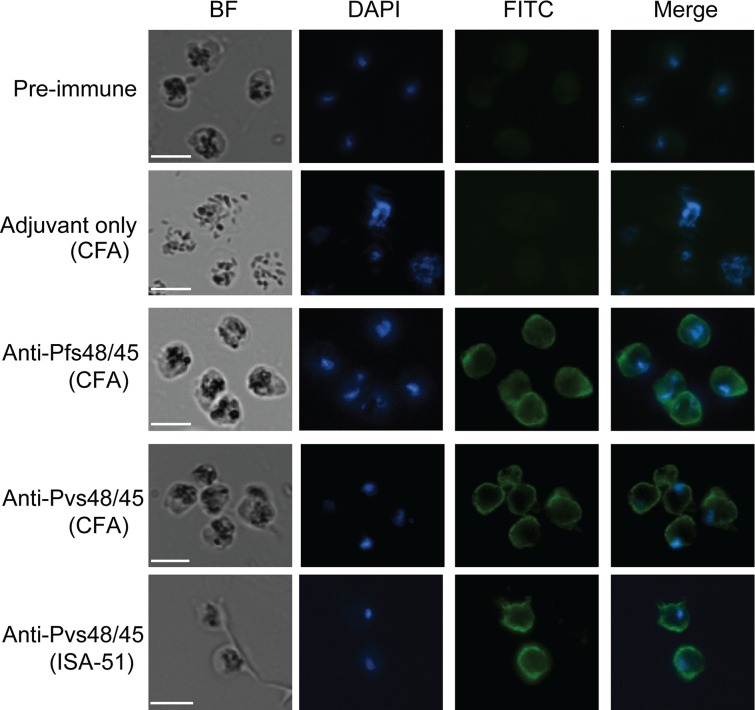
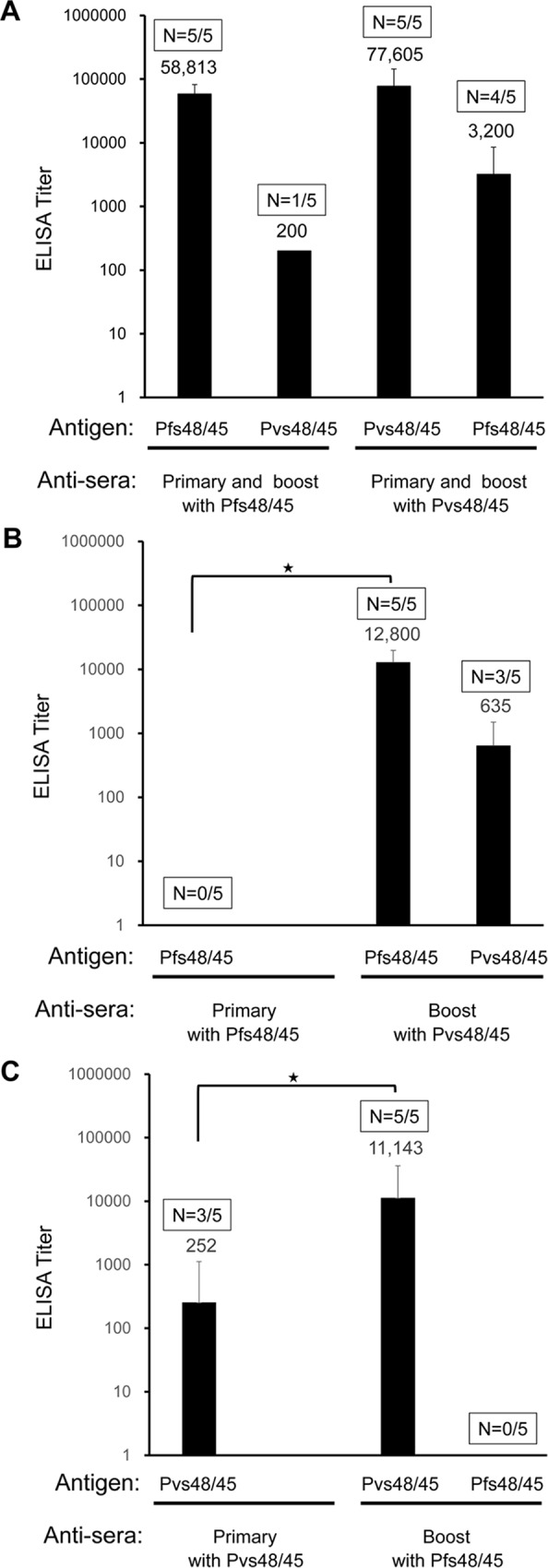
In general, malaria immunity has been suggested to be species specific with very little, if any, known cross-reactivity between Plasmodium vivax and P. falciparum, both of which are responsible for >90% of human malaria, and co-endemic in many countries. It is therefore believed that species-specific immunity may be needed to target different species of Plasmodium. Pfs48/45 and Pvs48/45 are well established targets in the sexual stages of the malaria parasites, and are being pursued for the development of transmission blocking vaccines. Comparison of their sequences reveals 61% and 55% identity at the DNA and protein level, respectively raising the possibility that these two target antigens might share cross-reacting epitopes. Having succeeded in expressing recombinant Pfs48/45 and Pvs48/45 proteins, we hypothesized that these proteins will not only exhibit immunological cross-reactivity but also cross-boost immune responses. Mice were immunized with purified recombinant proteins using CFA, Montanide ISA-51 and alum as adjuvants, and the sera were analyzed by ELISA, Western blotting and indirect fixed and live IFA to address the hypothesis. Our studies revealed that Pvs48/45-immune sera showed strong cross-reactivity to full length Pfs48/45 protein, and the majority of this cross reactivity was in the amino-terminal and carboxyl-terminal sub-fragments of Pfs48/45. In cross-boosting experiments Pfs48/45 and Pvs48/45 antigens were able to cross-boost each other in mouse immunization studies. Additionally we also noticed an effect of adjuvants in the overall magnitude of observed cross-reactivity. These studies may have significant implications for immunity targeting transmission of both the species of malaria parasites.


Conflict of interest statement

Competing Interests: The authors have declared that no competing interests exist.

Figures

Fig 1
Fig 1. Cross-reactivity of Pvs48/45 and Pfs48/45 immune mouse sera in ELISA.
(A) Anti-Pvs48/45 sera from BALB/c mice after 3-dose protein immunization in three adjuvants (CFA, Montanide ISA-51 and Alum) were tested for reactivity against Pvs48/45 and Pfs48/45 antigens by ELISA. (B) Anti-Pfs48/45 sera from BALB/c mice after 3-dose protein immunization in three adjuvants were tested for reactivity against Pfs48/45 and Pvs48/45 antigens by ELISA. (C) ELISA results using anti-Pfs48/45 sera from C57BL/6 mice after 3-dose protein immunization in CFA tested for reactivity against Pfs48/45 and Pvs48/45 antigens. Antigens used for ELISA analysis are identified below each column. The geometric mean ELISA titers are shown above each column. The error bars indicate SD. The numbers within rectangles above each column show number of positive mouse sera / total mice tested.
Fig 2
Fig 2. Western blotting analysis of mouse anti-sera with the full-length Pfs48/45 and Pvs48/45 recombinant antigens.
Left panels show Western blotting results with anti-Pvs48/45 sera from BALB/c mice (N = 5 per group, M1 –M5) immunized respectively with 3 adjuvants (CFA, Montanide ISA-51 and Alum). Each serum was tested at 1:1,000 dilution for recognition of Pfs48/45 antigen and at 1:10,000 dilution for recognition of Pvs48/45 antigen. The right panels show Western blotting results with anti-Pfs48/45 sera from BALB/c mice (M1 –M5) immunized using CFA, Montanide ISA-51 and Alum. The dilution of each serum was 1:1,000 for recognition of Pvs48/45 antigen, and 1:10,000 for recognition of Pfs48/45 antigen.
Fig 3
Fig 3. Analysis of antibody isotypes.
Anti-Pvs48/45 sera showing positive reactivity to Pfs48/45 in ELISA and Western blotting (4 out of 5 from CFA group and 2 out of 5 from Montanide ISA-51 group) were individually tested to compare immunoglobulin isotypes. ELISA plates coated with Pvs48/45 (panel A) or Pfs48/45 (panel B) were incubated with sera (1:10,000 dilution for Fig 3A and 1:100 dilution for Fig 3B). The plates were then incubated with peroxidase-conjugated goat anti-mouse IgG1, IgG2a, IgG2b and IgG3 (1:2,500 dilution) and processed as in standard ELISA. Shown are mean absorbance values for each isotype and the insets in panels A and B show relative proportions of IgG2a, IgG2b and IgG3 isotypes compared to IgG1. The error bars indicate SD.
Fig 4
Fig 4. Analysis of avidity.
Anti-Pvs48/45 sera showing positive reactivity to Pfs48/45 in ELISA and Western blotting (4 out of 5 from CFA group and 2 out of 5 from Montanide ISA-51 group) were individually tested to compare avidity of antibodies. ELISA plates coated with Pvs48/45 or Pfs48/45 were incubated with sera at 1:10,000 dilution for Pvs48/45 and at 1:100 dilution for Pfs48/45. Absorbance values after NaSCN treatment were converted to percent of total binding (absorbance without NaSCN) and avidity index was deduced from the molar concentration of NaSCN resulting in 50% dissociation of bound antibody. (A) 4 sera in CFA group against Pvs48/45. (B) 2 sera in ISA-51group against Pvs48/45. (C) 4 sera in CFA group against Pfs48/45. (D) 2 sera in CFA group against Pfs48/45. The mean and standard deviation (X¯±SD) of avidity index are shown in each panel. Statistical significance was determined by Student’s t-test with p values <0.05.
Fig 5
Fig 5. Reactivity of anti-Pvs48/45 sera to recombinant sub-fragments of Pfs48/45.
(A) Schematic representation of Pfs48/45 and the amino acid boundaries of five sub-fragments (F1, F2, F3, F4 and F5) of Pfs48/45. S, secretory signal sequence; A, anchor sequence; CRD, cysteine-rich domain. Bars show the relative positions of cysteine residues. (B) Western blotting analysis of the six anti-Pvs48/45 sera from CFA (CFA-M1, CFA-M2, CFA-M4 and CFA-M5) and Montanide ISA-51 (ISA-M1, ISA-M2) adjuvant groups with five overlapping fragments of Pfs48/45. All the anti-Pvs48/45 sera were tested at 1:1,000 dilution. The two anti-Pfs48/45 sera (CFA-M1, CFA-M2) were employed as positive control at a dilution of 1:10,000.
Fig 6
Fig 6. Recognition of Pfs48/45 in fixed P. falciparum parasites in indirect immunofluorescence assays.
The representative results for fixed parasites incubated with pooled pre-immune sera, pooled sera from CFA adjuvant alone immunized mice, a representative anti-Pfs48/45 serum, a representative ELISA and WB positive anti-Pvs48/45 serum (CFA group), and a representative ELISA and WB positive anti-Pvs48/45 (Montanide ISA-51 group) at a dilution of 1:100. BF, bright field; DAPI, the nuclei stained by NucBlue reagent; FITC, antibody reactivity to the parasite visualized with FITC-conjugated anti-mouse IgG antibody, and Merge of DAPI and FITC images. All images were visualized and captured at 1000X magnification. Scale bar (white line), 5μm.
Fig 7
Fig 7. Recognition of Pfs48/45 by anti-Pvs48/45 antisera on the surface of live P. falciparum gametes.
Individual mouse sera were tested (1:100 dilution) by live IFA. Panels a-b show reactivity patterns obtained with two different anti-Pfs48/45 sera. Panels c-f show patterns of positive reactivity of anti-Pvs48/45 sera in live IFA. Panel g shows a representative bright field image of live gamete preparation used and panel h shows the result with a pool of pre-immune mouse sera also tested at 1:100 dilution. The inset in panel h is included to show lack of any detectable reactivity even after altering brightness and contrast settings.
Fig 8
Fig 8. Recognition of Pfs48/45 by anti-Pvs48/45 antisera under non-reducing conditions by Western blotting using purified gametocytes of P. falciparum.
Approximately 1 million gametocytes (P. falciparum, NF54) per lane were used under non-reducing condition of SDS-PAGE for Western blotting. Lane 1 shows results with a pool of pre-immune mouse sera (1:1000 dilution). Lanes 2 and 3 show results with two different anti-Pfs48/45 sera tested at 1:5000 dilution. Immune sera from five mice (M1-M5 in Figs 1 and 2) immunized with Pvs48/45 using CFA as adjuvant were evaluated at 1:1000 dilution (lanes 4–8). The serum from M3 in this CFA group, which did not display any cross-reactivity in Western blotting and ELISA, also did not detect Pfs4845 antigen in the gametocytes (lane 6). Lanes 9–13 show results on the lack of recognition of Pfs47 by four (M1, M2, M3 and M4) out of these five anti-Pvs48/45 sera tested (1:1000 dilution) using reduced form of E. coli expressed Pfs47 protein. Pooled sera from mice immunized with rPfs47 using CFA as an adjuvant was used as a positive control (lane 9). Recombinant Pfs47 (unpublished) used was expressed in E.coli as 6X His tagged protein using pET (K-) expression vector and purified exactly as Pfs48/45 and Pvs48/45 proteins used in these studies.
Fig 9
Fig 9. Cross boosting of Pfs48/45 and Pvs48/45 antibody responses.
(A) Groups of mice were primed and boosted with the same antigen, either Pfs48/45 or Pvs48/45. Antibody titers were determined against both antigens by ELISA. The data shown are from mice after the booster immunization. (B) Shows results of mice primed with Pfs48/45 and boosted with Pvs48/45. Antibody titers were determined both after the primary and booster immunizations using ELISA plates coated with either Pfs48/45 or Pvs48/45. Antibody titers against Pfs48/45 between prime and boost were statistically significant (p = 0.0055). As shown, Pfs48/45 specific antibody titers after priming with Pfs48/45 were ≤ 100, we used a value of 100 for statistical comparison with titers after boost with Pvs48/45. (C) Shows results of mice primed with Pvs48/45 and boosted with Pfs48/45. Antibody titers were determined both after the primary and booster immunizations using plates coated with either antigen. Antibody titers against Pvs48/45 between prime and boost were statistically significant (p = 0.036). The geometric mean ELISA titer of each group is shown. Boxes above each column show number of positive mice / total mice tested. Statistical significance of the antibody titers between prime and boost were determined by Mann Whitney test with p value < 0.05, indicated by an asterisk (*).

Similar articles

Cited by

References

    1. Nikolaeva D, Draper SJ, Biswas S. Toward the development of effective transmission-blocking vaccines for malaria. Expert Review of Vaccines. 2015;14(5):653–80. 10.1586/14760584.2015.993383 . - DOI - PubMed
    1. Rener J, Graves P, Carter R, Williams J, Burkot T. Target antigens of transmission-blocking immunity on gametes of Plasmodium falciparum. J Exp Med. 1983;158:976–81. 10.1084/jem.158.3.976 - DOI - PMC - PubMed
    1. Quakyi IA, Carter R, Rener J, Kumar N, Good MF, Miller LH. The 230-kDa gamete surface protein of Plasmodium falciparum is also a target for transmission-blocking antibodies. J Immunol. 1987;139(12):4213–7. - PubMed
    1. Arévalo-Herrera M, Vallejo AF, Rubiano K, Solarte Y, Marin C, Castellanos A, et al. Recombinant Pvs48/45 Antigen Expressed in E. coli Generates Antibodies that Block Malaria Transmission in Anopheles albimanus Mosquitoes. PloS one. 2015;10(3):e0119335 10.1371/journal.pone.0119335 - DOI - PMC - PubMed
    1. Kaslow DC, Quakyi IA, Syin C, Raum MG, Keister DB, Coligan JE, et al. A vaccine candidate from the sexual stage of human malaria that contains EGF-like domains. Nature. 1988;333(6168):74–6. - PubMed

MeSH terms

LinkOut - more resources