Review
. 2016 Jul 16;21(7):860.
doi: 10.3390/molecules21070860.

Applications of (19)F-NMR in Fragment-Based Drug Discovery

Affiliations
Review

Applications of (19)F-NMR in Fragment-Based Drug Discovery

Raymond S Norton et al. Molecules. .

Abstract

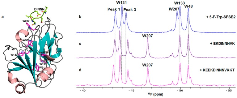
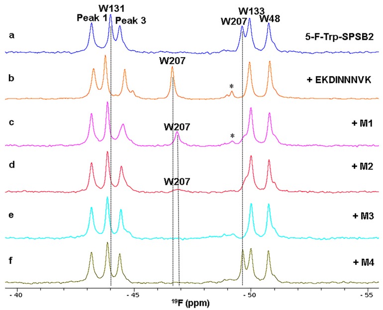
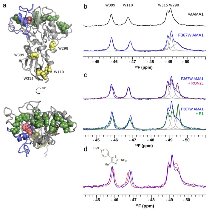
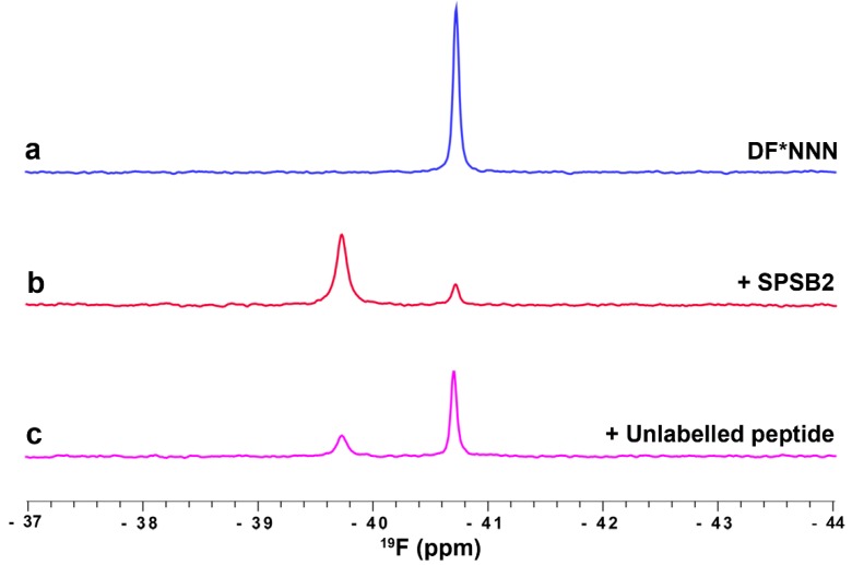
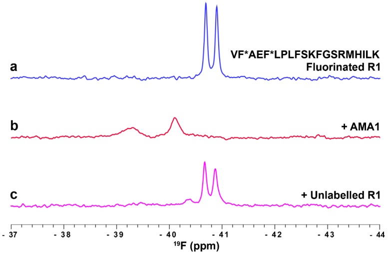
(19)F-NMR has proved to be a valuable tool in fragment-based drug discovery. Its applications include screening libraries of fluorinated fragments, assessing competition among elaborated fragments and identifying the binding poses of promising hits. By observing fluorine in both the ligand and the target protein, useful information can be obtained on not only the binding pose but also the dynamics of ligand-protein interactions. These applications of (19)F-NMR will be illustrated in this review with studies from our fragment-based drug discovery campaigns against protein targets in parasitic and infectious diseases.

Keywords: 19F-NMR; chemical shift; fragment-based drug design; labelling; ligand; linewidth; peptide; protein.


Conflict of interest statement

The authors declare no conflict of interest.

Figures

Figure 1
Figure 1
(a) Crystal structure of SPSB2 with bound DINNN peptide (PDB id: 3EMW). The six Trp residues of SPSB2 (W48, W91, W95, W131, W133 and W207) are shown in stick representation and coloured magenta. Four Trp resonances were successfully assigned using site-directed mutagenesis, (W48, W131, W133, and W207) but the completely buried W91 and W95 could not be assigned by this means and are labelled as peak 1 or 3. W207 is closest to DINNN; 19F-NMR spectra of 5-F-Trp-SPSB2 in the absence (b) and presence (c,d) of 9- and 13-residue peptides containing the key binding epitope from iNOS, DINNN. All 19F spectra were acquired at 30 °C with 100 µM 5-F-Trp-SPSB2 in 50 mM sodium phosphate, pH 7.4, 50 mM NaCl, 2 mM EDTA, 2 mM DTT, 0.02% sodium azide. The W207 resonance was significantly perturbed by DINNN-containing peptides, indicating that these peptides target the iNOS binding site [31].
Figure 2
Figure 2
19F-NMR spectra of 5-F-Trp-SPSB2 in the absence and presence of peptidomimetics M1, M2, M3 and M4. Chemical structures of M1M4 are shown in Figure S1. The peptide EKDINNNVK was included as a control. Each experiment contains 50 µM of 5-F-Trp-SPSB2 in the presence of either 9-residue long DINNN-containing peptide or mimetic at a SPSB2: peptide/mimetic molar ratio of 1:1.5. All six 5-F-Trp residues in SPSB2 were observed in the 19F-NMR spectrum. Most 5-F-Trp resonances (W48, W131, W133 and W207) were assigned except the completely buried Trp residues, W91 and W95 [31], which are labelled as peaks 1 and 3. The W207 resonance was significantly perturbed by both the peptide and the mimetics. In some instances, the presence of mimetic (e.g., M1M3) caused the appearance of some minor peaks, which appear to be minor conformers of peak 3 and W48. The peaks marked with asterisks are from denatured protein; all six 5-F-Trp resonances converged to a single peak at −49.2 ppm when the temperature was raised to 50 °C. 19F-NMR spectra were recorded at 30 °C in 50 mM sodium phosphate, pH 7.4, 50 mM NaCl, 2 mM DTT, 2 mM EDTA, 0.02% sodium azide at 564 MHz on a Bruker Avance 600 spectrometer equipped with a cryoprobe tuned to 19F and without 1H decoupling.
Figure 3
Figure 3
19F-NMR as a probe of ligand binding to AMA1. (a) The structure of AMA1 (domains I and II; PDB id 2Z8V) showing the RON2 binding site (green), the DII loop (blue), the site of the introduced 5-F-Trp 367 (red) and the four native Trp residues (yellow) (b) 19F-NMR spectrum of 5-F-Trp labelled F367W AMA1 (blue) contains signals from the four Trp residues in wt AMA1 (black; assigned by mutagenesis), plus an additional broad signal attributed to the introduced 5-F-Trp367. Deconvolution of the spectrum as the sum of five Lorentzian signals is shown (grey); (c) Addition of the peptide ligands RON2L (purple) or R1 (green) causes the 5-F-Trp367 resonance to become sharp; (d) A series of amino-thiophene fragments at 0 (blue), 1 (purple) and 3 mM (red) bind to AMA1 and cause concentration-dependent sharpening of the 5-F-Trp367 resonance. Samples contained ~100 µM 5-F-Trp F367W AMA1 in 20 mM sodium phosphate, pH 7.4, and spectra were recorded at 25 °C and a 19F frequency of 564 MHz without 1H decoupling [52].
Figure 4
Figure 4
Fluorinated peptide ligand binding to SPSB2. 19F-NMR spectra of 100 µM fluorinated DF*NNN peptide in the absence (blue) and presence (red) of 100 µM SPSB2. Addition of 100 µM 13-residue iNOS peptide resulted in partial displacement of DF*NNN from the binding site (magenta). Spectra were recorded at 25 °C in 50 mM sodium phosphate, pH 7.4, 50 mM NaCl, at 564 MHz without 1H decoupling.
Figure 5
Figure 5
Monitoring ligand binding to AMA1 using a fluorinated R1 peptide probe. 19F-NMR spectrum of 10 µM 4-F-Phe R1 peptide in the absence (a) and presence (b) of 35 µM AMA1. 125 µM unlabelled R1 peptide was used to compete out the fluorinated R1 peptide from the hydrophobic cleft on AMA1 (c). Spectra were recorded at 30 °C in 20 mM sodium phosphate, pH 7.4, at 564 MHz without 1H decoupling.

References

    1. Osborne R. Fresh from the biotech pipeline—2012. Nat. Biotechnol. 2013;31:100–103. doi: 10.1038/nbt.2498. - DOI - PubMed
    1. Butler M.S., Cooper M.A. Screening strategies to identify new antibiotics. Curr. Drug Targets. 2012;13:373–387. doi: 10.2174/138945012799424624. - DOI - PubMed
    1. Rees D.C., Congreve M., Murray C.W., Carr R. Fragment-based lead discovery. Nat. Rev. Drug Discov. 2004;3:660–672. doi: 10.1038/nrd1467. - DOI - PubMed
    1. Murray C.W., Rees D.C. The rise of fragment-based drug discovery. Nat. Chem. 2009;1:187–192. doi: 10.1038/nchem.217. - DOI - PubMed
    1. Lipinski C.A., Lombardo F., Dominy B.W., Feeney P.J. Experimental and computational approaches to estimate solubility and permeability in drug discovery and development settings. Adv. Drug Del. Rev. 1997;23:3–25. doi: 10.1016/S0169-409X(96)00423-1. - DOI - PubMed

MeSH terms

LinkOut - more resources