. 2016 Jul 18;21(7):933.
doi: 10.3390/molecules21070933.

Aberrant Expression of Novel Cytokine IL-38 and Regulatory T Lymphocytes in Childhood Asthma

Affiliations

Aberrant Expression of Novel Cytokine IL-38 and Regulatory T Lymphocytes in Childhood Asthma

Man Chu et al. Molecules. .

Abstract

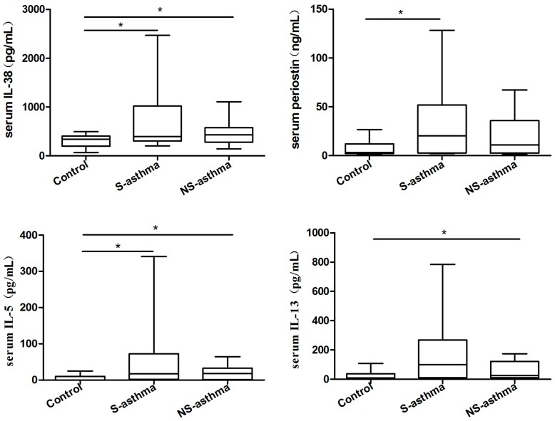
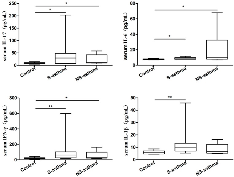
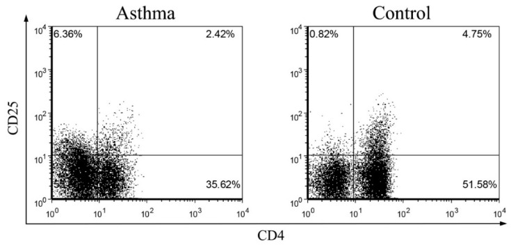
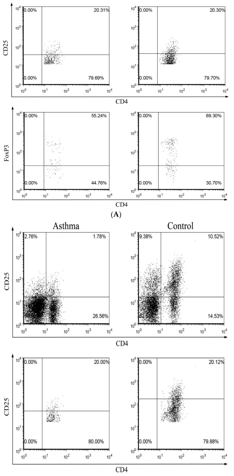
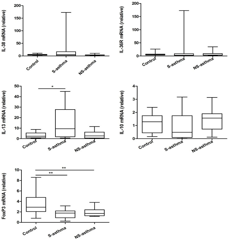
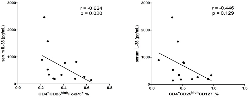
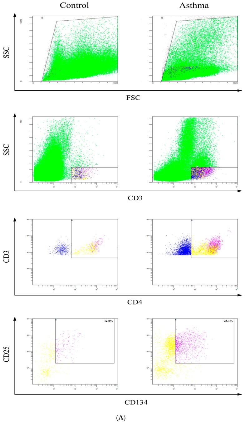
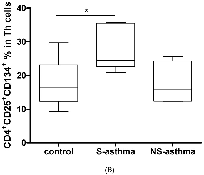
We investigated the expression of novel anti-inflammatory interleukin (IL)-38 and regulatory T (Treg) lymphocytes in childhood asthma patients. The protein and mRNA expression level of IL-38, periostin, peripheral CD4⁺CD25⁺CD134⁺ T lymphocytes as well as CD4⁺CD25(high)FoxP3⁺ and CD4⁺CD25(high)CD127(-) Treg lymphocytes from 40 asthmatic patients and 20 normal control (NC) subjects were studied using ELISA, qPCR and flow cytometry. Serum and supernatant cytokines/chemokines were determined by multiplex assay. Serum IL-38, IL-5, IL-17, IL-6, interferon-γ, periostin, IL-1β and IL-13 concentrations were significantly higher in asthmatic patients with or without steroid treatment than those in controls (all p < 0.05). The percentages of both CD4⁺CD25(high)FoxP3⁺ and CD4⁺CD25(high)CD127(-) Treg lymphocytes were markedly decreased in asthmatic patients with and without steroid treatment than those in controls (all p < 0.05). The elevated IL-38 concentration negatively correlated with the percentage of Treg lymphocytes in asthmatic patients with high level (>40 ng/mL) of periostin (p < 0.05). Although the comparable mRNA levels of IL-38 and its receptor IL-36R were found between patients and controls, the mRNA level of IL-38 positively correlated with IL-36R and negatively correlated with IL-10 in all asthmatic patients (both p < 0.05). The percentage of CD4⁺CD25⁺CD134⁺ activated T lymphocytes was also significantly higher in asthmatic patients with steroid treatment than those in controls (p < 0.05). This cross-sectional study demonstrated that the overexpression of circulating IL-38 may play a role in the immunopathogenesis in asthma.

Keywords: IL-38; childhood asthma; cytokines; regulatory T lymphocytes.


Conflict of interest statement

The authors declare no conflict of interest.

Figures

Figure 1
Figure 1
Comparison of serum concentrations of IL-38, periostin, IL-5, IL-13, IL-17, IL-6, IFN-γ and IL-1β between asthmatic patients with or without steroid treatment, and control subjects. Serum IL-38, periostin, IL-5, IL-13, IL-17, IL-6, IFN-γ and IL-1β were measured using ELISA and multiplex assay kit. Results are presented as box and whisker plots with median (interquartile range). Statistical significances were indicated by * p < 0.05 and ** p < 0.01 (Mann-Whitney U test). S-asthma: steroid-treated asthmatic patients, NS-asthma: non-steroid-treated asthmatic patients, control: control subjects.
Figure 1
Figure 1
Comparison of serum concentrations of IL-38, periostin, IL-5, IL-13, IL-17, IL-6, IFN-γ and IL-1β between asthmatic patients with or without steroid treatment, and control subjects. Serum IL-38, periostin, IL-5, IL-13, IL-17, IL-6, IFN-γ and IL-1β were measured using ELISA and multiplex assay kit. Results are presented as box and whisker plots with median (interquartile range). Statistical significances were indicated by * p < 0.05 and ** p < 0.01 (Mann-Whitney U test). S-asthma: steroid-treated asthmatic patients, NS-asthma: non-steroid-treated asthmatic patients, control: control subjects.
Figure 2
Figure 2
Comparison of circulating CD4+CD25highFoxP3+ and CD4+CD25highCD127 Treg lymphocytes percentages between asthmatic patients with or without steroid treatment, and control subjects. Representative dot plots and gating strategy are shown for the (A) CD4+CD25highFoxP3+ and (B) CD4+CD25highCD127 Treg lymphocytes. Briefly, the CD4+CD25+ cells were gated from total lymphocytes, and then the FoxP3+ or CD127 cells were gated from the top 20% high CD25+ cells of CD4+CD25+ cells; (C,D) The proportion of circulating Treg lymphocytes in total lymphocytes from asthmatic patients with or without steroid treatment, and control subjects were determined by flow cytometry. Results are presented as box and whisker plots with median (interquartile range). Statistical significances were indicated by * p < 0.05 and ** p < 0.01 (Mann-Whitney U test). S-asthma: steroid-treated asthmatic patients, NS-asthma: non-steroid-treated asthmatic patients, control: control subjects.
Figure 2
Figure 2
Comparison of circulating CD4+CD25highFoxP3+ and CD4+CD25highCD127 Treg lymphocytes percentages between asthmatic patients with or without steroid treatment, and control subjects. Representative dot plots and gating strategy are shown for the (A) CD4+CD25highFoxP3+ and (B) CD4+CD25highCD127 Treg lymphocytes. Briefly, the CD4+CD25+ cells were gated from total lymphocytes, and then the FoxP3+ or CD127 cells were gated from the top 20% high CD25+ cells of CD4+CD25+ cells; (C,D) The proportion of circulating Treg lymphocytes in total lymphocytes from asthmatic patients with or without steroid treatment, and control subjects were determined by flow cytometry. Results are presented as box and whisker plots with median (interquartile range). Statistical significances were indicated by * p < 0.05 and ** p < 0.01 (Mann-Whitney U test). S-asthma: steroid-treated asthmatic patients, NS-asthma: non-steroid-treated asthmatic patients, control: control subjects.
Figure 3
Figure 3
The differences of mRNA expression of IL-38, IL-36R, IL-13, IL-10 and FoxP3 between asthmatic patients and controls. PBMC was purified and total RNA was extracted, reverse transcribed and analyzed by real time PCR. Results are presented as box and whisker plots with median (interquartile range). Statistical significances were indicated by * p < 0.05 and ** p < 0.01 (Mann-Whitney U test). S-asthma: steroid-treated asthmatic patients; NS-asthma: non-steroid-treated asthmatic patients, control: control subjects.
Figure 4
Figure 4
Correlations between serum IL-38 concentrations with peripheral regulatory T lymphocytes in asthmatic patients with high level of serum periostin (>40 ng/mL). n = 13. Correlation was determined by non-parametric Spearman’s correlation test.
Figure 5
Figure 5
Determination of activated mitogen-specific CD4+CD25+CD134+ T cells from the in vitro whole blood in asthmatic patients and control. (A) The representative dot plot and gating strategy of activated CD4+CD25+CD134+ T cells in whole blood were shown; (B) The percentage of CD4+CD25+CD134+ T cells in asthmatic patients and control subjects was determined by flow cytometry (n = 8 in each group). Statistical significances were indicated by * p < 0.05 (Mann-Whitney U test). S-asthma: steroid-treated asthmatic patients; NS-asthma: non-steroid-treated asthmatic patients, control: control subjects.
Figure 5
Figure 5
Determination of activated mitogen-specific CD4+CD25+CD134+ T cells from the in vitro whole blood in asthmatic patients and control. (A) The representative dot plot and gating strategy of activated CD4+CD25+CD134+ T cells in whole blood were shown; (B) The percentage of CD4+CD25+CD134+ T cells in asthmatic patients and control subjects was determined by flow cytometry (n = 8 in each group). Statistical significances were indicated by * p < 0.05 (Mann-Whitney U test). S-asthma: steroid-treated asthmatic patients; NS-asthma: non-steroid-treated asthmatic patients, control: control subjects.

Similar articles

Cited by

References

    1. Geering B., Stoeckle C., Conus S., Simon H.U. Living and dying for inflammation: Neutrophils, eosinophils, basophils. Trends Immunol. 2013;34:398–409. doi: 10.1016/j.it.2013.04.002. - DOI - PubMed
    1. Eder W., Ege M.J., von Mutius E. The asthma epidemic. N. Engl. J. Med. 2006;355:2226–2235. doi: 10.1056/NEJMra054308. - DOI - PubMed
    1. Illi S., von Mutius E., Lau S., Niggemann B., Grüber C., Wahn U. Perennial allergen sensitisation early in life and chronic asthma in children: A birth cohort study. Lancet. 2006;368:763–770. doi: 10.1016/S0140-6736(06)69286-6. - DOI - PubMed
    1. Lloyd C.M., Hessel E.M. Functions of T cells in asthma: More than just TH2 cells. Nat. Rev. Immunol. 2010;10:838–848. doi: 10.1038/nri2870. - DOI - PMC - PubMed
    1. Strickland D.H., Holt P.G. T regulatory cells in childhood asthma. Trends Immunol. 2011;32:420–427. doi: 10.1016/j.it.2011.06.010. - DOI - PubMed

MeSH terms