

Pet serine protease from enteroaggregative Escherichia coli stimulates the inflammatory response activating human macrophages
- PMID: 27439312
- PMCID: PMC4955197
- DOI: 10.1186/s12866-016-0775-7
Pet serine protease from enteroaggregative Escherichia coli stimulates the inflammatory response activating human macrophages
Abstract
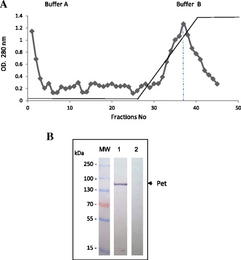
Background: Pet is a toxin from the family of Serine Protease Autotransporters of Enterobacteriaceae which was initially identified in Enteroaggregative Escherichia coli strains. This protease exhibits enterotoxin properties, damages the cell cytoskeleton and induces intestinal epithelium alterations, which are associated with a severe inflammatory process. An in-vitro study was conducted to evaluate the effect of Pet on the migration of human peripheral blood monocytes-derived macrophages and its participation in the activation of the early inflammatory response and cytokine expression.
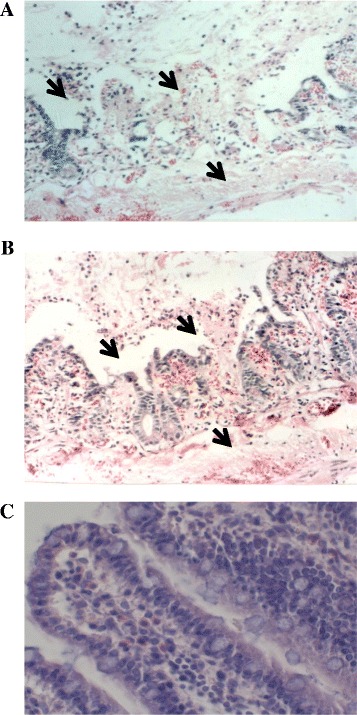
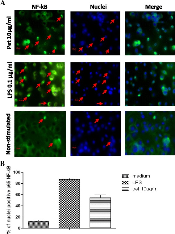
Results: In the macrophage migration activation assay, Pet produced a similar effect to that induced by opsonized zymosan (ZAS). Regarding the cytokine expression, an increase of IL-8, TNF-α (pro-inflammatory) and IL-10 (anti-inflammatory) was identified. In addition to the above results, the nuclear translocation of NF-kB pp65 was also identified. These events are probably related to the inflammatory response identified in the histological examination of intestine rat samples inoculated with Pet during a ligated loop assay.
Conclusion: The results showed that Pet participates as an immunostimulant molecule for macrophages, which activates both their mobility and cytokine expression. These observations suggest that the toxin participates in the inflammatory process that is observed during the host infection by EAEC Pet producing.
Keywords: Cytokines; Escherichia coli; Innate immune response; Serin protease.
Figures

Similar articles
-
The serine protease motif of EspC from enteropathogenic Escherichia coli produces epithelial damage by a mechanism different from that of Pet toxin from enteroaggregative E. coli.Infect Immun. 2004 Jun;72(6):3609-21. doi: 10.1128/IAI.72.6.3609-3621.2004. Infect Immun. 2004. PMID: 15155671 Free PMC article.
-
Curcumin Blocks Cytotoxicity of Enteroaggregative and Enteropathogenic Escherichia coli by Blocking Pet and EspC Proteolytic Release From Bacterial Outer Membrane.Front Cell Infect Microbiol. 2019 Sep 25;9:334. doi: 10.3389/fcimb.2019.00334. eCollection 2019. Front Cell Infect Microbiol. 2019. PMID: 31681620 Free PMC article.
-
Cytoskeletal effects induced by pet, the serine protease enterotoxin of enteroaggregative Escherichia coli.Infect Immun. 1999 May;67(5):2184-92. doi: 10.1128/IAI.67.5.2184-2192.1999. Infect Immun. 1999. PMID: 10225873 Free PMC article.
-
Enteroaggregative Escherichia coli plasmid-encoded toxin.Future Microbiol. 2010 Jul;5(7):1005-13. doi: 10.2217/fmb.10.69. Future Microbiol. 2010. PMID: 20632801 Review.
-
Escherichia coli STb toxin and colibacillosis: knowing is half the battle.FEMS Microbiol Lett. 2008 Jan;278(2):137-45. doi: 10.1111/j.1574-6968.2007.00967.x. Epub 2007 Nov 6. FEMS Microbiol Lett. 2008. PMID: 17995951 Review.
Cited by
-
Bacteria from gut microbiota associated with diarrheal infections in children promote virulence of Shiga toxin-producing and enteroaggregative Escherichia coli pathotypes.Front Cell Infect Microbiol. 2022 Aug 9;12:867205. doi: 10.3389/fcimb.2022.867205. eCollection 2022. Front Cell Infect Microbiol. 2022. PMID: 36017363 Free PMC article.
-
Moringa isothiocyanate-1 inhibits LPS-induced inflammation in mouse myoblasts and skeletal muscle.PLoS One. 2022 Dec 16;17(12):e0279370. doi: 10.1371/journal.pone.0279370. eCollection 2022. PLoS One. 2022. PMID: 36525453 Free PMC article.
-
Analysis of inflammatory cytokine expression in the urinary tract of BALB/c mice infected with Proteus (P.) mirabilis and enteroaggregative Escherichia (E.) coli (EAEC) strains.Folia Microbiol (Praha). 2020 Feb;65(1):133-142. doi: 10.1007/s12223-019-00714-2. Epub 2019 May 19. Folia Microbiol (Praha). 2020. PMID: 31104302
-
The efficacy of anti-proteolytic peptide R7I in intestinal inflammation, function, microbiota, and metabolites by multi-omics analysis in murine bacterial enteritis.Bioeng Transl Med. 2022 Nov 8;8(2):e10446. doi: 10.1002/btm2.10446. eCollection 2023 Mar. Bioeng Transl Med. 2022. PMID: 36925697 Free PMC article.
-
Polyvalent Bacterial Lysate with Potential Use to Treatment and Control of Recurrent Urinary Tract Infections.Int J Mol Sci. 2024 Jun 3;25(11):6157. doi: 10.3390/ijms25116157. Int J Mol Sci. 2024. PMID: 38892345 Free PMC article.
References
-
- Sainz T, Perez J, Fresan MC, Flores V, Jimenez L, Hernandez U, Herrera I, Eslava C. Histological alterations and immune response induced by Pet toxin during colonization with enteroaggregative Escherichia coli (EAEC) in a mouse model infection. J Microbiol. 2002;40:91–7.
MeSH terms
Substances
LinkOut - more resources
Full Text Sources
Other Literature Sources
