. 2016 Jul 21;15(1):85.
doi: 10.1186/s12938-016-0207-9.

Selective laser melting of titanium alloy enables osseointegration of porous multi-rooted implants in a rabbit model

Affiliations

Selective laser melting of titanium alloy enables osseointegration of porous multi-rooted implants in a rabbit model

Wei Peng et al. Biomed Eng Online. .

Abstract

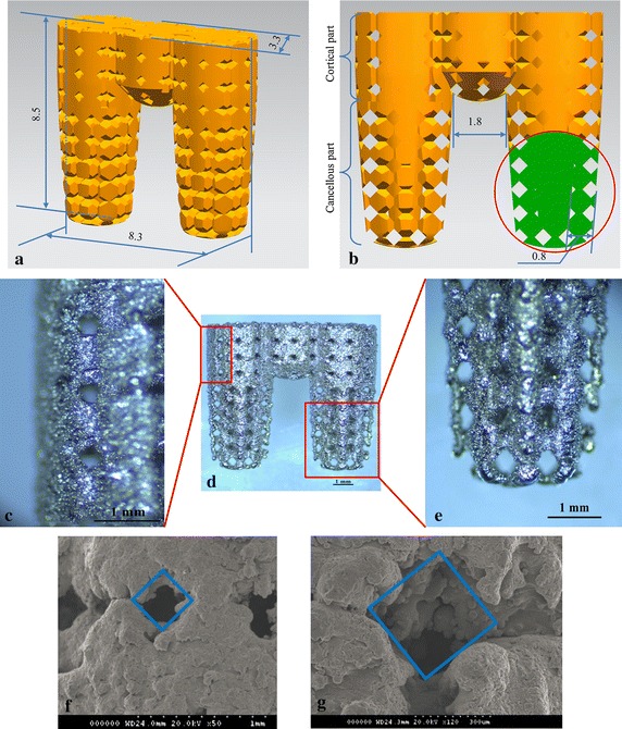
Background: Osseointegration refers to the direct connection between living bone and the surface of a load-bearing artificial implant. Porous implants with well-controlled porosity and pore size can enhance osseointegration. However, until recently implants were produced by machining solid core titanium rods. The aim of this study was to develop a multi-rooted dental implant (MRI) with a connected porous surface structure to facilitate osseointegration.

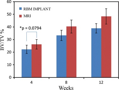
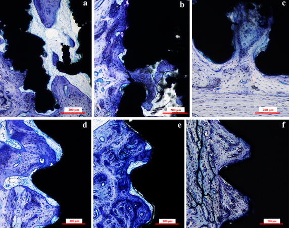
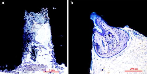
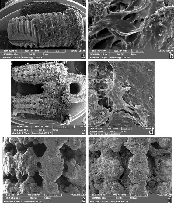
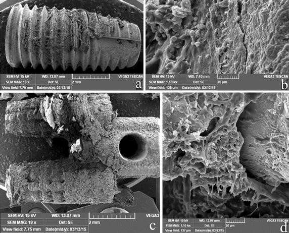
Methods: MRIs manufactured by selective laser melting (SLM) and commercial implants with resorbable blasting media (RBM)-treated surfaces were inserted into the hind limbs of New Zealand white rabbits. Osseointegration was evaluated periodically over 12 weeks by micro-computerized tomography (CT) scanning, histological analysis, mechanical push-out tests, and torque tests.

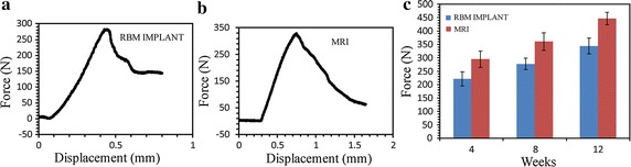
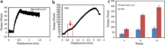
Results: Bone volume densities were consistently higher in the MRI group than in the RBM group throughout the study period, ultimately resulting in a peak value of 48.41 % for the MRI group. Histological analysis revealed denser surrounding bone growth in the MRIs; after 4 and 8 weeks, bone tissue had grown into the pore structures and root bifurcation areas, respectively. Biomechanics tests indicated binding of the porous MRIs to the neobone tissues, as push-out forces strengthened from 294.7 to 446.5 N and maximum mean torque forces improved from 81.15 to 289.57 N (MRI), versus 34.79 to 87.8 N in the RBM group.

Conclusions: MRIs manufactured by SLM possess a connected porous surface structure that improves the osteogenic characteristics of the implant surface.

Keywords: Biomechanics; Implant design; Multi-rooted implant; Osseointegration; Titanium (alloys).


Figures

Fig. 1
Fig. 1
Multi-rooted implant (MRI). a Overall implant dimensions. b Partial cross-section of the MRI, illustrating the pore structure in detail. c The surface of the cortical bone region of fabricated MRI. d The overall profile of the fabricated MRI. e The surface of the cancellous bone region of the fabricated MRI. f Scanning electron microscopy (SEM) image of the cortical bone region of the implant; the pore structure width was approximately 290 µm. g SEM image of the cancellous bone region; the pore structure width was approximately 390 µm
Fig. 2
Fig. 2
Bone volume per total volume (BV/TV) values of the MRIs and resorbable blasting media (RBM) implants after 4, 8, and 12 weeks. A repeated measures analysis with analysis of variance (ANOVA) and Bonferroni post hoc test showed significant differences (p < 0.05) in all cases, except between 8 and 12 weeks within the RBM implant group (p = 0.0583); (n = 6, ±SD). *No significance at 95 % (t-test)
Fig. 3
Fig. 3
Histological sections of the MRIs and RBM implants. Representative sections of the MRIs in rabbit hind limbs at a 4 weeks, b 8 weeks, and c 12 weeks after implantation, and RBM implants in rabbit hind limbs at d 4 weeks, e 8 weeks, and f 12 weeks after implantation. The sections were stained with toluidine blue
Fig. 4
Fig. 4
Bone formation at the root furcation area of MRI. a A histological section of an MRI, 8 weeks after operation, shows bone growth between the root areas. b A representative histological section of an MRI, 12 weeks after implantation, exhibits bone growth at the root furcation
Fig. 5
Fig. 5
Result of push-out test for the RBM implant and MRI. a Representative force–displacement curves for the RBM implant after 8 weeks. b Representative force–displacement curves for the MRI after 8 weeks. c Maximum push-out forces required for the removal of MRIs and RBM implants. The graph plots the average maximum push-out forces of the MRIs and RBM implants after 4, 8, and 12 weeks (n = 6, ±SD). A repeated-measures analysis with ANOVA and Bonferroni post hoc test showed significant differences (p < 0.05), except between 4 and 8 weeks in the RBM implant group (p = 0.1188) and the MRI group (p = 0.1707)
Fig. 6
Fig. 6
SEM images of the push-out implants 8 weeks after implantation. a A global image of an RBM implant. b A high-resolution image (×1.1 K) of an RBM implant. c A global image of an MRI. d A high-resolution image (×1.1 K) of an MRI. e An image of the cancellous part of an MRI before implantation. f An image of the part at (e) 8 weeks after implantation
Fig. 7
Fig. 7
Result of torque test for the RBM implant and MRI. The displacement is measured from the movement of load-cell. a Representative torque-displacement curves for the RBM implant after 8 weeks. b Representative torque-displacement curves for the MRI after 8 weeks. c Maximum torque forces for the MRIs and RBM implants. The graph shows the average maximum torque forces of the MRIs and RBM implants over the 4, 8, and 12 week evaluation period (n = 6, ±SD). A repeated measures analysis with ANOVA and Bonferroni post hoc test showed significant differences for all groups (p < 0.05), with the exception of 8 and 12 weeks within the RBM implant group (p = 0.3463)
Fig. 8
Fig. 8
SEM images of the torque test-removed implants 8 weeks after implantation. a A global image of an RBM implant, b high-resolution image (×1.1 K) of an RBM implant, c global image of an MRI, d high-resolution image (×1.1 K) of an MRI implant

References

    1. Chappuis V, Buser R, Brägger U, Bornstein MM, Salvi GE, Buser D. Long-term outcomes of dental implants with a titanium plasma-sprayed surface: a 20-year prospective case series study in partially edentulous patients. Clin Implant Dent Relat Res. 2013;15:780–790. doi: 10.1111/cid.12056. - DOI - PubMed
    1. Papaspyridakos P, Chen CJ, Singh M, Weber HP, Gallucci GO. Success criteria in implant dentistry: a systematic review. J Dent Res. 2012;91:242–248. doi: 10.1177/0022034511431252. - DOI - PubMed
    1. Fugazzotto PA, Vlassis J. Long-term success of sinus augmentation using various surgical approaches and grafting materials. Int J Oral Maxillofac Implant. 1998;13:52–58. - PubMed
    1. Sisti A, Canullo L, Mottola MP, Covani U, Barone A, Botticelli D. Clinical evaluation of a ridge augmentation procedure for the severely resorbed alveolar socket: multicenter randomized controlled trial, preliminary results. Clin Oral Implant Res. 2012;23:526–535. doi: 10.1111/j.1600-0501.2011.02386.x. - DOI - PubMed
    1. Jackson BJ. Small diameter implants: specific indications and considerations for the posterior mandible: a case report. J Oral Implantol. 2011;37:156–164. doi: 10.1563/AAID-JOI-D-09-00142.1. - DOI - PubMed

LinkOut - more resources