Fluorescent vinblastine probes for live cell imaging
- PMID: 27439765
- PMCID: PMC4970878
- DOI: 10.1039/c6cc04129a
Fluorescent vinblastine probes for live cell imaging
Abstract
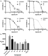
Herein we describe the synthesis of several fluorescent analogues of the clinically approved microtubule destabilizing agent vinblastine. The evaluated probes are the most potent described and provides the first example of uptake, distribution and live cell imaging using this well known antimitotic agent.
Figures


Similar articles
-
Interaction of tubulin with a new fluorescent analogue of vinblastine.Biochemistry. 2002 Nov 26;41(47):14010-8. doi: 10.1021/bi026182m. Biochemistry. 2002. PMID: 12437358
-
Griseofulvin stabilizes microtubule dynamics, activates p53 and inhibits the proliferation of MCF-7 cells synergistically with vinblastine.BMC Cancer. 2010 May 19;10:213. doi: 10.1186/1471-2407-10-213. BMC Cancer. 2010. PMID: 20482847 Free PMC article.
-
Correlation of cytotoxicity and mitotic spindle dissolution by vinblastine in mammalian cells.Cancer Res. 1977 Dec;37(12):4346-51. Cancer Res. 1977. PMID: 922725
-
Fluorescence spectroscopic methods to analyze drug-tubulin interactions.Methods Cell Biol. 2010;95:301-29. doi: 10.1016/S0091-679X(10)95017-6. Methods Cell Biol. 2010. PMID: 20466142 Review.
-
Structural basis for the interaction of tubulin with proteins and drugs that affect microtubule dynamics.Annu Rev Cell Dev Biol. 2000;16:89-111. doi: 10.1146/annurev.cellbio.16.1.89. Annu Rev Cell Dev Biol. 2000. PMID: 11031231 Review.
Cited by
-
SMALL MOLECULE IMAGING AGENT FOR MUTANT KRAS G12C.Adv Ther (Weinh). 2021 May;4(5):2000290. doi: 10.1002/adtp.202000290. Epub 2021 Mar 12. Adv Ther (Weinh). 2021. PMID: 33997272 Free PMC article.
-
Targeted Nanotherapy by Vinblastine-Loaded Chitosan-Coated PLA Nanoparticles to Improve the Chemotherapy via Reactive Oxygen Species to Hamper Hepatocellular Carcinoma.ACS Omega. 2024 Dec 20;10(1):170-180. doi: 10.1021/acsomega.4c02983. eCollection 2025 Jan 14. ACS Omega. 2024. PMID: 39829490 Free PMC article.
-
Quantitating drug-target engagement in single cells in vitro and in vivo.Nat Chem Biol. 2017 Feb;13(2):168-173. doi: 10.1038/nchembio.2248. Epub 2016 Dec 5. Nat Chem Biol. 2017. PMID: 27918558 Free PMC article.
-
Measurement of drug-target engagement in live cells by two-photon fluorescence anisotropy imaging.Nat Protoc. 2017 Jul;12(7):1472-1497. doi: 10.1038/nprot.2017.043. Epub 2017 Jun 29. Nat Protoc. 2017. PMID: 28686582 Free PMC article.
-
Imaging of Tie2 with a Fluorescently Labeled Small Molecule Affinity Ligand.ACS Chem Biol. 2020 Jan 17;15(1):151-157. doi: 10.1021/acschembio.9b00724. Epub 2019 Dec 13. ACS Chem Biol. 2020. PMID: 31809013 Free PMC article.
References
-
- Noble RL, Beer CT, Cutts JH. Ann NY Acad Sci. 1958;76:882–894. - PubMed
-
- Ferrara R, Pilotto S, Peretti U, Caccese M, Kinspergher S, Carbognin L, Karachaliou N, Rosell R, Tortora G, Bria E. Expert Opin Pharmacother. 2016:1–17. 2016. - PubMed
-
- Bellmunt J, Théodore C, Demkov T, Komyakov B, Sengelov L, Daugaard G, Caty A, Carles J, Jagiello-Gruszfeld A, Karyakin O, Delgado FM, Hurteloup P, Winquist E, Morsil N, Salhi Y, Culine S, Maase HVD. J Clin Oncol. 2009;27:4454–4461. - PubMed
MeSH terms
Substances
Grants and funding
LinkOut - more resources
Full Text Sources
Other Literature Sources

