. 2016 Sep 9;291(37):19261-73.
doi: 10.1074/jbc.M116.732529. Epub 2016 Jul 20.

IQGAP1 Binds to Yes-associated Protein (YAP) and Modulates Its Transcriptional Activity

Affiliations

IQGAP1 Binds to Yes-associated Protein (YAP) and Modulates Its Transcriptional Activity

Samar Sayedyahossein et al. J Biol Chem. .

Abstract

During development, the Hippo signaling pathway regulates key physiological processes, such as control of organ size, regeneration, and stem cell biology. Yes-associated protein (YAP) is a major transcriptional co-activator of the Hippo pathway. The scaffold protein IQGAP1 interacts with more than 100 binding partners to integrate diverse signaling pathways. In this study, we report that IQGAP1 binds to YAP and modulates its activity. IQGAP1 and YAP co-immunoprecipitated from cells. In vitro analysis with pure proteins demonstrated a direct interaction between IQGAP1 and YAP. Analysis with multiple fragments of each protein showed that the interaction occurs via the IQ domain of IQGAP1 and the TEAD-binding domain of YAP. The interaction between IQGAP1 and YAP has functional effects. Knock-out of endogenous IQGAP1 significantly increased the formation of nuclear YAP-TEAD complexes. Transcription assays were performed with IQGAP1-null mouse embryonic fibroblasts and HEK293 cells with IQGAP1 knockdown by CRISPR/Cas9. Quantification demonstrated that YAP-TEAD-mediated transcription in cells lacking IQGAP1 was significantly greater than in control cells. These data reveal that IQGAP1 binds to YAP and modulates its co-transcriptional function, suggesting that IQGAP1 participates in Hippo signaling.

Keywords: Hippo pathway; IQGAP1; cell signaling; protein complex; transcription factor; yes-associated protein (YAP).


Figures

FIGURE 1.
FIGURE 1.
IQGAP1 associates with YAP in cells. A, GST-IQGAP1 (IQGAP1) or GST alone was incubated with equal amounts of protein from HeLa cell lysates. Complexes were isolated, washed, and analyzed by SDS-PAGE. The gel was cut at the 100-kDa region. The lower portion of the gel was transferred to PVDF, and the blot was probed with anti-YAP antibody (upper panel). The top part of the gel was stained with Coomassie Blue (lower panel). B and C, HeLa cells were lysed, and equal amounts of protein were loaded directly onto the gel (Lysate) or immunoprecipitated with either polyclonal anti-IQGAP1 or rabbit IgG antibodies in (B) or monoclonal anti-YAP or mouse IgG antibodies (as a negative control) in (C). Immune complexes were analyzed by SDS-PAGE and transferred to PVDF membrane. Blots were probed with anti-IQGAP1 and anti-YAP antibodies. All data are representative of at least three independent experiments. D, to activate the Hippo pathway, HEK293T cells were cultured in medium containing 10% FBS (+) or starved of serum (−) for 16 h. Equal amounts of protein lysate were resolved by SDS-PAGE and Western blotting. The blots were probed with the indicated antibodies. E, cells, treated as described for D, were lysed, and equal amounts of protein were loaded onto the gel (Lysate) or immunoprecipitated with either polyclonal anti-YAP or rabbit IgG (negative control) antibodies. Immune complexes were analyzed by SDS-PAGE and transferred to PVDF membrane. The blots were probed with anti-IQGAP1 and anti-YAP antibodies. The IQGAP1 bands were quantified with Image Studio 2.0 (LI-COR Biosciences) and corrected for the amount of immunoprecipitated YAP in the corresponding sample. The data are expressed as means ± S.E. (error bars) (n = 2) with FBS-treated samples set as 1.
FIGURE 2.
FIGURE 2.
The IQ region of IQGAP1 binds to the TEAD-binding region of YAP. A and C, schematic representation of IQGAP1 (A) and YAP (C) constructs. Full-length (FL) and deletion mutants are depicted. The specific amino acid residues in each construct are indicated. CHD, calponin homology domain; WW, tryptophan containing domain; GRD, RasGAP related domain; RGCT, RasGAP C terminus; TBD, TEAD-binding; TAD, transcription activation domain. B, [35S]methionine-labeled IQGAP1 constructs generated by TnT were incubated with equal amounts of GST-YAP (top panel) or GST alone (middle panel). Samples were run on SDS-PAGE, and gels were dried and analyzed by autoradiography. The bottom panel depicts 5% input of TnT peptides used for pulldown. D, [35S]methionine-labeled YAP constructs generated by TnT were incubated with equal amounts of GST-IQGAP1 (top panel) or GST alone (middle panel). Samples were run on SDS-PAGE, and gels were dried before being analyzed by autoradiography. Input represents 1% of the protein included in the pulldown. The data are representative of three independent experiments.
FIGURE 3.
FIGURE 3.
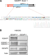
Generation of IQGAP1-null HEK293 cells using CRISPR/Cas9. A, diagram of the regions of IQGAP1 exon 1 targeted by the paired guide RNA. HEK293 transfected with pSPCas9n(BB)-2A-Puro and the paired sgRNA were selected with puromycin. Under the diagram is the sequence for IQGAP1 Exon 1 and the nearby surrounding sequences. Underlining indicates the targeted sgRNA sequences, and double underlining indicates the PAM sequence used by Cas9n. Colors indicate individual nucleotides in the sequence. Individual clones were selected by puromycin, and IQGAP1 genomic mutation was verified. B, equal amounts of protein lysate from HEK293 control and IQGAP1-null cells generated using the CRISPR/Cas9 system were resolved by SDS-PAGE. Western blotting was performed using the indicated antibodies. Actin was used as a loading control.
FIGURE 4.
FIGURE 4.
IQGAP1 modulates YAP-TEAD transcriptional activity. A and B, control (+/+) and IQGAP1-null (−/−) HEK293 (A) and MEF (B) cells were transfected with TEAD reporter luciferase plasmid and Renilla luciferase-polymerase III constructs. Equal numbers of cells were lysed and were processed using a Dual-Luciferase assay. The graphs depict the luciferase signal corrected for Renilla. The values were normalized to IQGAP1+/+ cells. The data are expressed as means ± S.E. from four separate experiments, each performed in duplicate. *, p < 0.01; **, p < 0.001. Western blots show the whole cell lysates run on SDS-PAGE, transferred to PVDF, and probed with the specified antibodies. Actin was used as a loading control. pYAP, phosphorylated YAP. C, IQGAP1−/− HEK293 cells were transfected with either empty vector (V), full-length IQGAP1 (FL), or IQGAP1ΔIQ (ΔIQ). IQGAP1+/+ HEK293 cells transfected with empty vector were used as control. After 24 h, the cells were transfected with luciferase and Renilla plasmids for an additional 24 h. Whole cell lysates were processed using Dual-Luciferase assay. The graphs depict luciferase activity from four independent experiments, each performed in duplicate. The data were normalized to IQGAP1−/− cells transfected with empty vector. The data represent the means ± S.E. *, p < 0.05, analysis of variance. The lower panels depict equal amounts of protein from cell lysates analyzed by immunoblot probed with the specified antibodies. D, total RNA was extracted from control and IQGAP1-null MEFs. Quantitative RT-PCR was performed to measure AMOTL2 and CTGF hnRNA. The amount of hnRNA was corrected for β-actin hnRNA in the same sample. hnRNA in IQGAP1+/+ MEF cells was set as 1. The data represent the means ± S.E. (error bars) of three independent experiments, each performed in triplicate. *, p < 0.01; **, p < 0.001 compared with control cells. E and F, total RNA was extracted from control MEFs transfected with empty vector (V) and IQGAP1-null MEFs transfected with empty vector (V), full-length IQGAP1 (FL), or IQGAP1ΔIQ (ΔIQ). Quantitative RT-PCR was performed to measure AMOTL2 (E) and CTGF (F) hnRNA. The amount of hnRNA was corrected for β-actin hnRNA in the same sample. hnRNA in IQGAP1−/− MEFs transfected with empty vector was set as 1. The data represent the means ± S.E. (error bars) of at least four independent experiments, each performed in triplicate. *, p < 0.05 compared with IQGAP1−/− cells transfected with empty vector. Western blots show whole cell lysates probed with anti-IQGAP1 polyclonal antibodies. Actin was used as a loading control.
FIGURE 5.
FIGURE 5.
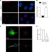
Increased YAP-TEAD nuclear interaction in the absence of IQGAP1. A, IQGAP1+/+ and IQGAP1−/− MEFs were fixed and stained with both anti-YAP and anti-TEAD antibodies. PLA was performed using Duolink in situ detection reagents as described under “Experimental Procedures.” Red spots indicate positive PLA. DNA is stained by Hoescht (blue). Dotted lines outline the nuclei. Scale bar, 10 μm. B, PLA spots in the nucleus were quantified from confocal images of 130 cells. The number of spots per nucleus in control (black bar) and IQGAP1-null (white bar) MEFs were quantified using IMARIS software. The data are expressed as means ± S.E. (error bars) with the number of spots in control cells set to 1. *, p < 0.001 Student's t test. C, IQGAP1−/− MEFs were transfected with GFP-tagged IQGAP1 or GFP alone (control). Samples were processed for PLA as described for A. Red spots indicate positive PLA. DNA is stained by Hoescht (blue). Scale bar, 10 μm. D, PLA spots in the nucleus were quantified from confocal images of 35 cells. The number of spots per nucleus in GFP control and GFP-IQGAP1 expressing MEFs were quantified using IMARIS software. The data are expressed as means ± S.E. with the number of spots in GFP transfected cells set to 1. *, p < 0.001 Student's t test.
FIGURE 6.
FIGURE 6.
IQGAP1 does not alter YAP subcellular localization. A, IQGAP1+/+ and IQGAP1−/− MEFs were lysed and fractionated as described under “Experimental Procedures.” Equal amounts of cytoplasmic (Cyt) and nuclear (Nuc) proteins were resolved by SDS-PAGE, and Western blotting was performed with the indicated antibodies. HSP90 and histone H3 (H3) were used as controls for cytoplasmic and nuclear fractions, respectively. B, the YAP bands were quantified with Image Studio 2.0. The data are expressed as means ± S.E. (n = 3) with IQGAP1+/+ samples set to 1. C, IQGAP1+/+ and IQGAP1−/− MEFs were fixed and stained with anti-YAP monoclonal antibodies (green). Actin was visualized using Alexa Fluor® 568 Phalloidin (red), and DNA was stained with Hoescht (blue). Scale bar, 20 μm. Representative fields are shown. D, to activate the Hippo pathway, MEF cells were cultured in medium containing 10% FBS (+) or starved of serum (−) for 16 h. Cell lysates were obtained, and equal amounts of protein were resolved by SDS-PAGE. The blots were probed with the antibodies indicated. E, IQGAP1+/+ MEFs, treated with FBS (+) or starved (−) for 16 h, were fractionated as described for A. The resultant Western blots were probed with the antibodies indicated. The data for C–E are representative of two independent experiments.
FIGURE 7.
FIGURE 7.
A model of IQGAP1 in the regulation of YAP nuclear activity. The Hippo pathway is regulated by multiple signals that activate or inhibit YAP transcriptional activity. Several mechanisms can attenuate YAP function. These include: i) Cytoplasmic retention. YAP interactions with other proteins, such as AMOT or possibly IQGAP1, sequester YAP in the cytoplasm, inhibiting its nuclear translocation and function. ii) Nuclear competition. IQGAP1 in the nucleus attenuates the formation of YAP-TEAD complexes. iii) Phosphorylation. LATS1/2 and other kinases phosphorylate YAP at Ser127 and Ser381. Phosphorylation at Ser127 promotes YAP interaction with 14-3-3, which retains YAP in the cytoplasm, whereas phosphorylation at Ser381 augments ubiquitination and proteasomal degradation.

Similar articles

Cited by

References

    1. Hansen C. G., Moroishi T., and Guan K. L.(2015) YAP and TAZ: a nexus for Hippo signaling and beyond. Trends Cell Biol. 25, 499–513 - PMC - PubMed
    1. Moroishi T., Hansen C. G., and Guan K. L.(2015) The emerging roles of YAP and TAZ in cancer. Nat. Rev. Cancer 15, 73–79 - PMC - PubMed
    1. Sudol M., Bork P., Einbond A., Kastury K., Druck T., Negrini M., Huebner K., and Lehman D. (1995) Characterization of the mammalian YAP (Yes-associated protein) gene and its role in defining a novel protein module, the WW domain. J. Biol. Chem. 270, 14733–14741 - PubMed
    1. Zhao B., Ye X., Yu J., Li L., Li W., Li S., Yu J., Lin J. D., Wang C. Y., Chinnaiyan A. M., Lai Z. C., and Guan K. L. (2008) TEAD mediates YAP-dependent gene induction and growth control. Genes Dev. 22, 1962–1971 - PMC - PubMed
    1. Liu-Chittenden Y., Huang B., Shim J. S., Chen Q., Lee S. J., Anders R. A., Liu J. O., and Pan D. (2012) Genetic and pharmacological disruption of the TEAD-YAP complex suppresses the oncogenic activity of YAP. Genes Dev. 26, 1300–1305 - PMC - PubMed

MeSH terms

Substances

LinkOut - more resources