. 2016 Sep 12;90(19):8768-79.
doi: 10.1128/JVI.01369-16. Print 2016 Oct 1.

Concurrence of Iridovirus, Polyomavirus, and a Unique Member of a New Group of Fish Papillomaviruses in Lymphocystis Disease-Affected Gilthead Sea Bream

Affiliations

Concurrence of Iridovirus, Polyomavirus, and a Unique Member of a New Group of Fish Papillomaviruses in Lymphocystis Disease-Affected Gilthead Sea Bream

Alberto López-Bueno et al. J Virol. .

Abstract

Lymphocystis disease is a geographically widespread disease affecting more than 150 different species of marine and freshwater fish. The disease, provoked by the iridovirus lymphocystis disease virus (LCDV), is characterized by the appearance of papillomalike lesions on the skin of affected animals that usually self-resolve over time. Development of the disease is usually associated with several environmental factors and, more frequently, with stress conditions provoked by the intensive culture conditions present in fish farms. In gilthead sea bream (Sparus aurata), an economically important cultured fish species in the Mediterranean area, a distinct LCDV has been identified but not yet completely characterized. We have used direct sequencing of the virome of lymphocystis lesions from affected S. aurata fish to obtain the complete genome of a new LCDV-Sa species that is the largest vertebrate iridovirus sequenced to date. Importantly, this approach allowed us to assemble the full-length circular genome sequence of two previously unknown viruses belonging to the papillomaviruses and polyomaviruses, termed Sparus aurata papillomavirus 1 (SaPV1) and Sparus aurata polyomavirus 1 (SaPyV1), respectively. Epidemiological surveys showed that lymphocystis disease was frequently associated with the concurrent appearance of one or both of the new viruses. SaPV1 has unique characteristics, such as an intron within the L1 gene, and as the first member of the Papillomaviridae family described in fish, provides evidence for a more ancient origin of this family than previously thought.

Importance: Lymphocystis disease affects marine and freshwater fish species worldwide. It is characterized by the appearance of papillomalike lesions on the skin that contain heavily enlarged cells (lymphocysts). The causative agent is the lymphocystis disease virus (LCDV), a large icosahedral virus of the family Iridoviridae In the Mediterranean area, the gilthead sea bream (Sparus aurata), an important farmed fish, is frequently affected. Using next-generation sequencing, we have identified within S. aurata lymphocystis lesions the concurrent presence of an additional LCDV species (LCDV-Sa) as well as two novel viruses. These are members of polyomavirus and papillomavirus families, and here we report them to be frequently associated with the presence of lymphocysts in affected fish. Because papillomaviruses have not been described in fish before, these findings support a more ancient origin of this virus family than previously thought and evolutionary implications are discussed.


Figures

FIG 1
FIG 1
Virome of lymphocystis disease-affected gilthead sea bream. (A) Picture of a sample juvenile gilthead sea bream with lymphocystis collected from a Mediterranean Sea farm (southern Spain) in 2001. (B) Transmission electron micrographs of an ultrathin section of skin with lymphocystis lesions showing arrays of LCDV-Sa particles. (C) Taxonomic profile based on a BLASTx comparison of 10,000 metagenomic reads obtained from lesions of a lymphocystis-affected juvenile gilthead sea bream against GenBank-nr database. (D) Transmission electron micrographs of a negatively stained semipurified viral sample derived from gilthead sea bream lymphocysts. LCDV-Sa particles (L) and putative additional icosahedral particles (arrows) are indicated. Scale bars are shown.
FIG 2
FIG 2
Genome analysis and phylogeny of LCDV-Sa. (A) Graphical circular map of the LCDV-Sa genome. The outer scale is numbered clockwise in kilobase pairs. Predicted putative genes are denoted by rectangles, and those in red correspond to iridovirus core genes. The internal histogram shows the coverage in logarithmic scale along the LCDV-Sa genome achieved by Illumina metagenomic sequencing. (B) Dot plot comparison of LCDV-Sa versus LCDV-1 and LCDV-C. Scale is numbered in kilobase pairs. (C) Phylogenetic analysis showing the relationship of LCDV-Sa to representative iridoviruses. The unrooted phylogenetic tree is based on aligned amino acid sequences encoded by 26 concatenated core genes. The evolutionary relationships were inferred through the maximum-likelihood method using R with 1,000 bootstrap replicates. Gray and black circles represent branch nodes with >75% and >90% bootstrap values, respectively. The scale bar indicates the number of amino acid substitutions per residue. The green rectangle indicates members of the genus Lymphocystivirus.
FIG 3
FIG 3
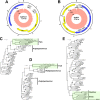
Genome analyses and phylogeny of SaPyV1 and SaPV1. (A and B) Circular graphs of SaPyV1 (A) and SaPV1 (B) genomes are shown. The outer scales are numbered clockwise in kilobase pairs. Predicted genes are indicated by arrows. Yellow, blue, and gray colors indicate structural, nonstructural, and unknown-function proteins, respectively. The internal histograms show the coverage in logarithmic scale along the genomes achieved by Illumina metagenomic sequencing. (C and D) Phylogenetic trees of polyomaviruses based on the amino acid sequences of the large T antigen (C) and VP1 (D) proteins. (E) Phylogenetic analysis of papillomaviruses based on aligned amino acid sequences of the SaPV1 L1 protein and the L1 protein of other representative papillomaviruses. The evolutionary relationships were inferred through the maximum-likelihood method using R with 1,000 bootstrap replicates. The trees were arbitrarily rooted. Gray and black circles represent branch nodes with >75% and >90% bootstrap values, respectively. The scale bar indicates the number of amino acid substitutions per residue. Green rectangles indicate fish polyomaviruses and bird-reptile papillomaviruses.
FIG 4
FIG 4
The SaPV1 L1 major structural protein is expressed from a spliced mRNA. (A) Diagram showing the predicted structure of the SaPV1 L1-encoding gene containing two exons (L1a and L1b). The intron sequence is shown, with splice donor and acceptor consensus sequences in red and the splice site indicated. Relevant nucleotide positions and translation are included. (B) HEK293 cells were transfected with plasmids as indicated on the left, and fluorescence was observed at 48 h after transfection. (C) RT-PCR analysis of RNA from Vero cells transfected with the plasmid bearing the SaPV1 L1 region using L1-specific oligonucleotides performed in the absence (−) or presence (+) of reverse transcriptase. For reference, the PCR using the same oligonucleotide pair was performed using the purified plasmid as the template or water as a negative control. Arrows on the right indicate the positions corresponding to the sizes of the unprocessed (top) and spliced (bottom) SaPV1 L1 messenger. Sizes (in base pairs) of selected markers are indicated on the left.

References

    1. Smail DA, Munro ES. 2012. The virology of teleosts, p 186–291. In Roberts RJ. (ed), Fish pathology, 4th ed, Wiley-Blackwell, Oxford, United Kingdom.
    1. Weissenberg R. 1949. Studies on lymphocystis tumor cells of fish; the osmiophilic granules of the cytoplasmic inclusions and their interpretation as elementary bodies of the lymphocystis virus. Cancer Res 9:537–542. - PubMed
    1. Weissenberg R. 1914. Über infectiöse Zellhypertrophie bei Fischen (Lymphocystiserkrankung). Sitzungsber Kgl Preuss Akad Wiss Sitz Physik Mathem 16:792.
    1. Wolf K. 1962. Experimental propagation of lymphocystis disease. Virology 18:249–256. doi: 10.1016/0042-6822(62)90011-9. - DOI - PubMed
    1. Walker R. 1962. Fine structure of lymphocystis virus of fish. Virology 18:503–505. doi: 10.1016/0042-6822(62)90047-8. - DOI - PubMed

Publication types

MeSH terms