. 2016 Sep 12;90(19):8478-86.
doi: 10.1128/JVI.00705-16. Print 2016 Oct 1.

Viral Macro Domains Reverse Protein ADP-Ribosylation

Affiliations

Viral Macro Domains Reverse Protein ADP-Ribosylation

Changqing Li et al. J Virol. .

Abstract

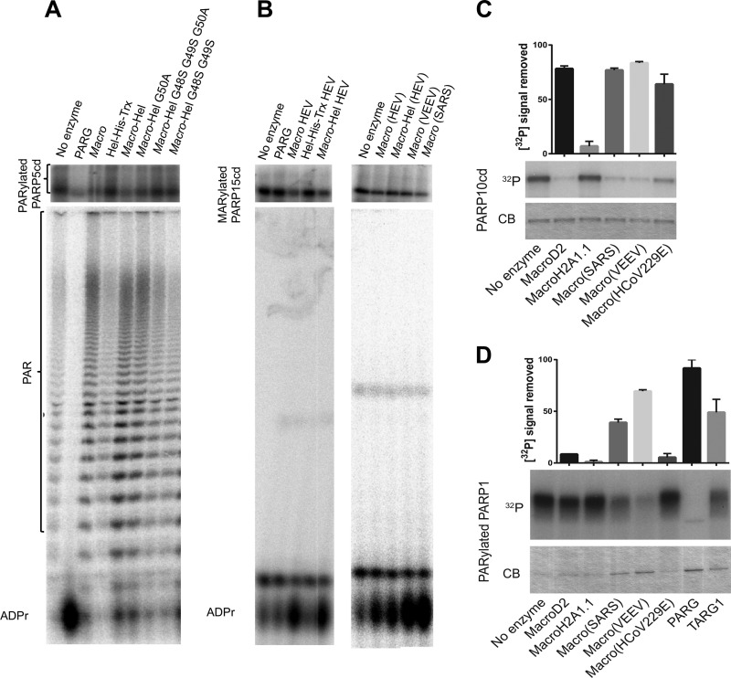
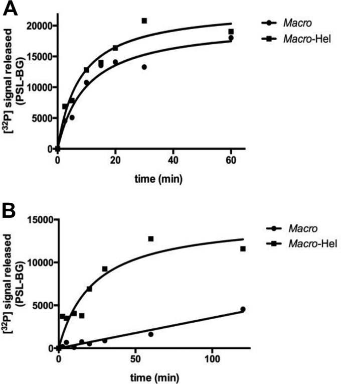
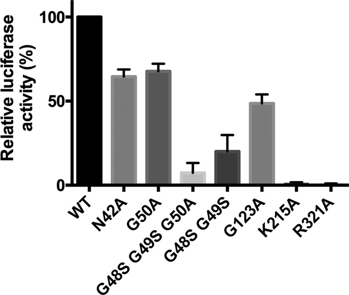
ADP-ribosylation is a posttranslational protein modification in which ADP-ribose is transferred from NAD(+) to specific acceptors to regulate a wide variety of cellular processes. The macro domain is an ancient and highly evolutionarily conserved protein domain widely distributed throughout all kingdoms of life, including viruses. The human TARG1/C6orf130, MacroD1, and MacroD2 proteins can reverse ADP-ribosylation by acting on ADP-ribosylated substrates through the hydrolytic activity of their macro domains. Here, we report that the macro domain from hepatitis E virus (HEV) serves as an ADP-ribose-protein hydrolase for mono-ADP-ribose (MAR) and poly(ADP-ribose) (PAR) chain removal (de-MARylation and de-PARylation, respectively) from mono- and poly(ADP)-ribosylated proteins, respectively. The presence of the HEV helicase in cis dramatically increases the binding of the macro domain to poly(ADP-ribose) and stimulates the de-PARylation activity. Abrogation of the latter dramatically decreases replication of an HEV subgenomic replicon. The de-MARylation activity is present in all three pathogenic positive-sense, single-stranded RNA [(+)ssRNA] virus families which carry a macro domain: Coronaviridae (severe acute respiratory syndrome coronavirus and human coronavirus 229E), Togaviridae (Venezuelan equine encephalitis virus), and Hepeviridae (HEV), indicating that it might be a significant tropism and/or pathogenic determinant.

Importance: Protein ADP-ribosylation is a covalent posttranslational modification regulating cellular protein activities in a dynamic fashion to modulate and coordinate a variety of cellular processes. Three viral families, Coronaviridae, Togaviridae, and Hepeviridae, possess macro domains embedded in their polyproteins. Here, we show that viral macro domains reverse cellular ADP-ribosylation, potentially cutting the signal of a viral infection in the cell. Various poly(ADP-ribose) polymerases which are notorious guardians of cellular integrity are demodified by macro domains from members of these virus families. In the case of hepatitis E virus, the adjacent viral helicase domain dramatically increases the binding of the macro domain to PAR and simulates the demodification activity.


Figures

FIG 1
FIG 1
Proteins relevant to this study. Predicted macro domain (amino acids [aa] 789 to 902) and Hel domain (amino acids 974 to 1185) inside ORF1 of the HEV genome (MeT, methyltransferase; Y, Y domain; PCP, papain-like cysteine protease; V, hypervariable proline-rich hinge; macro: macro domain; Hel, helicase; RdRP, RNA-dependent RNA polymerase). Rectangles show the produced proteins, as indicated. (B) Coomassie blue-stained SDS-PAGE gel showing the purified macro domain (macro-HEV), Hel (Hel-His-Trx HEV), macro-Hel, VEEV macro domain, SARS-CoV macro domain, Tetrahymena thermophila PARG, PARP5a catalytic domain (PARP5 human), and PARP15 catalytic domain (PARP15 human). (C) Structure-based sequence alignment of selected macro domains. Red rectangles, confirmed A1″Pase (I) or de-MARylation (II) activity; gray rectangles, no activity; blank rectangle, not tested. Activity data are based on the results of this study and previously published data (2). Macro domains carrying both A1″Pase and de-MARylation activities are further divided into MacroD2- and c6orf130-like subgroups, based on sequence conservation and catalytic mechanisms. Asterisks indicate important amino acids for PAR binding and/or de-MARylation activity of the MacroD2-like subgroup. E. coli, Escherichia coli; Trx, thioredoxin.
FIG 2
FIG 2
PAR binding studies. 32P-labeled PARylated PARP5a or free PAR was incubated with decreasing concentrations of proteins, dot blotted, and imaged. (A) Binding of the macro domain, Hel, and macro-Hel of HEV to PARylated PARP5a and free PAR. (B) Effects of mutations on the binding of macro-Hel to free PAR. (C) Binding of the HEV macro domain, macro-Hel (HEV), and the VEEV macro domain to free PAR. (D) Poly(ADP-ribose) competitively inhibits the binding of Cy3-labeled RNA to macro-Hel. WT, wild type.
FIG 3
FIG 3
Effects of HEV macro-Hel and other macro domains on PARylated PARP5 and PARP 1 and on MARylated PARP15 and PARP10. SDS–15% PAGE (upper) and 7 M urea–20% PAGE (lower) autoradiography showing the following: de-PARylation of the PARP5a catalytic domain (PARP5cd) by T. thermophila PARG, the macro domain, Hel (Hel-His-Trx), macro-Hel wild type and mutants G50A, G48S-G49S-G50A, and G48S-G49S (A); de-MARylation of the PARP15 catalytic domain by the human PARG, the macro domain, Hel, macro-Hel as well as by the VEEV and SARS macro domains on mono-ADP-ribosylated. Identification of ADP-ribose was based on comigration with cold ADP-ribose detected using UV shadowing. (C) De-MARylation of the PARP10 catalytic domain by different macro domains as observed by SDS-PAGE with Coomassie blue staining (CB) or autoradiography (32P). (D) De-PARylation of PARP1 by different macro domains as observed by SDS-PAGE with Coomassie blue staining (CB) or autoradiography (32P). Quantifications of 32P signal removed are shown above. Error bars indicate standard deviations (n = 2). ADPr, ADP-ribose.
FIG 4
FIG 4
Time course of macro domain and macro-Hel activity on mono-ADP-ribosylated PARP15 (A) and poly(ADP)-ribosylated PARP5a (B) catalytic domains. The auto-ADP-ribosylated PARP5a or -15 catalytic domain was incubated with 100 nM macro domain or macro-Hel at 30°C for 1 h or 2 h. PSL-BG, photo-stimulated luminescence counts minus background counts.
FIG 5
FIG 5
Effect on HEV replication of mutations within the macro-Hel domain. Huh7 cells were transfected with HEV genotype 1 Sar55/S17/luc wild-type (WT) or capped mutant replicon RNA. Luciferase activity was measured after 72 h of incubation at 35°C.
FIG 6
FIG 6
The mode of ADP-ribose coordination in the VEEV macro domain pocket and proposed catalytic mechanism for de-MARylation activity. (A) Superimposition of VEEV macro domain (Protein Data Bank [PDB] accession number 3GQO) with human MacroD2 (PDB accession number 4IQY). (B) Close-up view of the 2.6Å-resolution X-ray structure of the VEEV macro domain in complex with ADP-ribose. ADP-ribose is shown with the corresponding electron density map (light gray). Hydrogen bonds between protein and ADP-ribose are indicated by dashed lines (dark gray). The H2O molecule is coordinated by hydrogen bonds to the 1″-OH, and oxygen from the α-phosphate is shown. (C) Proposed scheme for a catalytic mechanism (see the text for details). The 2mFo − DFc electron density map is contoured at 1.0 σ. The figure was generated using PyMOL.

References

    1. Gibson BA, Kraus WL. 2012. New insights into the molecular and cellular functions of poly(ADP-ribose) and PARPs. Nat Rev Mol Cell Biol 13:411–424. doi:10.1038/nrm3376. - DOI - PubMed
    1. Karlberg T, Langelier MF, Pascal JM, Schuler H. 2013. Structural biology of the writers, readers, and erasers in mono- and poly(ADP-ribose) mediated signaling. Mol Aspects Med 34:1088–1108. doi:10.1016/j.mam.2013.02.002. - DOI - PMC - PubMed
    1. Barkauskaite E, Jankevicius G, Ladurner AG, Ahel I, Timinszky G. 2013. The recognition and removal of cellular poly(ADP-ribose) signals. FEBS J 280:3491–3507. doi:10.1111/febs.12358. - DOI - PubMed
    1. Krietsch J, Rouleau M, Pic E, Ethier C, Dawson TM, Dawson VL, Masson JY, Poirier GG, Gagne JP. 2013. Reprogramming cellular events by poly(ADP-ribose)-binding proteins. Mol Aspects Med 34:1066–1087. doi:10.1016/j.mam.2012.12.005. - DOI - PMC - PubMed
    1. Sharifi R, Morra R, Appel CD, Tallis M, Chioza B, Jankevicius G, Simpson MA, Matic I, Ozkan E, Golia B, Schellenberg MJ, Weston R, Williams JG, Rossi MN, Galehdari H, Krahn J, Wan A, Trembath RC, Crosby AH, Ahel D, Hay R, Ladurner AG, Timinszky G, Williams RS, Ahel I. 2013. Deficiency of terminal ADP-ribose protein glycohydrolase TARG1/C6orf130 in neurodegenerative disease. EMBO J 32:1225–1237. doi:10.1038/emboj.2013.51. - DOI - PMC - PubMed

Publication types

MeSH terms

LinkOut - more resources