. 2016 Sep 12;90(19):8705-19.
doi: 10.1128/JVI.01229-16. Print 2016 Oct 1.

Hepatitis B Virus Polymerase Localizes to the Mitochondria, and Its Terminal Protein Domain Contains the Mitochondrial Targeting Signal

Affiliations

Hepatitis B Virus Polymerase Localizes to the Mitochondria, and Its Terminal Protein Domain Contains the Mitochondrial Targeting Signal

Nuruddin Unchwaniwala et al. J Virol. .

Abstract

To understand subcellular sites of hepatitis B virus (HBV) replication, we visualized core (Cp), polymerase (Pol), and pregenomic RNA (pgRNA) in infected cells. Interestingly, we found that the majority of Pol localized to the mitochondria in cells undergoing viral replication. The mitochondrial localization of Pol was independent of both the cell type and other viral components, indicating that Pol contains an intrinsic mitochondrial targeting signal (MTS). Neither Cp nor pgRNA localized to the mitochondria during active replication, suggesting a role other than DNA synthesis for Pol at the mitochondria. The Pol of duck hepatitis B virus (DHBV) also localized to the mitochondria. This result indicates that localization of Pol to mitochondria is likely a feature of all hepadnaviruses. To map the MTS within HBV Pol, we generated a series of Pol-green fluorescent protein (Pol-GFP) fusions and found that a stretch spanning amino acids (aa) 141 to 160 of Pol was sufficient to target GFP to the mitochondria. Surprisingly, deleting aa 141 to 160 in full-length Pol did not fully ablate Pol's mitochondrial localization, suggesting that additional sequences are involved in mitochondrial targeting. Only by deleting the N-terminal 160 amino acids in full-length Pol was mitochondrial localization ablated. Crucial residues for pgRNA packaging are contained within aa 141 to 160, indicating a multifunctional role of this region of Pol in the viral life cycle. Our studies show an unexpected Pol trafficking behavior that is uncoupled from its role in viral DNA synthesis.

Importance: Chronic infection by HBV is a serious health concern. Existing therapies for chronically infected individuals are not curative, underscoring the need for a better understanding of the viral life cycle to develop better antiviral therapies. To date, the most thoroughly studied function of Pol is to package the pgRNA and reverse transcribe it to double-stranded DNA within capsids. This study provides evidence for mitochondrial localization of Pol and defines the MTS. Recent findings have implicated a non-reverse transcription role for Pol in evading host innate immune responses. Mitochondria play an important role in controlling cellular metabolism, apoptosis, and innate immunity. Pol may alter one or more of these host mitochondrial functions to gain a replicative advantage and persist in chronically infected individuals.


Figures

FIG 1
FIG 1
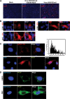
HBV subcellular localization during infection. (A) Cp immunofluorescence in HepG2-NTCP12 cells infected by WT viruses. (B) Cellular distribution of Cp in infected HepG2-NTCP12 cells. At least 100 cells were scored for this analysis. (C) (Top) pgRNA FISH analysis of infected HepG2-NTCP12 cells. The histogram depicts the distribution of the integrated fluorescence intensities of the RNA in a cell. a.u, arbitrary units. (Bottom) Simultaneous Cp and Pol immunofluorescence in infected HepG2-NTCP12 cells. (D) HepG2-NTCP12 cells were transfected with two different plasmids for detection of Pol. One plasmid expressed the pgRNA from the endogenous (ENDO) HBV promoter and the other from the CMV immediate early (CMV) promoter. In addition, a second plasmid was transfected that expressed the fluorescent protein EBFP2 with a C-terminal nuclear localization signal (NLS) to identify the transfected cells.
FIG 2
FIG 2
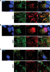
Pol localizes to mitochondria. Plasmid HBVEp+Cp+PolY63F+X+env+ was cotransfected with Mito-GFP into Huh7 cells to label mitochondria. Cells were then processed for detection of various viral markers. (A) Pol localizes to mitochondria. Pol immunofluorescence was observed by using a combination of the 2C8 and 8D5 antibodies. (B) Cp immunofluorescence. (C) FISH analysis of pgRNA.
FIG 3
FIG 3
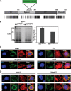
Pol-GFP is functional for intracellular viral replication and localizes to the mitochondria independently of cell type and other viral components. (A) Schematic representation of the four distinct domains of the human HBV Pol protein sequence. The boundaries of each domain are marked by amino acid numbers at the top. A G4SG4-GFP-G4SG4 sequence insertion was made between amino acids 292 and 293 within the spacer domain. Below the protein representation is an alignment of the Pol protein sequences from 7 different hepadnaviruses: human (UniProt accession no. P03156), chimpanzee (UniProt accession no. Q9QAW8), woolly monkey (UniProt accession no. O71304), woodchuck (UniProt accession no. P03160), duck (UniProt accession no. P0C691), snow goose (UniProt accession no. Q9WFB5), and heron (UniProt accession no. P13846) HBVs. The alignment was generated by Jalview by using the ClustalW multiple-sequence alignment program; each position was given a conservation score of 1 to 10 (1 is least conserved and 10 is fully conserved), and then each position's score was represented by a vertical line, using a grayscale range from white (least conserved) to black (highly conserved). TP, terminal protein; RT, reverse transcriptase. (B) Southern blot analysis of intracellular viral DNA (icDNA). Replication of HBVCp+Pol+X+ or HBVCp+PolGFP+X+ was complemented with HBVEp+. Minus-strand oligonucleotide probes were used to detect various intracellular viral replicative intermediates. Duplicate transfections are shown for each condition. For total icDNA measurement, all the signals between and including RC to SS DNA were measured. RC, relaxed circular DNA; DL, duplex linear DNA; SS, single-stranded DNA. (C) Pol-GFP localization in various cell types under replication-competent conditions. For each condition, HBVCp+PolGFP+X+ and HBVEp+ were cotransfected. Images of individual channels for DAPI, GFP, and Mitotracker Red are shown, followed by merged images showing all three channels. (D) Pol-GFP localizes to the mitochondria in a replication-independent manner. The “replication +” condition represents the HBVCp+PolGFP+X+ plasmid complemented with HBVEp+ to engender intracellular viral replication. The “replication −” condition represents the Pol-GFPonly plasmid alone.
FIG 4
FIG 4
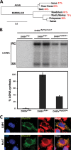
Mitochondrial targeting of Pol is conserved in duck hepadnaviruses. (A) Phylogenetic tree for Pol amino acid sequences across 4 mammalian and 3 avian hepadnaviruses. MUSCLE multiple-sequence alignment, PhyML phylogenetic analysis, and TreeDyn tree rendering were used to generate the phylogenetic tree (www.phylogeny.fr). The scale bar represents the number of amino acid substitutions per site. The percentages in red indicate the percent identities of the species-specific viral Pol to human Pol. The percentages for the pairwise alignment were obtained using the BLOSUM62 global alignment matrix. (B) Southern blot analysis of intracellular viral DNA (icDNA). Plasmid DHBVEp+Cp+env+ was complemented with either the DHBVPol−, DHBVPol+, or DHBVPolGFP+ plasmid. The total signal intensity was measured from RC to SS DNA and then plotted. Duplicate transfections are shown for each condition. RC, relaxed circular DNA; SS, single-stranded DNA. (C) Subcellular localization of DHBV GFP-labeled Pol (DPol-GFP) in LMH and Huh7 cells.
FIG 5
FIG 5
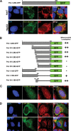
Defining the MTS. (A) Schematic representation of the Pol 1-292-GFP construct and cellular localization of Pol 1-292-GFP in Huh7 and HepG2 cells. (B) Schematic representation of the various Pol truncations studied for mitochondrial localization in Huh7 cells. ++, highly mitochondrial; +, partially mitochondrial; −, not mitochondrial. (C) Subcellular localization of Pol 141-160-GFP in Huh7 cells. (D) Mitochondrial localization of deletion mutants of full-length Pol in Huh7 cells. The Δ141–160 and Δ1–160 constructs are described in Materials and Methods and Table 1.
FIG 6
FIG 6
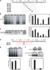
Pol's minimal targeting signal contains residues important for pgRNA packaging and is overlapped by a predicted alpha-helix. (A) Pol amino acid sequence from aa 130 to 170. Di-alanine variants generated from WT Pol are shown below the protein sequence. In the case of a preexisting alanine in the WT sequence, the alanine was replaced by a glycine instead (KA-AG). (B) Intracellular viral DNA replication of di-alanine mutants of Pol in Huh7 cells. All di-alanine mutants were generated from the Pol-GFP plasmid and complemented with NL84 to study intracellular viral replication and icDNA synthesis. Duplicate transfections are shown for each condition. (C) Native gel analysis to study plus-strand nucleic acid levels within capsids. ND, not detectable. (D) Amino acids highlighted in red and underlined were predicted to form an alpha-helix by the XtalPRED server, using the PSIPRED algorithm. The Pol amino acid sequences with di-alanine and di-proline substitutions at Q143 and T144 are highlighted. (E) icDNA levels for the QT-AA and QT-PP Pol constructs. Duplicate transfections are shown for each condition. (F) The QT-PP replication defect is at the level of pgRNA packaging. (Top) Western blot for Cp. (Middle) Native gel analysis of pgRNA levels in QT-PP Pol compared to those in DNA synthesis-null Y63F Pol generated in the HBVCp+PolGFP+X+ background. Pol plasmids were complemented with the HBVEp+ plasmid to provide packaging-competent Ep-pgRNA. Duplicate transfections are shown.

References

    1. Beasley RP. 1988. Hepatitis B virus. The major etiology of hepatocellular carcinoma. Cancer 61:1942–1956. - PubMed
    1. GBD 2013 Mortality and Causes of Death Collaborators. 2015. Global, regional, and national age-sex specific all-cause and cause-specific mortality for 240 causes of death, 1990–2013: a systematic analysis for the Global Burden of Disease Study 2013. Lancet 385:117–171. doi:10.1016/S0140-6736(14)61682-2. - DOI - PMC - PubMed
    1. Schweitzer A, Horn J, Mikolajczyk RT, Krause G, Ott JJ. 2015. Estimations of worldwide prevalence of chronic hepatitis B virus infection: a systematic review of data published between 1965 and 2013. Lancet 386:1546–1555. doi:10.1016/S0140-6736(15)61412-X. - DOI - PubMed
    1. Block TM, Gish R, Guo H, Mehta A, Cuconati A, Thomas London W, Guo J-T. 2013. Chronic hepatitis B: what should be the goal for new therapies? Antiviral Res 98:27–34. doi:10.1016/j.antiviral.2013.01.006. - DOI - PMC - PubMed
    1. Ganem D, Varmus HE. 1987. The molecular biology of the hepatitis B viruses. Annu Rev Biochem 56:651–693. doi:10.1146/annurev.bi.56.070187.003251. - DOI - PubMed

Publication types

MeSH terms

LinkOut - more resources