. 2016 Sep 12;90(19):8464-77.
doi: 10.1128/JVI.00471-16. Print 2016 Oct 1.

Human Cathelicidin Compensates for the Role of Apolipoproteins in Hepatitis C Virus Infectious Particle Formation

Affiliations

Human Cathelicidin Compensates for the Role of Apolipoproteins in Hepatitis C Virus Infectious Particle Formation

Francesc Puig-Basagoiti et al. J Virol. .

Abstract

Exchangeable apolipoproteins (ApoA, -C, and -E) have been shown to redundantly participate in the formation of infectious hepatitis C virus (HCV) particles during the assembly process, although their precise role in the viral life cycle is not well understood. Recently, it was shown that the exogenous expression of only short sequences containing amphipathic α-helices from various apolipoproteins is sufficient to restore the formation of infectious HCV particles in ApoB and ApoE double-gene-knockout Huh7 (BE-KO) cells. In this study, through the expression of a small library of human secretory proteins containing amphipathic α-helix structures, we identified the human cathelicidin antimicrobial peptide (CAMP), the only known member of the cathelicidin family of antimicrobial peptides (AMPs) in humans and expressed mainly in bone marrow and leukocytes. We showed that CAMP is able to rescue HCV infectious particle formation in BE-KO cells. In addition, we revealed that the LL-37 domain in CAMP containing amphipathic α-helices is crucial for the compensation of infectivity in BE-KO cells, and the expression of CAMP in nonhepatic 293T cells expressing claudin 1 and microRNA miR-122 confers complete propagation of HCV. These results suggest the possibility of extrahepatic propagation of HCV in cells with low-level or no expression of apolipoproteins but expressing secretory proteins containing amphipathic α-helices such as CAMP.

Importance: Various exchangeable apolipoproteins play a pivotal role in the formation of infectious HCV during the assembly of viral particles, and amphipathic α-helix motifs in the apolipoproteins have been shown to be a key factor. To the best of our knowledge, we have identified for the first time the human cathelicidin CAMP as a cellular protein that can compensate for the role of apolipoproteins in the life cycle of HCV. We have also identified the domain in CAMP that contains amphipathic α-helices crucial for compensation and show that the expression of CAMP in nonhepatic cells expressing claudin 1 and miR-122 confers complete propagation of HCV. We speculate that low levels of HCV propagation might be possible in extrahepatic tissues expressing secretory proteins containing amphipathic α-helices without the expression of apolipoproteins.


Figures

FIG 1
FIG 1
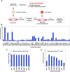
Construction of the protein library and screening. (A) Schematic diagram of the protein library screening process. cDNAs from the 44 selected proteins were individually amplified and cloned into a lentivirus expression vector (pCSII-EF-RfA) that was transfected into 293T cells to produce lentiviruses. BE-KO cells were infected with HCV at a multiplicity of infection of 1, 72 h after infection with lentiviruses. Infectious titers of HCV in the supernatants were determined by a focus-forming assay at 72 h postinfection. (B) Infectious titers in the culture supernatants from BE-KO cells expressing the library target proteins were determined by a focus-forming assay in Huh7.5.1 cells. Average titers were plotted as fold differences compared to the titers obtained from control cells. (C) BE-KO cells expressing each of the proteins identified by screening were infected with JFH1-Nluc at a multiplicity of infection of 1, and luciferase activity in the supernatant was determined at 2 days postinfection. (D) The supernatants were further inoculated into Huh7.5.1 cells, and luciferase activity in the culture supernatants was determined at 2 days postinfection.
FIG 2
FIG 2
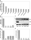
Expression of CAMP in various tissues and cell lines. (A) The relative mRNA expression level of CAMP in different human tissues was determined by using the NextBio Body Atlas application. The median expression level was calculated across all 128 human tissues from 1,068 arrays by using the Affymetrix GeneChip Human Genome U133 Plus 2.0 array. The mRNA expression level for each gene was log10 transformed. (B) Endogenous expression of CAMP mRNA. Total RNA was extracted from PBMCs and 293T, Huh7, Hep3B, and HepG2 cells, and the expression of CAMP mRNA was determined by qRT-PCR. ND, not determined. (C) Endogenous expression of ApoE and CAMP was determined by immunoblotting. (D and E) Expression levels of ApoE and CAMP in plasma and supernatants from 293T, Huh7, Hep3B, and HepG2 cells were determined by using ELISA kits.
FIG 3
FIG 3
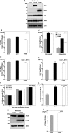
CAMP expression increases the production of infectious HCV particles in BE-KO cells. (A) Expression levels of ApoE and CAMP were determined by immunoblotting 48 h after transduction of lentiviruses into BE-KO cells. Expression levels of HCV core and NS5A proteins were determined 72 h after infection with HCV JFH1 at a multiplicity of infection of 1. (B and C) Intracellular HCV RNA levels (B) as well as intracellular and extracellular infectious titers (C) were determined 72 h after infection with HCV JFH1 at a multiplicity of infection of 1 by qRT-PCR and a focus-forming assay, respectively. (D and E) Intracellular HCV RNA levels (D) as well as extracellular infectious titers (E) were determined 72 h after infection with HCV Con1/JFH1 at a multiplicity of infection of 2 by qRT-PCR and a focus-forming assay, respectively. (F) BE-KO cells expressing either ApoE or CAMP were electroporated with JFH1-Nluc RNA, and luciferase activities in the supernatants collected every 24 h for 3 days were determined. (G) The supernatants at 3 days postelectroporation were further inoculated into Huh7.5.1 cells, and luciferase activity in the culture supernatants was determined at 2 days postinoculation. (H) Expression levels of ApoE and CAMP were determined by immunoblotting 48 h after transduction of lentiviruses into Huh7 cells. (I) Infectious titers in the supernatant were determined 72 h after infection with HCV JFH1 at a multiplicity of infection of 1. In all cases, asterisks indicate significant differences (*, P < 0.05; **, P < 0.01) versus the results for control cells.
FIG 4
FIG 4
CAMP expression does not increase production of infectious DENV or JEV particles in BE-KO cells. Infectious titers in the supernatants of BE-KO cells expressing either ApoE or CAMP were determined by a focus-forming assay 48 h after infection with either DENV (A) or JEV (B) at a multiplicity of infection of 1.
FIG 5
FIG 5
CAMP compensates for the role of apolipoproteins in BE-KO cells. (A and B) BE-KO cells expressing either ApoE or CAMP were inoculated with pseudoparticles bearing HCV envelope proteins E1 and E2 (HCVpp) (A) or VSV G protein (VSVpp) (B), and luciferase activity was determined at 24 h postinfection. (C) BE-KO cells expressing either ApoE or CAMP were infected with HCV at a multiplicity of infection of 1, and expression of the HCV core protein was determined by immunoblot analysis at 72 h postinfection. (D) Expression of core protein, lipid droplets (LDs), and cell nuclei was examined by immunofluorescence analysis after staining with anticore antibody, BODIPY, and DAPI, respectively. Pearson's correlation coefficients (r), quantifying the degree of colocalization of the HCV core protein with LDs, are presented in the merged images. The boxed areas in the merged images are magnified. (E) Specific infectivity in the supernatants of BE-KO cells expressing either ApoE or CAMP upon infection with HCV at a multiplicity of infection of 1 was determined by comparing the infectious titers with HCV RNA levels. (F and G) The supernatants of BE-KO cells expressing either ApoE or CAMP upon infection with HCV at a multiplicity of infection of 1 were subjected to density gradient fractionation, and infectious titers (G) and HCV RNA levels (F) for each fraction were determined by a focus-forming assay and qRT-PCR, respectively. (H) BE-KO cells expressing either ApoE or CAMP were lysed 72 h after infection with HCVcc and subjected to a proteinase K digestion protection assay. Cell lysates were split into 3 parts and incubated on ice for 1 h in the presence or absence of 50 μg/ml proteinase K with or without pretreatment with 5% Triton X-100. After treatment, the samples were subjected to immunoblotting using anticore antibody. (I and J) HCV RNA levels in the supernatants (I) and exosomes (J) of BE-KO cells expressing either ApoE or CAMP upon infection with HCV at a multiplicity of infection of 1 were determined by qRT-PCR.
FIG 6
FIG 6
CAMP activity is located in the LL-37 domain. (A) Schematics of CAMP and its deletion mutant. aa, amino acids. (B) Deletion mutant with an HA tag expressed in BE-KO cells by a lentiviral vector detected by immunoblotting. (C and D) BE-KO cells expressing either ApoE, CAMP, or LL-37 were infected with HCV at a multiplicity of infection of 1, and intracellular HCV RNA levels (C) and infectious titers (D) in the supernatants at 72 h postinfection were determined by qRT-PCR and a focus-forming assay, respectively. In all cases, asterisks indicate significant differences (*, P < 0.05; **, P < 0.01) versus the results for control cells.
FIG 7
FIG 7
Amphipathic α-helices in the LL-37 domain are necessary for CAMP activity. (A) Structure of LL-37 of CAMP and amino acid sequences of the LL-37 domain deletion mutants. (B) The expression of deletion mutants with HA tags in BE-KO cells by a lentiviral vector was determined by immunoblotting. (C and D) BE-KO cells expressing either ApoE, CAMP, KE-23, or LL-15 were infected with HCV at a multiplicity of infection of 1, and intracellular HCV RNA levels (C) and infectious titers (D) in the supernatants at 72 h postinfection were determined by qRT-PCR and a focus-forming assay, respectively. In all cases, asterisks indicate significant differences (*, P < 0.05; **, P < 0.01) versus the results for control cells.
FIG 8
FIG 8
CAMP expression confers production of infectious HCV particles in nonhepatic cells. (A) Expression of ApoE and CAMP in 293T-CLDN1-miR-122 cells was determined by immunoblot analysis. (B and C) Cells were infected with HCV at a multiplicity of infection of 10, and intracellular HCV RNA levels (B) and infectious titers (C) in the supernatants at 72 h postinfection were determined by qRT-PCR and a focus-forming assay, respectively. (D) Expression of ApoE and CAMP in 293T-CLDN1 cells was determined by immunoblot analysis. (E and F) Cells were infected with HCV containing a G28A mutation at a multiplicity of infection of 10, and intracellular HCV RNA levels (E) and infectious titers (F) in the supernatants at 72 h postinfection were determined by qRT-PCR and a focus-forming assay, respectively. (G) 293T-CLDN1-miR-122 cells expressing either CAMP or ApoE were electroporated with JFH1 RNA, and viral titers in the supernatant were determined by a focus-forming assay at days 3 and 6 posttransfection. In all cases, asterisks indicate significant differences (*, P < 0.05; **, P < 0.01) versus the results for control cells.

Similar articles

Cited by

References

    1. Maasoumy B, Wedemeyer H. 2012. Natural history of acute and chronic hepatitis C. Best Pract Res Clin Gastroenterol 26:401–412. doi:10.1016/j.bpg.2012.09.009. - DOI - PubMed
    1. Hajarizadeh B, Grebely J, Dore GJ. 2013. Epidemiology and natural history of HCV infection. Nat Rev Gastroenterol Hepatol 10:553–562. doi:10.1038/nrgastro.2013.107. - DOI - PubMed
    1. Negro F, Forton D, Craxì A, Sulkowski MS, Feld JJ, Manns MP. 2015. Extrahepatic morbidity and mortality of chronic hepatitis C. Gastroenterology 149:1345–1360. doi:10.1053/j.gastro.2015.08.035. - DOI - PubMed
    1. Elbaz T, El-Kassas M, Esmat G. 2015. New era for management of chronic hepatitis C virus using direct antiviral agents: a review. J Adv Res 6:301–310. doi:10.1016/j.jare.2014.11.004. - DOI - PMC - PubMed
    1. Bartosch B, Dubuisson J, Cosset FL. 2003. Infectious hepatitis C virus pseudo-particles containing functional E1-E2 envelope protein complexes. J Exp Med 197:633–642. doi:10.1084/jem.20021756. - DOI - PMC - PubMed

LinkOut - more resources