. 2016 Sep 12;90(19):8739-53.
doi: 10.1128/JVI.00797-16. Print 2016 Oct 1.

HIV-1 Vpr Inhibits Kaposi's Sarcoma-Associated Herpesvirus Lytic Replication by Inducing MicroRNA miR-942-5p and Activating NF-κB Signaling

Affiliations

HIV-1 Vpr Inhibits Kaposi's Sarcoma-Associated Herpesvirus Lytic Replication by Inducing MicroRNA miR-942-5p and Activating NF-κB Signaling

Qin Yan et al. J Virol. .

Abstract

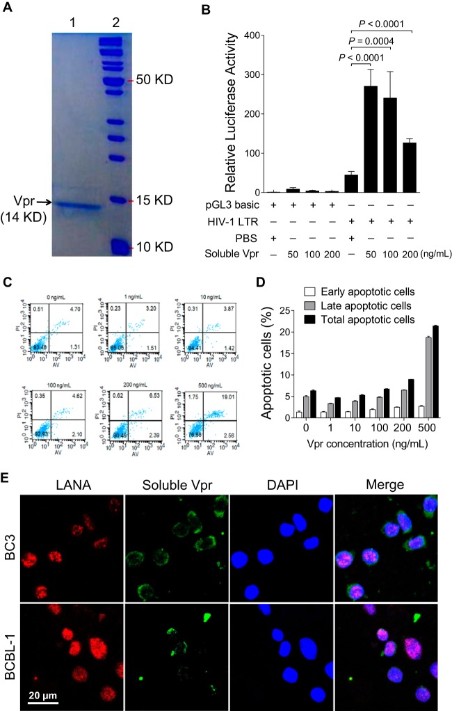
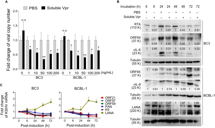
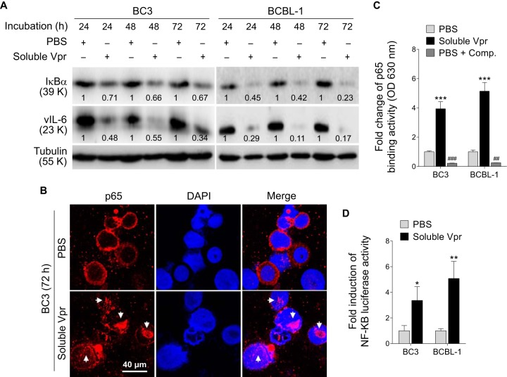
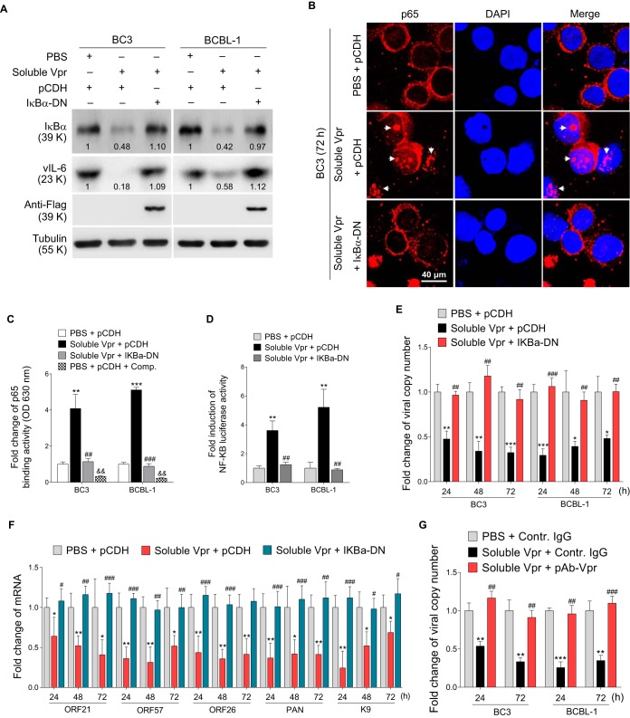
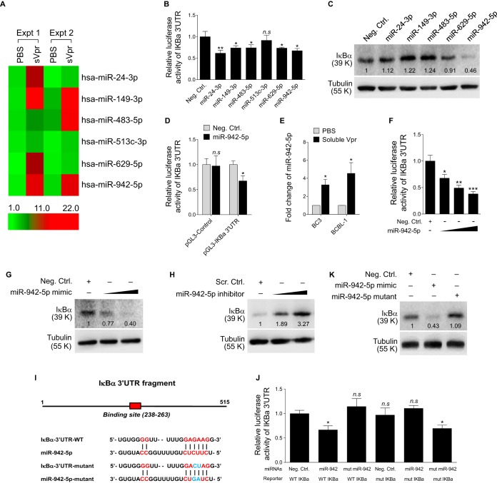
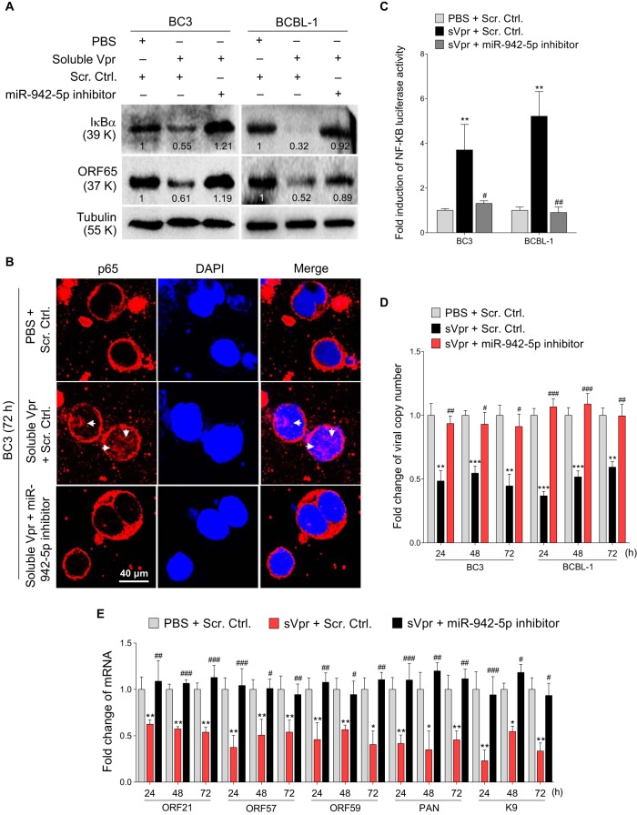
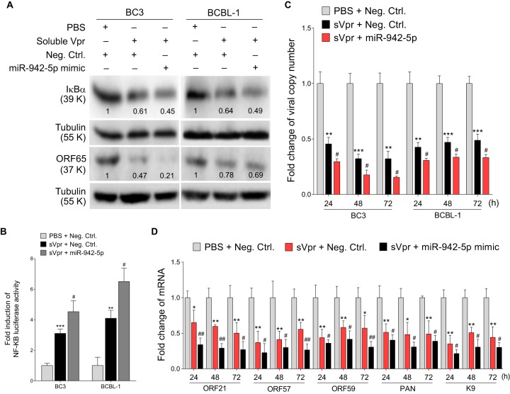
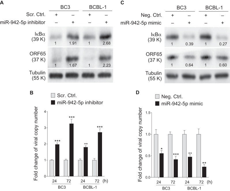
Kaposi's sarcoma-associated herpesvirus (KSHV) infection is required for the development of several AIDS-related malignancies, including Kaposi's sarcoma (KS) and primary effusion lymphoma (PEL). The high incidence of AIDS-KS has been ascribed to the interaction of KSHV and HIV-1. We have previously shown that HIV-1-secreted proteins Tat and Nef regulate the KSHV life cycle and synergize with KSHV oncogenes to promote angiogenesis and tumorigenesis. Here, we examined the regulation of KSHV latency by HIV-1 viral protein R (Vpr). We found that soluble Vpr inhibits the expression of KSHV lytic transcripts and proteins, as well as viral particle production by activating NF-κB signaling following internalization into PEL cells. By analyzing the expression profiles of microRNAs combined with target search by bioinformatics and luciferase reporter analyses, we identified a Vpr-upregulated cellular microRNA (miRNA), miR-942-5p, that directly targeted IκBα. Suppression of miR-942-5p relieved the expression of IκBα and reduced Vpr inhibition of KSHV lytic replication, while overexpression of miR-942-5p enhanced Vpr inhibition of KSHV lytic replication. Our findings collectively illustrate that, by activating NF-κB signaling through upregulating a cellular miRNA to target IκBα, internalized HIV-1 Vpr inhibits KSHV lytic replication. These results have demonstrated an essential role of Vpr in the life cycle of KSHV.

Importance: Coinfection by HIV-1 promotes the aggressive growth of Kaposi's sarcoma-associated herpesvirus (KSHV)-related malignancies, including Kaposi's sarcoma (KS) and primary effusion lymphoma (PEL). In this study, we have shown that soluble HIV-1 Vpr inhibits KSHV lytic replication by activating NF-κB signaling following internalization into PEL cells. Mechanistic studies revealed that a cellular microRNA upregulated by Vpr, miR-942-5p, directly targeted IκBα. Suppression of miR-942-5p relieved IκBα expression and reduced Vpr inhibition of KSHV replication, while overexpression of miR-942-5p enhanced Vpr inhibition of KSHV replication. These results indicate that by activating NF-κB signaling through upregulating a cellular miRNA to target IκBα, internalized Vpr inhibits KSHV lytic replication. This work illustrates a molecular mechanism by which HIV-1-secreted regulatory protein Vpr regulates KSHV latency and the pathogenesis of AIDS-related malignancies.


Figures

FIG 1
FIG 1
Soluble Vpr is internalized by PEL cells but does not induce apoptosis. (A) Purified Vpr protein was stained with Coomassie brilliant blue R-250. Lane 1, purified soluble Vpr protein; lane 2, molecular mass marker. KD, kilodalton. (B) Effects of soluble Vpr on HIV-1 LTR transactivation detected by luciferase reporter assay in 293T cells transfected with an HIV-1 LTR luciferase reporter or a pGL3 basic construct in the absence (PBS) or presence of soluble Vpr at 50, 100, and 200 ng/ml. Data represent means ± SEM from three independent experiments (n = 3), each with four technical replicates. (C) Effects of soluble Vpr on apoptosis in BC3 cells. BC3 cells were treated with Vpr at 0, 1, 10, 100, 200, and 500 ng/ml for 24 h and examined for apoptosis. The x and y axes indicate annexin V (AV) and propidium iodide (PI) staining intensities, respectively. (D) Quantification of early, late, and total apoptotic cells in BC3 cells treated with Vpr as described for panel C. (E) Confocal microscopy of BC3 cells and BCBL-1 cells incubated with 50 ng/ml soluble Vpr (tagged with 6× His) for 24 h and then stained for KSHV LANA (green) and Vpr (red) with an anti-LANA MAb and an anti-His MAb, respectively. Nuclei were stained with DAPI (blue).
FIG 2
FIG 2
Soluble Vpr inhibits KSHV lytic replication in PEL cells. (A) Effects of soluble Vpr on the production of KSHV virions. Supernatants from BC3 cells and BCBL-1 cells incubated with PBS or 0, 1, 10, 50, 100, or 200 ng/ml soluble Vpr for 24 h were treated with DNase to eliminate unspecific attachment of DNA. Virion DNA was extracted and quantified by real-time DNA-PCR. The viral copy number was normalized to the PBS group as a control. Results shown are from one experiment representative of three independent experiments with similar results. Each experiment contains four technical replicates. *, P < 0.05; **, P < 0.01 by Student's t test. n.s, not significant. (B) Effects of soluble Vpr on the expression of KSHV proteins. Western blotting was performed to detect the expression of KSHV lytic proteins RTA, ORF65, and vIL-6 and latent protein LANA in BC3 or BCBL-1 cells incubated with PBS or 50 ng/ml soluble Vpr for 6, 24, 48, and 72 h. The relative intensities of the bands were quantified and normalized to housekeeping protein α-tubulin. The values are labeled under or above the protein bands. The relative values of proteins in the PBS group were set as “1” for comparison. (C) Effects of soluble Vpr on the expression of KSHV mRNAs. RT-qPCRs were performed to detect the expression of KSHV lytic transcripts ORF21, ORF57, ORF59, PAN, and ORF-K9 and latent transcript LANA in BC3 cells (left panel) and BCBL-1 cells (right panel) treated as described for panel B. Results shown are from one experiment representative of three independent experiments with similar results, each experiment containing four technical replicates.
FIG 3
FIG 3
Soluble Vpr activates the NF-κB signaling pathway in PEL cells. (A) Effects of soluble Vpr on the expression of IκBα and KSHV lytic protein vIL-6 in PEL cells. Western blotting was performed to detect the expression of IκBα and KSHV vIL-6 in BC3 cells and BCBL-1 cells incubated with PBS or soluble Vpr for 24, 48, and 72 h. (B) Effects of soluble Vpr on the expression and nuclear translocation of NF-κB p65 in BC3 cells observed by confocal microscopy. BC3 cells were incubated with PBS or soluble Vpr for 72 h and then stained for NF-κB p65 (red) and nuclei (blue) with an anti-p65 MAb and DAPI, respectively. (C) Effects of soluble Vpr on NF-κB p65 DNA binding activity detected by ELISA in PEL cells. Nuclear proteins extracted from BC3 cells and BCBL-1 cells incubated with PBS or soluble Vpr for 72 h were examined in an ELISA. Competitive oligonucleotide (Comp.) was used as a negative control. Data represent means ± SEM determined from three independent experiments (n = 3), each with three technical replicates. ***, P < 0.001 by Student's t test versus the PBS group; ##, P < 0.01, and ###, P < 0.001 by Student's t test versus the soluble Vpr group. (D) Effects of soluble Vpr on NF-κB activity detected by luciferase reporter assay in PEL cells treated as described for panel C. Data represent means ± SEM determined from three independent experiments (n = 3), each with four technical replicates. *, P < 0.05; **, P < 0.01 by Student's t test.
FIG 4
FIG 4
The NF-κB signaling pathway mediates soluble Vpr inhibition of KSHV lytic replication. (A) Effects of lentiviral IκBα-DN transduction on Vpr inhibition of KSHV lytic protein vIL-6 in PEL cells. Western blotting was performed to detect the expression of IκBα and KSHV vIL-6 in BC3 cells and BCBL-1 cells incubated with PBS or soluble Vpr and transduced with lentiviral IκBα-DN (IκBα-DN) or the empty control (pCDH) for 72 h. (B) Effects of lentiviral IκBα-DN transduction on the expression and nuclear translocation of NF-κB p65 in Vpr-treated BC3 cells observed by confocal microscopy. BC3 cells treated as described for panel A were stained for NF-κB p65 (red) and nuclei (blue) with anti-p65 MAb and DAPI, respectively. (C) Effects of lentiviral IκBα-DN transduction on NF-κB p65 DNA binding activity in Vpr-treated PEL cells detected by ELISA. Nuclear proteins extracted from BC3 cells and BCBL-1 cells treated as described for panel A were examined by ELISA. Data represent means ± SEM determined from three independent experiments (n = 3), each with three technical replicates. **, P < 0.01; ***, P < 0.001, by Student's t test versus the PBS + pCDH group; ##, P < 0.01; ###, P < 0.001. by Student's t test versus the Soluble Vpr + pCDH group; &&, P < 0.01 by Student's t test versus the PBS + pCDH group. (D) Effects of lentiviral IκBα-DN transduction on NF-κB activity detected by luciferase reporter assay in PEL cells treated as described for panel A. Data represent means ± SEM determined from three independent experiments (n = 3), each with four technical replicates. **, P < 0.01, and ##, P < 0.01 by Student's t test versus the PBS + pCDH group and the Soluble Vpr + pCDH group, respectively. (E) Effects of lentiviral IκBα-DN transduction on the production of KSHV virions in Vpr-treated PEL cells. Supernatants from BC3 cells and BCBL-1 cells treated as described for panel A for 24, 48, and 72 h were extracted for virion DNA quantification by real-time DNA PCR. Results shown are from one experiment representative of three independent experiments with similar results, each with four technical replicates. *, P < 0.05; **, P < 0.01; ***, P < 0.001 by Student's t test versus the PBS + pCDH group; ##, P < 0.01; ###, P < 0.001 by Student's t test versus the soluble Vpr + pCDH group. (F) Effects of lentiviral IκBα-DN transduction on the expression of KSHV lytic mRNAs in Vpr-treated PEL cells. RT-qPCRs were performed to detect the expression of KSHV lytic transcripts ORF21, ORF57, ORF59, PAN, and ORF-K9 in BC3 cells treated as described for panel E. Results shown are from one experiment representative of three independent experiments with similar results, each with four technical replicates. *, P < 0.05; and **, P < 0.01 by Student's t test versus the PBS + pCDH group; #, P < 0.05; ##, P < 0.01; ###, P < 0.001 by Student's t test versus the soluble Vpr + pCDH group. (G) Effects of an anti-Vpr antibody on soluble Vpr inhibition of KSHV virion production in BC-3 and BCBL-1 cells incubated with PBS or soluble Vpr in the presence of goat IgG (Contr. IgG) or an anti-Vpr antibody (pAb-Vpr), respectively. Supernatants from BC3 cells and BCBL-1 cells treated for 24 and 72 h were extracted for virion DNA quantification by real-time DNA PCR. Results shown are from one experiment representative of three independent experiments with similar results, each with four technical replicates. **, P < 0.01; ***, P < 0.001 by Student's t test versus the PBS + Contr. IgG group; ##, P < 0.01; ###, P < 0.001 by Student's t test versus the soluble Vpr + Contr. IgG group.
FIG 5
FIG 5
Identification of miR-942-5p that targets IκBα 3′UTR. (A) Heat map representation of six microRNAs differentially induced in BC3 cells incubated with PBS or soluble Vpr (sVpr) for 72 h. The expression levels of miRNAs were examined with miRNA microarrays. The experiments were performed with two technical replicates, and the data shown represent the means. The color scale from green to red represents low-to-high expression intensity of miRNAs. (B) Verification of six microRNAs identified by miRNA microarray screen by luciferase reporter assay. Mimics of miRNAs (10 nM) as predicted by data in panel A or miRNA negative control (Neg. Ctrl.) were cotransfected with pGL3-IκBα 3′UTR luciferase reporter into HEK293T cells for luciferase reporter assays. Data represent the means ± SEM from three independent experiments (n = 3), each with three technical replicates. *, P < 0.05; **, P < 0.01 by Student's t test; n.s, not significant. (C) Effects of six microRNAs identified by miRNA microarray screen on the expression of endogenous IκBα in BC3 cells transfected with mimics of miRNAs (20 nM) as predicted by the data in panel A or miRNA negative control (Neg. Ctrl.) for 24 h. (D) Effects of miR-942-5p on the reporter activity of the pGL-3-IκBα 3′UTR and the control reporter pGL3-Control. HEK293T cells were transfected with miR-942-5p (miR-942-5p) mimics (10 nM) or miRNA negative control (Neg. Ctrl.) along with pGL3-Control or pGL-3-IκBα 3′UTR reporter plasmid for 24 h. Data represent the means ± SEM from three independent experiments (n = 3), each with four technical replicates. *, P < 0.05 by Student's t test versus the Neg. Ctrl. group; n.s, not significant. (E) Expression of miR-942-5p detected by RT-qPCR in BC3 cells and BCBL-1 cells incubated with PBS or soluble Vpr for 24 h. Data represent the means ± SEM from three independent experiments (n = 3), each with four technical replicates. *, P < 0.05 by Student's t test versus the PBS group. (F) Dose-dependent luciferase reporter assay in HEK293T cells transfected with miR-942-5p mimics (10, 20, and 50 nM) or a negative control along with pGL-3-IκBα 3′UTR reporter plasmid for 24 h. Data represent the means ± SEM from three independent experiments (n = 3), each with three technical replicates. *, P < 0.05; **, P < 0.01; ***, P < 0.001 by Student's t test versus the Neg. Ctrl. group. (G) Dose-dependent Western blot for endogenous IκBα in BC3 cells transfected with miR-942-5p mimics (10 and 20 nM) or a negative control for 24 h. (H) Dose-dependent Western blot for endogenous IκBα in BC3 cells transfected with miR-942-5p inhibitor (10 and 20 nM) or a negative control (Scr. Ctrl.) for 24 h. (I) Schematic illustration of the putative seed sequences of miR-942-5p target in the IκBα 3′UTR. Binding sites are marked in red, while mutated nucleotides are marked in blue. (J) Effect of seed mutation in miR-942-5p or the putative miR-942-5p binding site in the IκBα 3′UTR on the reporter activity. IκBα 3′UTR wild type (WT IκBα) was cotransfected with miRNA mimic negative control (Neg. Ctrl.), natural (miR-942), or mutant miR-942-5p (mut miR-942) into HEK293T cells for 24 h, while mutant IκBα 3′UTR construct (mut IκBα) was also cotransfected with miRNA mimic negative control (Neg. Ctrl.), natural (miR-942), or mutant miR-942-5p (mut miR-942). Data represent the means ± SEM from three independent experiments (n = 3), each with four technical replicates. *, P < 0.05 by Student's t test versus the Neg. Ctrl. plus WT IκBα group; n.s, not significant. (K) Effects of natural and mutant miR-942-5p on the expression of endogenous IκBα in BC3 cells transfected with miR-942-5p mimics (20 nM), miR-942-5p mutant, and a negative control for 24 h.
FIG 6
FIG 6
Inhibition of miR-942-5p inactivates the NF-κB signaling pathway and disrupts KSHV latency in PEL cells. (A) Effects of the miR-942-5p inhibitor on the expression of IκBα and KSHV lytic protein ORF65 in Vpr-treated PEL cells. Western blotting was performed to detect the expression of IκBα and KSHV ORF65 in BC3 cells and BCBL-1 cells incubated with PBS or soluble Vpr and transfected with miR-942-5p inhibitor (miR-942-5p inhibitor) or a negative control (Scr. Ctrl.) for 72 h. The relative values of proteins in the PBS plus Scr. Ctrl. group were set as “1” for comparison. (B) Effects of the miR-942-5p inhibitor on the expression and nuclear translocation of NF-κB p65 in Vpr-treated PEL cells observed by confocal microscopy. BC3 cells treated as described for panel A were stained for NF-κB p65 (red) and nuclei (blue) with an anti-p65 MAb and DAPI, respectively. (C) Effects of the miR-942-5p inhibitor on NF-κB activity detected by luciferase reporter assay in PEL cells treated as described for panel A. Data represent means ± SEM determined from three independent experiments (n = 3), each with four technical replicates. **, P < 0.01 by Student's t test versus the PBS + Scr. Ctrl. group; #, P < 0.05; ##, P < 0.01 by Student's t test versus the sVpr + Scr. Ctrl. group. (D) Effects of the miR-942-5p inhibitor on the production of KSHV virions in Vpr-treated PEL cells. Supernatants from BC3 cells and BCBL-1 cells treated as described for panel A for 24, 48, and 72 h were extracted for virion DNA quantification by real-time DNA PCR. Results shown are from one experiment representative of three independent experiments with similar results, each with four technical replicates. **, P < 0.01; ***, P < 0.001 by Student's t test versus the PBS + Scr. Ctrl. group; #, P < 0.05; ##, P < 0.01; ###, P < 0.001 by Student's t test versus the sVpr + Scr. Ctrl. group. (E) Effects of the miR-942-5p inhibitor on the expression of KSHV lytic mRNAs in Vpr-treated PEL cells. RT-qPCRs were performed to detect the expression of KSHV lytic transcripts, ORF21, ORF57, ORF59, PAN, and K9 in BC3 cells treated as described for panel D. Results shown are from one experiment representative of three independent experiments with similar results, each with four technical replicates. *, P < 0.05; **, P < 0.01 by Student's t test versus the PBS + Scr. Ctrl. group. #, P < 0.05; ##, P < 0.01; ###, P < 0.001 by Student's t test versus the sVpr + Scr. Ctrl. group.
FIG 7
FIG 7
Overexpression of miR-942-5p activates the NF-κB signaling pathway and inhibits KSHV lytic replication in PEL cells. (A) Effects of miR-942-5p mimic on the expression of IκBα and KSHV lytic protein ORF65 in Vpr-treated PEL cells. Western blotting was performed to detect the expression of IκBα and KSHV ORF65 in BC3 cells and BCBL-1 cells incubated with PBS or soluble Vpr and transfected with miR-942-5p mimic or a negative control (Neg. Ctrl.) for 72 h. The relative values of proteins in the PBS plus Neg. Ctrl. group were set as “1” for comparison. (B) Effects of miR-942-5p mimic on NF-κB activity detected by luciferase reporter assay in PEL cells treated as described for panel A. Data represent means ± SEM determined from three independent experiments (n = 3), each with four technical replicates. **, P < 0.01; ***, P < 0.001 by Student's t test versus the PBS + Neg. Ctrl. group. #, P < 0.05 by Student's t test versus the sVpr + Neg. Ctrl. group. (C) Effects of miR-942-5p mimic on the production of KSHV virions in Vpr-treated PEL cells. Supernatants from BC3 cells and BCBL-1 cells treated as described for panel A for 24, 48, and 72 h were extracted for virion DNA quantification by real-time DNA-PCR. Results shown are from one experiment representative of three independent experiments with similar results, each with four technical replicates. **, P < 0.01; ***, P < 0.001 by Student's t test versus the PBS + Neg. Ctrl. group. #, P < 0.05 by Student's t test versus the sVpr + Neg. Ctrl. group. (D) Effects of miR-942-5p mimic on the expression of KSHV lytic mRNAs in Vpr-treated PEL cells. RT-qPCRs were performed to detect the expression of KSHV lytic transcripts ORF21, ORF57, ORF59, PAN, and ORF-K9 in BC3 cells treated as described for panel C. Results shown are from one experiment representative of three independent experiments with similar results, each with four technical replicates. *, P < 0.05; **, P < 0.01 by Student's t test versus the PBS + Neg. Ctrl. group. #, P < 0.05; ##, P < 0.01 by Student's t test versus the sVpr + Neg. Ctrl. group.
FIG 8
FIG 8
miR-942-5p inhibits KSHV lytic replication by targeting IκBα. (A) Effects of the miR-942-5p inhibitor on the expression of IκBα and KSHV lytic protein ORF65 in PEL cells. Western blotting was performed to detect the expression of IκBα and KSHV ORF65 in BC3 cells and BCBL-1 cells transfected with miR-942-5p inhibitor or a negative control (Scr. Ctrl.) for 72 h. The relative values of proteins in the Scr. Ctrl. group were set as “1” for comparison. (B) Effects of the miR-942-5p inhibitor on the production of KSHV virions in PEL cells. Supernatants from BC3 cells and BCBL-1 cells treated as described for panel A for 24 and 72 h were extracted for virion DNA quantification by real-time DNA-PCR. Results shown are from one experiment representative of three independent experiments with similar results, each with four technical replicates. **, P < 0.01; ***, P < 0.001 by Student's t test versus the Scr. Ctrl. group. (C) Effects of miR-942-5p mimic on the expression of IκBα and KSHV lytic protein ORF65 in PEL cells. Western blotting was performed to detect the expression of IκBα and KSHV ORF65 in BC3 cells and BCBL-1 cells transfected with miR-942-5p mimic or a negative control (Neg. Ctrl.) for 72 h. The relative values of proteins in the Neg. Ctrl. group were set as “1” for comparison. (D) Effects of miR-942-5p mimic on the production of KSHV virions in PEL cells. Supernatants from BC3 cells and BCBL-1 cells treated as described for panel C for 24 and 72 h were extracted for virion DNA quantification by real-time DNA-PCR. Results shown are from one experiment representative of three independent experiments with similar results, each with four technical replicates. *, P < 0.05; **, P < 0.01; ***, P < 0.001 by Student's t test versus the Neg. Ctrl. group.

Similar articles

Cited by

References

    1. Ganem D. 2010. KSHV and the pathogenesis of Kaposi sarcoma: listening to human biology and medicine. J Clin Invest 120:939–949. doi:10.1172/JCI40567. - DOI - PMC - PubMed
    1. Mesri EA, Cesarman E, Boshoff C. 2010. Kaposi's sarcoma and its associated herpesvirus. Nat Rev Cancer 10:707–719. doi:10.1038/nrc2888. - DOI - PMC - PubMed
    1. Casper C. 2011. The increasing burden of HIV-associated malignancies in resource-limited regions. Annu Rev Med 62:157–170. doi:10.1146/annurev-med-050409-103711. - DOI - PubMed
    1. Chang Y, Cesarman E, Pessin MS, Lee F, Culpepper J, Knowles DM, Moore PS. 1994. Identification of herpesvirus-like DNA sequences in AIDS-associated Kaposi's sarcoma. Science 266:1865–1869. doi:10.1126/science.7997879. - DOI - PubMed
    1. Cesarman E, Chang Y, Moore PS, Said JW, Knowles DM. 1995. Kaposi's sarcoma-associated herpesvirus-like DNA sequences in AIDS-related body-cavity-based lymphomas. N Engl J Med 332:1186–1191. doi:10.1056/NEJM199505043321802. - DOI - PubMed

Publication types

Substances

LinkOut - more resources