. 2016 Jul 19:22:827-35.
eCollection 2016.

Genetic analysis of Tunisian families with Usher syndrome type 1: toward improving early molecular diagnosis

Affiliations

Genetic analysis of Tunisian families with Usher syndrome type 1: toward improving early molecular diagnosis

Imen Ben-Rebeh et al. Mol Vis. .

Abstract

Purpose: Usher syndrome accounts for about 50% of all hereditary deaf-blindness cases. The most severe form of this syndrome, Usher syndrome type I (USH1), is characterized by profound congenital sensorineural deafness, vestibular dysfunction, and retinitis pigmentosa. Six USH1 genes have been identified, MYO7A, CDH23, PCDH15, USH1C, SANS, and CIB2, encoding myosin VIIA, cadherin-23, protocadherin-15, harmonin, scaffold protein containing ankyrin repeats and a sterile alpha motif (SAM) domain, and calcium- and integrin-binding member 2, respectively.

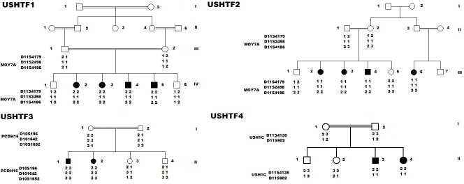
Methods: In the present study, we recruited four Tunisian families with a diagnosis of USH1, together with healthy unrelated controls. Affected members underwent detailed audiologic and ocular examinations. We used the North African Deafness (NADf) chip to search for known North African mutations associated with USH. Then, we selected microsatellite markers covering USH1 known loci to genotype the DNA samples. Finally, we performed DNA sequencing of three known USH1 genes: MYO7A, PCDH15, and USH1C.

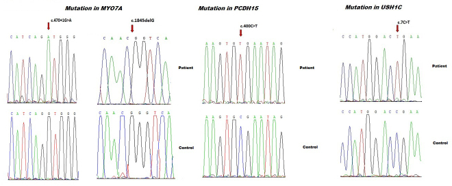
Results: Four biallelic mutations, all single base changes, were found in the MYO7A, USH1C, and PCDH15 genes. These mutations consist of a previously reported splicing defect c.470+1G>A in MYO7A, three novel variants, including two nonsense (p.Arg3X and p.Arg134X) in USH1C and PCDH15, respectively, and one frameshift (p.Lys615Asnfs*6) in MYO7A.

Conclusions: We found a remarkable genetic heterogeneity in the studied families with USH1 with a variety of mutations, among which three were novel. These novel mutations will be included in the NADf mutation screening chip that will allow a higher diagnosis efficiency of this extremely genetically heterogeneous disease. Ultimately, efficient molecular diagnosis of USH in a patient's early childhood is of utmost importance, allowing better educational and therapeutic management.


Figures

Figure 1
Figure 1
Pedigree and haplotype for four consanguineous Tunisian families who segregate Usher type 1. In the pedigree, the square symbol indicates male, the circle symbol denotes female, and the black squares represent affected individuals. Linkage analysis performed on all families showed that two were linked to USH1B, and the remaining families with USH1 were linked to USH1C and USH1F.
Figure 2
Figure 2
Visual field test results obtained for the right (RE) and the left eye (LE) of two patients (USHTF1IV5, USHTF4II3). A: Result of measurements of the visual fields. B: Result of eye fundus measurement. A series of random lights of different intensities were flashed in the peripheral field of vision of both patients. When they perceived the computer-generated light that suddenly appeared in their field of view, they pressed a button to indicate their responses, and then we saw this spot (Dot see). If the patient was unable to see the light in an appropriate portion of his field of view, then we saw on the computer a spot (Dot don’t see) indicating vision loss. In all patients, the nasal and temporal fields were not preserved, and only the central field was maintained. Fundus ophthalmoscopy showed pigmentary anomalies typical of retinitis pigmentosa (RP), attenuated arteriolar vessels and increased brightness of the internal limiting membrane. The visual field showed an anular scotoma reduced by 15°. C: Ganzfeld-Electroretinogram of the right and left eyes of two patients (USHTF1IV5, USHTF4II3). The electroretinogram (ERG) and the visual-evoked potential (VEP) test the function of the visual pathway from the retina (ERG) to the occipital cortex (VEP). These tests were conducted by placing a standard ERG device attached to the skin on 2 mm above the orbit. VEPs were recorded simultaneously from the electrode attached to the occipital scalp 2 mm above the region on the midsagittal plane. An electrode placed on the forehead provided a ground. The results are directly related to the part of a visual field that might be defective. This is based on the anatomic relationship of the retinal images and the visual field. After dark adaptation for 30 min, the doctor placed anesthetic drops in the patient’s eye and placed a contact lens on the surface of the eye. Once the contact lens was in place, a series of blue, red, and white lights were shown to the patient. The VEP is an evoked electrophysiological potential that can be extracted, using signal averaging, from the electroencephalographic activity recorded at the scalp. ERG and VEP were differentially amplified band pass filtered (0,1,30 Hz), recorded over 300 ms epochs, and the signals averaged. Two trials were given. The visual evoked potential to flash stimulation consists of a series of negative and positive waves. The earliest detectable response has a peak latency of approximately 30 ms post-stimulus. For the flash VEP, the most robust components are the N2 (negative) and P2 (positive) peaks. Measurements of the P2 amplitude should be made from the positive P2 peak at around 207.3 ms. The ERG recorded in BT189 showed an absence of responses although the VEP showed a normal responses in both eyes. These traces confirm evidence of significant bilateral global retinal degeneration. Only cone flicker responses of less than 15% of the normal mean were recordable under photopic conditions while all other responses were below noise level, a typical finding for patients with retinitis pigmentosa.
Figure 3
Figure 3
The direct sequencing of MYO7A, USH1C, and PCDH15 detected a total of three distinct novel pathogenic mutations: one frameshift mutation, two nonsense mutations, and one previously reported splicing defect-causing mutation. The known variant was a nucleotide substitution (c.470+1G>A), and the frameshift mutation is caused by nucleotide deletion c.1845delG causing USH1B. The molecular screening of USH1C and PCDH15 revealed two novel nonsense mutations occurring at the homozygous state: p.Arg3X in USH1C and p.Arg134X in PCDH15.

References

    1. Keats BJ, Corey DP. The Usher syndromes. Am J Med Genet. 1999;89:158–66. - PubMed
    1. Bonnet C, El-Amraoui A. Usher syndrome (sensorineural deafness and retinitis pigmentosa): Pathogenesis, molecular diagnosis and therapeutic approaches. Curr Opin Neurol. 2012;25:42–9. - PubMed
    1. Smith RJ, Berlin CI, Hejtmancik JF, Keats BJ, Kimberling WJ, Lewis RA, Moller CG, Pelias MZ, Tranebjaerg L. Clinical diagnosis of the Usher syndromes. Usher Syndrome Consortium. Am J Med Genet. 1994;50:32–8. - PubMed
    1. Kinga MB, Mark C, Emily P, Shyana H, Jaclyn L, Daniel GT, Joseph W, Daniel NG, Carol WD, Michael HF, Xiaowu G, Eliot LB. Targeted Exon Sequencing in Usher Syndrome Type I. Biochemistry and Molecular Biology. 2014;55:8488–96. - PubMed
    1. Jose MM, Elena A, Teresa J, Fiona BK, Ascension GP, Carmen A. An Update on the Genetics of Usher Syndrome. J Ophthalmol. 2011;•••:8. - PMC - PubMed

MeSH terms