Review
. 2016 Jul 14:10:67-83.
doi: 10.4137/CMPed.S40070. eCollection 2016.

Intrauterine Growth Restriction: Antenatal and Postnatal Aspects

Affiliations
Review

Intrauterine Growth Restriction: Antenatal and Postnatal Aspects

Deepak Sharma et al. Clin Med Insights Pediatr. .

Abstract

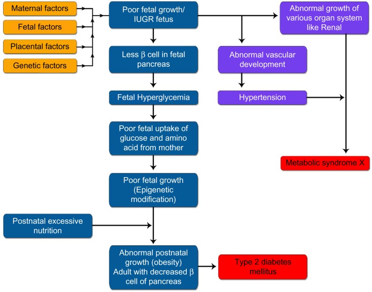
Intrauterine growth restriction (IUGR), a condition that occurs due to various reasons, is an important cause of fetal and neonatal morbidity and mortality. It has been defined as a rate of fetal growth that is less than normal in light of the growth potential of that specific infant. Usually, IUGR and small for gestational age (SGA) are used interchangeably in literature, even though there exist minute differences between them. SGA has been defined as having birth weight less than two standard deviations below the mean or less than the 10th percentile of a population-specific birth weight for specific gestational age. These infants have many acute neonatal problems that include perinatal asphyxia, hypothermia, hypoglycemia, and polycythemia. The likely long-term complications that are prone to develop when IUGR infants grow up includes growth retardation, major and subtle neurodevelopmental handicaps, and developmental origin of health and disease. In this review, we have covered various antenatal and postnatal aspects of IUGR.

Keywords: Intrauterine growth restriction (IUGR); asymmetrical IUGR; developmental origin of health and disease; fetal genes; maternal genes; placental genes; small for gestational age (SGA); symmetrical IUGR; thrifty phenotype (Barker hypothesis).


Figures

Figure 1
Figure 1
IUGR can be the result of maternal, fetal, placental, genetic cause or can be combination of either of the combination. (Copyright images Deepak Sharma).
Figure 2
Figure 2
Screening for Small–for–Gestational–Age (SGA) Fetus. Note: Reproduced from: Royal College of Obstetricians and Gynaecologists. The Investigation and Management of the Small–for–Gestational–Age Fetus. Green-top Guideline No. 31. London: RCOG; 2014, with the permission of the Royal College of Obstetricians and Gynaecologists.
Figure 3
Figure 3
The Management of the Small–for–Gestational–Age (SGA) Foetus. Notes: Reproduced from: Royal College of Obstetricians and Gynaecologists. The Investigation and Management of the Small–for–Gestational–Age Fetus. Green-top Guideline No. 31. London: RCOG; 2014, with the permission of the Royal College of Obstetricians and Gynaecologists. Weekly measurement of fetal size is valuable in predicting birthweight and determining size-for-gestational age. If two AC/EFW measurements are used to estimate growth, they should be at least 3 weeks apart. Use cCTG when DV Doppler is unavailable or results are inconsistent – recommend delivery if STV <3 ms. Abbreviations: AC, abdominal circumference; EFW, estimated fetal weight; Pl, pulsatility index; RI, resistance index; UA, umbilical artery; MCA, middle cerebral artery; DV, ducts venosus; FGR, fetal growth restriction; EDV, end-diastolic velocities.
Figure 4
Figure 4
This was a 36-week male neonate born to mother with severe pre-eclampsia with birth weight of 1600 grams. This baby was asymmetrical IUGR. Note loss of fat whole over the body, visible rib cage, excessive skin fold whole over the body and relatively large heads compared with rest of the body. The neonate also had excessive skin folds (more than 3) over inter-scapular and gluteal area with loss of underlying fats. There are loose folds of skin in the nape of neck and arms.
Figure 5
Figure 5
Clinical features of infants at birth that are having intrauterine growth restriction. Figure Copyright Deepak Sharma.
Figure 6
Figure 6
Immediate neonatal complications seen in intrauterine growth restricted neonates. Figure Copyright Deepak Sharma.
Figure 7
Figure 7
Increased risk for various physical and neurodevelopmental problems in intrauterine growth restricted neonates when they reach their childhood and adulthood. Figure Copyright Deepak Sharma.
Figure 8
Figure 8
Figure showing various adult disease the IUGR infant is prone to develop in his adulthood as per “Developmental origin of health and diseases (DoHaD)”. IUGR infants undergoes epigenetic modification in-utero and postnatally have abnormal nutrition and growth leading to various disease of adulthood in these infants. Figure Copyright Deepak Sharma.
Figure 9
Figure 9
Barker Hypothesis (Thrifty phenotype) explaining the Fetal Origin of Adult Disease (FOAD) or “Developmental origin of health and diseases (DoHaD)” in IUGR infants. Figure Copyright Deepak Sharma.
Figure 10
Figure 10
Follow up programme of infants who are born with intrauterine growth restriction. Figure copyright Deepak Sharma.

References

    1. Battaglia FC, Lubchenco LO. A practical classification of newborn infants by weight and gestational age. J Pediatr. 1967;71(2):159–63. - PubMed
    1. Sharma D, Shastri S, Farahbakhsh N, Sharma P. Intrauterine growth restriction–part 1. J Matern Fetal Neonatal Med. 2016;7:1–11. - PubMed
    1. Sharma D, Farahbakhsh N, Shastri S, Sharma P. Intrauterine growth restriction – part 2. J Matern Fetal Neonatal Med. 2016;0(0):1–12. - PubMed
    1. de Onis M, Blössner M, Villar J. Levels and patterns of intrauterine growth retardation in developing countries. Eur J Clin Nutr. 1998;52(Suppl 1):S5–15. - PubMed
    1. Lee PA, Chernausek SD, Hokken-Koelega ACS, Czernichow P, International Small for Gestational Age Advisory Board International Small for Gestational Age Advisory Board consensus development conference statement: management of short children born small for gestational age, April 24-October 1, 2001. Pediatrics. 2003;111(6 pt 1):1253–61. - PubMed

LinkOut - more resources