

Case Reports
doi: 10.4081/rt.2016.6241.
Nuclear Protein of the Testis Midline Carcinoma Masquerading as a Primary Mediastinal Seminoma
Affiliations
- PMID: 27441078
- PMCID: PMC4935827
- DOI: 10.4081/rt.2016.6241
Item in Clipboard
Case Reports
Nuclear Protein of the Testis Midline Carcinoma Masquerading as a Primary Mediastinal Seminoma
Rare Tumors.
.
Abstract
Nuclear protein of the testis (NUT) midline carcinomas are rare aggressive carcinomas characterized by chromosomal rearrangements that involve the gene encoding the NUT. This article reviews the clinicopathologic features and the differential diagnosis of these malignancies.
Keywords: Nuclear protein of the testis midline carcinoma; histone deacetylase inhibitors; primary mediastinal seminoma.
Conflict of interest statement
Conflict of interest: the authors declare no potential conflict of interest.
Figures
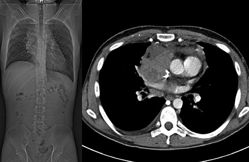
Coronal computed tomography image of the chest and abdomen before treatment.

Diagnosis of nuclear protein of the testis (NUT) midline carcinoma. A) Undifferentiated small cells with foci of squamous differentiation (Hematoxylin & Eosin, 20×). B) Immunohistochemistry of tumor cell nuclei showing speckled staining for NUT (250×) using anti-NUT rabbit polyclonal antibody (clone C52, 1:100).
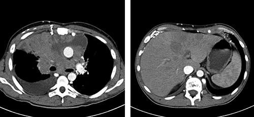
A) Coronal computed tomography (CT) image of the chest after treatment with histone deacetylase inhibitors, indicating marked adverse change from the time of diagnosis. There is encasement of major vessels, right atrium and right ventricle. B) Coronal CT image of the abdomen. There are metastases in liver that had significantly increased in size.
References
-
- Droz JP, Horwich A. Extragonadal germ cell tumors. Vogelzang NJ, Scardino PT, Shipley WU, Coffey DS. (eds). Comprehensive textbook of genitourinary oncology. 2nd ed. Philadelphia: Lippincott Williams and Wilkins; 2000.
-
- Bokemeyer C, Nichols CR, Droz JP, et al. Extragonadal germ cell tumors of the mediastinum and retroperitoneum: results from an international analysis. J Clin Oncol 2002;20:1864-73. - PubMed
-
- Chaganti RS, Houldsworth J. Genetics and biology of adult human male germ cell tumors. Cancer Res 2000;60:1475-82. - PubMed
-
- Gândara F, Leitao A, Bernardo M, Ceia F. Mediastinal seminoma: a case report. Internet J Intern Med 2011;9:1.
Publication types
LinkOut - more resources
Full Text Sources
Other Literature Sources
