

Novel molecular triggers underlie valproate-induced liver injury and its alleviation by the omega-3 fatty acid DHA: role of inflammation and apoptosis
- PMID: 27441301
- PMCID: PMC4946287
- DOI: 10.1016/j.heliyon.2016.e00130
Novel molecular triggers underlie valproate-induced liver injury and its alleviation by the omega-3 fatty acid DHA: role of inflammation and apoptosis
Abstract
Background/aim: Hepatic injury is a hallmark adverse reaction to Valproate (VPA), a common used drug in the management of numerous CNS disorders, including epilepsy. DHA has a myriad of health benefits, including renal- and hepato-protective effects. Unfortunately, however, the underpinnings of such liver-pertinent VPA- and DHA-actions remain largely undefined. Accordingly, this study attempted to unveil the cellular and molecular triggers whereby VPA evokes, while DHA abates, hepatotoxicity.
Methods: We evaluated activity and/or expression of cellular markers of oxidative stress, inflammation, and apoptosis in rat liver, following treatment with VPA (500 mg/kg/day) with and without concurrent treatment with DHA (250 mg/kg/day) for two weeks.
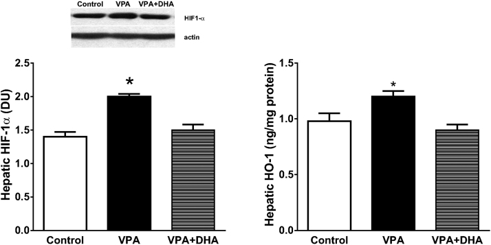
Results and conclusion: VPA promoted hepatic oxidative stress as evidenced by enhancing activity/expression of NADPH-oxidase and its subunits, a ROS-generator, and by accumulation of lipid-peroxides. Moreover, VPA enhanced hepatic phosphorylation/activation of mitogen-activated protein kinase (MAPK), and expression of cyclooxygenase-2(COX-2), as proinflammatory signals. Besides, VPA promoted hepatocellular apoptosis, as attested by enhanced expression of cleaved caspase-9 and increased number of TUNEL-positive hepatocytes. Lastly, VPA upregulated levels of hypoxia-inducible factor-1-alpha (HIF-1α), a multifaceted modulator of hepatocytic biology, and activity of its downstream antioxidant enzyme heme-oxygenase-1(HO-1). These changes were significantly blunted by co-administration of DHA. Our findings demonstrate that VPA activated NADPH-oxidase and HIF-1α to induce oxidative-stress and hypoxia as initiators of hepatic injury. These changes were further aggravated by up-regulation of inflammatory (MAPK and COX-2) and apoptotic cascades, but could be partly lessened by HO-1 activation. Concurrent administration of DHA mitigated all VPA-induced anomalies.
Keywords: Biochemistry; Cell biology; Medicine; Physiology; Systems biology.
Figures


Similar articles
-
Valproate-induced liver injury: modulation by the omega-3 fatty acid DHA proposes a novel anticonvulsant regimen.Drugs R D. 2014 Jun;14(2):85-94. doi: 10.1007/s40268-014-0042-z. Drugs R D. 2014. PMID: 24733439 Free PMC article.
-
Eicosapentaenoic acid ablates valproate-induced liver oxidative stress and cellular derangement without altering its clearance rate: dynamic synergy and therapeutic utility.Biochim Biophys Acta. 2011 Jul-Aug;1811(7-8):460-7. doi: 10.1016/j.bbalip.2011.04.014. Epub 2011 May 5. Biochim Biophys Acta. 2011. PMID: 21571092
-
Docosahexaenoic acid (DHA)-induced heme oxygenase-1 attenuates cytotoxic effects of DHA in vascular smooth muscle cells.Atherosclerosis. 2013 Oct;230(2):406-13. doi: 10.1016/j.atherosclerosis.2013.08.002. Epub 2013 Aug 13. Atherosclerosis. 2013. PMID: 24075775
-
Decoding cell death signals in liver inflammation.J Hepatol. 2013 Sep;59(3):583-94. doi: 10.1016/j.jhep.2013.03.033. Epub 2013 Apr 6. J Hepatol. 2013. PMID: 23567086 Review.
-
n-3 Polyunsaturated fatty acids for the management of alcoholic liver disease: A critical review.Crit Rev Food Sci Nutr. 2019;59(sup1):S116-S129. doi: 10.1080/10408398.2018.1544542. Epub 2018 Dec 22. Crit Rev Food Sci Nutr. 2019. PMID: 30580553 Review.
Cited by
-
Life factors acting on systemic lupus erythematosus.Front Immunol. 2022 Sep 15;13:986239. doi: 10.3389/fimmu.2022.986239. eCollection 2022. Front Immunol. 2022. PMID: 36189303 Free PMC article. Review.
-
The Effect of Coenzyme Q10 on Liver Injury Induced by Valproic Acid and Its Antiepileptic Activity in Rats.Biomedicines. 2022 Jan 13;10(1):168. doi: 10.3390/biomedicines10010168. Biomedicines. 2022. PMID: 35052847 Free PMC article.
-
Relationships between low-grade peripheral inflammation and psychotropic drugs in schizophrenia: results from the national FACE-SZ cohort.Eur Arch Psychiatry Clin Neurosci. 2018 Sep;268(6):541-553. doi: 10.1007/s00406-017-0847-1. Epub 2017 Nov 10. Eur Arch Psychiatry Clin Neurosci. 2018. PMID: 29127503
-
Celecoxib Decrease Seizures Susceptibility in a Rat Model of Inflammation by Inhibiting HMGB1 Translocation.Pharmaceuticals (Basel). 2021 Apr 19;14(4):380. doi: 10.3390/ph14040380. Pharmaceuticals (Basel). 2021. PMID: 33921725 Free PMC article.
-
Arecoline-Induced Hepatotoxicity in Rats: Screening of Abnormal Metabolic Markers and Potential Mechanisms.Toxics. 2023 Dec 4;11(12):984. doi: 10.3390/toxics11120984. Toxics. 2023. PMID: 38133385 Free PMC article.
References
-
- Baran O.P., Kervancioglu P., Akkus M., Nergiz Y. Ultrastructural investigation of the protective role of folic acid and vitamin E against toxic effects of valproic acid on maternal liver tissue during period of gestation. Saudi Med. J. 2006;27:407–409. - PubMed
-
- Bitman M., Vrzal R., Dvorak Z., Pavek P. Valproate activates ERK signaling pathway in primary human hepatocytes. Biomed. Pap. Med. Fac. Univ. Palacky Olomouc Czech. Repub. 2014;158:39–43. - PubMed
-
- Chang T.K., Abbott F.S. Oxidative stress as a mechanism of valproic acid-associated hepatotoxicity. Drug Metab. Rev. 2006;38:627–639. - PubMed
LinkOut - more resources
Full Text Sources
Other Literature Sources
Research Materials
