Review
. 2016 Aug;34(3):451-72.
doi: 10.1016/j.ccl.2016.04.005.

Pulmonary Hypertension in Children

Affiliations
Review

Pulmonary Hypertension in Children

Dunbar Ivy. Cardiol Clin. 2016 Aug.

Abstract

The prevalence of PH is increasing in the pediatric population, because of improved recognition and increased survival of patients, and remains a significant cause of morbidity and mortality. Recent studies have improved the understanding of pediatric PH, but management remains challenging because of a lack of evidence-based clinical trials. The growing contribution of developmental lung disease requires dedicated research to explore the use of existing therapies as well as the creation of novel therapies. Adequate study of pediatric PH will require multicenter collaboration due to the small numbers of patients, multifactorial disease causes, and practice variability.

Keywords: Bronchopulmonary dysplasia; Pulmonary arterial hypertension; Single-ventricle circulation.


Figures

Figure 1
Figure 1
Kaplan-Meier curves showing the survival pediatric PAH patients at 3 PH centers (NY, New York; NL, Netherlands): 1-, 3-, 5-, and 7-year transplantation-free survival rates were 96%, 89%, 81%, and 79%, respectively From Zijlstra WM, Douwes JM, Rosenzweig EB. Survival differences in pediatric pulmonary arterial hypertension: clues to a better understanding of outcome and optimal treatment strategies. J Am Coll Cardiol 2014;63(20):2159–69; with permission.
Figure 2
Figure 2
Venn diagram illustrating the heterogeneity and multifactorial elements in pediatric pulmonary hypertensive vascular disease. Adapted from Cerro MJ, Abman S, Diaz G, et al. A consensus approach to the classification of pediatric pulmonary hypertensive vascular disease: Report from the PVRI Pediatric Taskforce, Panama 2011. Pulm Circ 2011;1(2):286–98; with permission.
Figure 3
Figure 3
Annual incidence rates for pediatric pulmonary hypertension. PH indicates pulmonary hypertension; PAH, pulmonary arterial hypertension; PAH-CHD, PAH associated with congenital heart defects; and IPAH, idiopathic PAH. From van Loon RL, Roofthooft MT, Hillege HL, et al. Pediatric pulmonary hypertension in the Netherlands: epidemiology and characterization during the period 1991 to 2005. Circulation 2011;124(16):1755–64; with permission.
Figure 4
Figure 4
Survival curves for the subgroups within the associated pulmonary arterial hypertension (APAH) group from the UK pulmonary hypertension service. The number in each group (brackets) and the predicted survival out of a possible 5 years is depicted. Note the worse survival for children with post operative congenital heart disease. From Haworth SG, Hislop AA. Treatment and survival in children with pulmonary arterial hypertension: the UK Pulmonary Hypertension Service for Children 2001–2006. Heart 2009;95(4):312–7; with permission.
Figure 5
Figure 5
The incidence of pulmonary hypertension according to the degree of bronchopulmonary dysplasia severity. Numbers above the bars indicate the percentage of patients with pulmonary hypertension. Data from Refs
Figure 6
Figure 6
Measurement and normal values for tricuspid annular plane systolic excursion (TAPSE). From Koestenberger M, Ravekes W, Everett AD. Right ventricular function in infants, children and adolescents: reference values of the tricuspid annular plane systolic excursion (TAPSE) in 640 healthy patients and calculation of z score values. J Am Soc Echocardiogr 2009;22(6):715–9; with permission.
Figure 7
Figure 7
Parasternal short axis view of the right and left ventricles at the level of the papillary muscles. The RV/LV ratio is derived from RV diameter and LV diameter at end-systole. RV/LV end systole ratio is predictive of outcome. Estimated survival curves for four possible RV/LV ratios estimated from the Cox varying coefficients regression corresponding to a hazard ratio of 2.49 for RV/LV ratio. From Jone PN, Hinzman J, Wagner BD, et al. Right ventricular to left ventricular diameter ratio at end-systole in evaluating outcomes in children with pulmonary hypertension. J Am Soc Echocardiogr 2014;27(2):172–8; with permission.
Figure 8
Figure 8
The systolic (S) to diastolic (D) time ratio from tricuspid regurgitation velocity can be measured as a measure of right ventricular function. An increase in the S/D ratio predicts worse outcome in children with PH. Alkon et al in 2010 used an simple measure of systolic to diastolic time (S/D) ratio from the TR jet to evaluate pediatric PH patients and found that as the RV function worsens, the systolic portion of the cardiac cycle lengthens leading to an increased S:D ratio. S/D ratio was found to be higher in PH patients compared to controls and is associated with worse RV FAC change, worse hemodynamics by cath, shorter 6 minute walk test, and worse clinical outcomes independent of PVR or pressures. S/D ratio < 1 is associated with low risk of negative outcome and S/D ratio > 1.4 was associated with high risk of negative outcome. From Alkon J, Humpl T, Manlhiot C, et al. Usefulness of the right ventricular systolic to diastolic duration ration to predict functional capacity and survival in children with pulmonary arterial hypertension. Am J Cardiol 2010;106(3):430–6; with permission.
Figure 9
Figure 9
Tissue Doppler imaging of the right ventricle at the lateral annulus of the tricuspid valve measures the myocardial systolic wave (S’) which measures the systolic longitudinal function of the RV and two diastolic waves: early diastolic (E’) and late diastolic (A’), which denote the diastolic function of the ventricles. Low E’ velocity less than 8 cm/s is predictive of poor outcome in pediatric IPAH. From Takatsuki S, Nakayama T, Jone PN, et al. Tissue Doppler imaging predicts adverse outcome in children with idiopathic pulmonary arterial hypertension. J Pediatr 2012;161(6):1126–31; with permission.
Figure 10
Figure 10
Kaplan-Meier survival curves for children with IPAH and PAH associated with CHD. Survival curves are shown for all patients (left) and for the subgroup of IPAH patients (right) categorized with either brain natriuretic peptide (BNP) > 180 pg/ml or < 180 pg/ml. From Bernus A, Wagner BD, Accurso F, et al. Brain natriuretic peptide levels in managing pediatric patients with pulmonary arterial hypertension. Chest 2009;135(3):745–51; with permission.
Figure 11
Figure 11
High levels of tissue inhibitors of metalloproteinases-1 (TIMP-1), which is overexpressed by proinflammatory cytokines, and low levels of apolipoprotein-A1, which reduces levels of oxidized lipids and improves vascular disease are strongly associated with outcome in pediatric PH. Patients with high TIMP-1 and low apolipoprotein-AI values (red) had lower survival (log-rank test p-value, 0.01). The number of subjects at-risk are displayed along the x-axis. From Wagner BD, Takatsuki S, Accurso FJ, et al. Evaluation of circulating proteins and hemodynamics towards predicting mortality in children with pulmonary arterial hypertension. PLoS One 2013;8(11):e80235; with permission.
Fig 12
Fig 12
The number of acute pulmonary vasodilator responders according to the three criteria in use, in children vs. adults with idiopathic pulmonary arterial hypertension (iPAH)/hereditary pulmonary arterial hypertension (HPAH), iPAH/HPAH vs. pulmonary arterial hypertension associated with congenital heart disease, and patients without vs. with post-tricuspid shunt, respectively. Data presented as percentage of patient group (%) and patient numbers (indicated in bars). Comparison between groups performed using Fisher’s exact test. Note the few % of patients with PAH-CHD responding to acute vasodilator challenge. From Douwes JM, van Loon RL, Hoendermis ES, et al. Acute pulmonary vasodilator response in paediatric and adult pulmonary arterial hypertension: occurrence and prognostic value when comparing three response criteria. Eur Heart J 2011;32(24):3137–46; with permission.
Figure 13
Figure 13
Risk factors that should be considered when planning therapeutic management options in pulmonary hypertension. CI, cardiac index; mPAp, mean pulmonary artery pressure; mSAp, mean systemic aortic pressure; NT-proBNP, N-terminal-pro-brain natriuretic peptide; PVRI, indexed pulmonary vascular resistance; RAP, right atrial pressure; RV, right ventricle; SBNP, serum brain natriuretic peptide. From Ivy DD, Abman SH, Barst RJ, et al. Pediatric pulmonary hypertension. J Am Coll Cardiol 2013;62(25 Suppl):D117–26; with permission.
Figure 14
Figure 14
Treatment algorithm proposed in the management of pediatric patients with idiopathic or heritable pulmonary arterial hypertension. This may be translatable to other patients with pulmonary hypertension. CCB, calcium channel blocker; ERA, endothelin receptor antagonist; PDE-5i, phosphodiesterase 5 inhibitor. Adapted from Ivy DD, Abman SH, Barst RJ, et al. Pediatric pulmonary hypertension. J Am Coll Cardiol 2013;62(25 Suppl):D117–26; with permission.
Figure 15
Figure 15
Kaplan-Meier survival curve in 77 children with PH treated with epoprostenol, treprostinil, and those who transitioned, with 95% confidence intervals (CI) depicted. Transplant-free 5-year survival was 70% (95% CI, 56%–80%). From Siehr SL, Ivy DD, Miller-Reed K, et al. Children with pulmonary arterial hypertension and prostanoid therapy: long-term hemodynamics. J Heart Lung Transplant 2013;32(5):546–52; with permission.
Figure 16
Figure 16
Kaplan-Meier estimated survival from start of sildenafil treatment in Sildenafil in Treatment-Naive Children, Aged 1 to 17 Years, With Pulmonary Arterial Hypertension (STARTS-1) and STARTS-2. Patients were censored at the last date they were known to be alive; if a patient received a transplant, he or she was censored the day before transplant. Patients at risk are those who are ongoing in the study or known to be alive at the specified time (i.e., not dead, not lost to follow-up, or not in study long enough to reach time point). From Barst RJ, Beghetti M, Pulido T, et al. STARTS-2: long-term survival with oral sildenafil monotherapy in treatment-naïve pediatric pulmonary arterial hypertension. Circulation 2014;129(19):1914–23; with permission.
Figure 17
Figure 17
Survival according to extent of pulmonary hypertension therapy in 275 recently diagnosed consecutive pediatric PAH patients at 3 referral centers between 2000 and 2010. Survival improves on combination therapy for pulmonary hypertension over monotherapy. CCB, calcium channel blocker. From Zijlstra WM, Douwes JM, Rosenzweig EB. Survival differences in pediatric pulmonary arterial hypertension: clues to a better understanding of outcome and optimal treatment strategies. J Am Coll Cardiol 2014;63(20):2159–69; with permission.
Figure 18
Figure 18
Echocardiogram with color compare of the Potts shunt in a patient with severe IPAH. LPA, left pulmonary artery.

References

    1. D'Alonzo GE, Barst RJ, Ayres SM, et al. Survival in patients with primary pulmonary hypertension. Results from a national prospective registry. Ann Intern Med. 1991;115:343–349. - PubMed
    1. Barst RJ, Maislin G, Fishman AP. Vasodilator therapy for primary pulmonary hypertension in children. Circulation. 1999;99:1197–1208. - PubMed
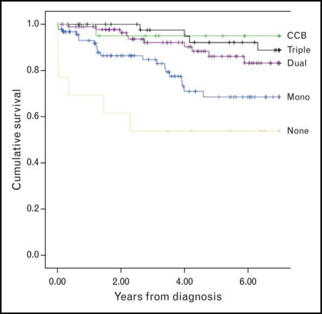
    1. Zijlstra WM, Douwes JM, Rosenzweig EB, et al. Survival differences in pediatric pulmonary arterial hypertension: clues to a better understanding of outcome and optimal treatment strategies. J Am Coll Cardiol. 2014;63:2159–2169. - PubMed
    1. Ivy DD, Abman SH, Barst RJ, et al. Pediatric pulmonary hypertension. J Am Coll Cardiol. 2013;62:D117–D126. - PubMed
    1. Barst RJ, Ertel SI, Beghetti M, Ivy DD. Pulmonary arterial hypertension: a comparison between children and adults. Eur Respir J. 2011;37:665–677. - PMC - PubMed

MeSH terms