. 2016 Jul 22;16(1):44.
doi: 10.1186/s12880-016-0147-7.

Reliability of radiographic measurements for acute distal radius fractures

Affiliations

Reliability of radiographic measurements for acute distal radius fractures

Narelle J Watson et al. BMC Med Imaging. .

Abstract

Background: The management of distal radial fractures is guided by the interpretation of radiographic findings. The aim of this investigation was to determine the intra- and inter-observer reliability of eight traditionally reported anatomic radiographic parameters in adults with an acute distal radius fracture.

Methods: Five observers participated. All were routinely involved in making treatment decisions based on distal radius fracture radiographs. Observers performed independent repeated measurements on 30 radiographs for eight anatomical parameters: dorsal shift (mm), intra-articular gap (mm), intra-articular step (mm), palmar tilt (degrees), radial angle (degrees), radial height (mm), radial shift (mm), ulnar variance (mm). Intraclass correlation coefficients (ICCs) and the magnitude of retest errors were calculated.

Results: Measurement reliability was summarised as high (ICC > 0.80), moderate (0.60-0.80) or low (<0.60). Intra-observer reliability was high for dorsal shift and palmar tilt; moderate for radial angle, radial height, ulnar variance and radial shift; and low for intra-articular gap and step. Inter-observer reliability was high for palmar tilt; moderate for dorsal shift, ulnar variance, radial angle and radial height; and low for radial shift, intra-articular gap and step. Error magnitude (95 % confidence interval) was within 1-2 mm for intra-articular gap and step, 2-4 mm for ulnar variance, 4-6 mm for radial shift, dorsal shift and radial height, and 6-8° for radial angle and palmar tilt.

Conclusions: Based on previous reports of critical values for palmar tilt, ulnar variance and radial angle, error margins appear small enough for measurements to be useful in guiding treatment decisions. Our findings indicate that clinicians cannot reliably measure values ≤1 mm for intra-articular gap and step when interpreting radiographic parameters using the standardised methods investigated in this study. As a guide for treatment selection, palmar tilt, ulnar variance and radial angle measurements may be useful, but intra-articular gap and step appear unreliable.

Keywords: Distal radius fracture; Radiographs; Reliability.


Figures
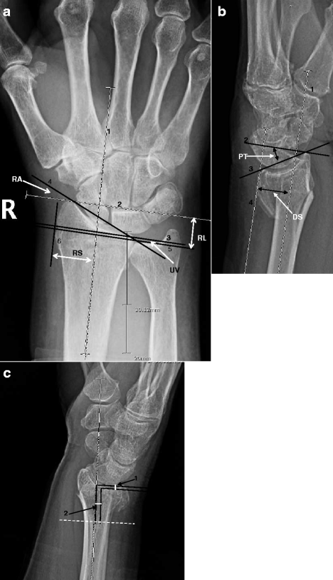
Fig. 1
Fig. 1
a Posterioanterior measurement guidelines as described in Kreder et al. [6]. b Lateral measurement guidelines as described in Kreder et al. [6]. c Step and gap measurement as described in Kreder et al. [6]. a RA, radial angle; RL, radial length; UV, ulnar variance; RS, radial shift. 1) This line represents the long axis of the radius. The center of the radius shaft is determined at 3 cm and 5 cm below the mid-region of the proximal lunate articular surface. 2) A line perpendicular to the center long axis of the radius is drawn at the level of the most distal aspect of the radial articular surface. 3) A line perpendicular to the central long axis of the radius is drawn at the level of the ulnar margin of the distal radial articular surface. 4) The radial and ulnar margins of the distal radial articular surface are connected. 5) A line perpendicular to the central long axis of the radius is drawn at the level of the distal ulnar articular surface. 6) A line tangential to the most radial point on the radial metaphysis is drawn parallel to the central long axis of the radius. b PT palmar tilt angle (dorsal tilt=negative palmar tilt); DS, dorsal shift. 1) This line represents the long axis of the radius. The center of the radial shaft is determined at 3 cm and 5 cm below the mid-region of the proximal lunate articular surface. 2) A line perpendicular to the central long axis of the radius is drawn at a convenient level. 3) The dorsal and anterior margins of the distal radial articular surface are connected. 4) A line tangential to the most dorsal point on the radial metaphysis is drawn parallel to the central long axis of the radius. c 1) Step-off at the distal radius articular cortical margin is measured by drawing lines perpendicular with the central long axis of the radius from the most distal margin of each side of the cortical discontinuity. 2) Gap deformity is measured by dropping lines that are parallel from the central long axis of the radius from the most distal margin of each side of the cortical deformity. The gap distance is measured along a line perpendicular to the central long axis of the radius.
Fig. 2
Fig. 2
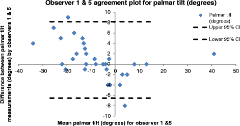
Bland and Altman [20] distribution plot for palmar tilt, showing the difference between measurements by observers 1 & 5 plotted against the average measurement for the two observers using data from the first set of measurements
Fig. 3
Fig. 3
Bland and Altman [20] distribution plot showing for palmar tilt, the difference between observer 2 & 4 measurements plotted against the average measurement for the two observers using data from the first set of measurements

Similar articles

Cited by

References

    1. Larsen CF, Lauritsen J. Epidemiology of acute wrist trauma. Int J Epidemiol. 1993;22(5):911–6. doi: 10.1093/ije/22.5.911. - DOI - PubMed
    1. Cummings SR, Black DM, Rubin SM. Lifetime risks of hip, Colles’, or vertebral fracture and coronary heart disease among white postmenopausal women. Arch Intern Med. 1989;149:2445–8. doi: 10.1001/archinte.1989.00390110045010. - DOI - PubMed
    1. Byl NN, Kohlhase W, Engel G. Functional limitation immediately after cast immobilization and closed reduction of distal radius fractures: Preliminary report. J Hand Ther. 1999;12(3):201–11. doi: 10.1016/S0894-1130(99)80047-7. - DOI - PubMed
    1. Beaule PE, Dervin GF, Giachino AA, et al. Self-reported disability following distal radius fractures: The influence of hand dominance. J Hand Surg [Am] 2000;25(3):476–82. doi: 10.1016/S0363-5023(00)70027-X. - DOI - PubMed
    1. Foldhazy Z, Tornkvist H, Elmstedt E, et al. Long-term outcome of nonsurgically treated distal radius fractures. J Hand Surg [Am] 2007;32(9):1374–84. doi: 10.1016/j.jhsa.2007.08.019. - DOI - PubMed

Publication types