. 2016 Jul 22:6:30182.
doi: 10.1038/srep30182.

A critical role for the self-assembly of Amyloid-β1-42 in neurodegeneration

Affiliations

A critical role for the self-assembly of Amyloid-β1-42 in neurodegeneration

Karen E Marshall et al. Sci Rep. .

Abstract

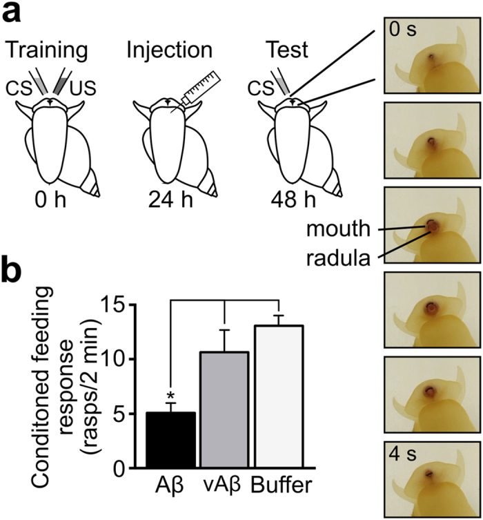
Amyloid β1-42 (Aβ1-42) plays a central role in Alzheimer's disease. The link between structure, assembly and neuronal toxicity of this peptide is of major current interest but still poorly defined. Here, we explored this relationship by rationally designing a variant form of Aβ1-42 (vAβ1-42) differing in only two amino acids. Unlike Aβ1-42, we found that the variant does not self-assemble, nor is it toxic to neuronal cells. Moreover, while Aβ1-42 oligomers impact on synaptic function, vAβ1-42 does not. In a living animal model system we demonstrate that only Aβ1-42 leads to memory deficits. Our findings underline a key role for peptide sequence in the ability to assemble and form toxic structures. Furthermore, our non-toxic variant satisfies an unmet demand for a closely related control peptide for Aβ1-42 cellular studies of disease pathology, offering a new opportunity to decipher the mechanisms that accompany Aβ1-42-induced toxicity leading to neurodegeneration.


Conflict of interest statement

The variant peptide, a kit including the variant peptide, and their uses, are covered by an International (PCT) patent application PCT/GB2015/052242 filed by the University of Sussex.

Figures

Figure 1
Figure 1. The graph produced using WALTZ shows two peaks that indicate two amyloidogenic regions (residues 16–21 and residues 37–42) in Aβ1-42 compared to the trace for vAβ1-42 peptide design showing abolition of the amyloidogenic regions.
Figure 2
Figure 2
(a) Negative stain transmission electron microscopy images of Aβ1-42 (left panels) and vAβ1-42 peptide (right panels) showing assembly of Aβ1-42 into fibrils at around 24 hours, preceded by small spherical structures (both shown by arrows). Conversely the vAβ1-42 peptide does not form fibres even up to 7 days. All peptides were prepared as described in the methods at 50 μM. Scale bars 200 nm. (b) CD spectra of Aβ1-42 and vAβ1-42 over time showing the formation of β-sheet structures for Aβ1-42 after around 24 hours whilst vAβ1-42 remains as random coil structure up to the final time point of 7 days. (c) Thioflavin T fluorescence showing increasing fluorescence at 483 nm of Aβ1-42 over 7 days, compared to no change in fluorescence of vAβ1-42. (d) Sequence of Aβ1-42 (top) and vAβ1-42 (bottom), showing epitope regions for 4G8 and 6E10 antibodies. The amino acid substitutions are underlined. Dot blots using anti-oligomer antibody, NU1 and anti-Aβ antibodies 4G8 and 6E10 show oligomer reactive species only in Aβ1-42 samples and not vAβ1-42. Similarly, 4G8 does not detect vAβ1-42 due to the F19S substitution in the epitope region. 6E10 binds both Aβ1-42 and vAβ1-42 as the epitope is the same in both peptides. (e) Western blot of Aβ1-42 (top) and vAβ1-42 (bottom) with 6E10 over time shows monomers (M), dimers (D), trimers (T), higher molecular weight species and fibres (F) are only formed by the wild-type peptide. vAβ1-42 runs as a monomer.
Figure 3
Figure 3
(a) DIC widefield images of neurons live in culture following treatment with either Aβ1-42 oligomers or vAβ1-42 after 3 or 7 days. Some live neurons are still clearly visible in the Aβ1-42 culture after 3 days but by 7 days none appear healthy. Scale bar 20 μm. (b) Measurement of proportion of dead cells compared to the total counted in culture by Readyprobes assay following 3 and 7 days exposure to Aβ1-42 or vAβ1-42 or buffer only (total number of cells counted (number of dead cells in brackets) at 3 days: n = 1163 (545), 1012 (220) and 1661 (279) and 7 days: n = 1032 (610), 684 (37) and 1112 (97) for Aβ1-42, vAβ1-42 and buffer respectively). (c) MTT assay (24 hours: n = 19, 9 and 20, 48 hours: n = 14, 9 and 13 for Aβ1-42, vAβ1-42 and buffer respectively) and (d) CTB (24 hours: n = 12, 11, and 16, 48 hours: n = 12, 9, and 18 for Aβ1-42, vAβ1-42 and buffer respectively) assay using SH-SY5Y cells. 10 μM oligomeric Aβ1-42 has a significant effect on the cells after 24 hours whilst vAβ1-42 is the same as buffer only. Unpaired parametric student’s t test, only significant differences are shown, where p = < 0.05 (*), <0.01 (**), <0.0001 (****) and >0.05 was not significant. Error bars are expressed as ±SEM.
Figure 4
Figure 4
Hippocampal neurons treated with untagged oligomeric Aβ1-42 or vAβ1-42 (a,b) or Alexa fluor 555 tagged peptides (c) and imaged by confocal microscopy. Cells in a and b were fixed and stained with anti-oligomer antibody NU1 (a) or 6E10 (b). Maximum projection images of six 0.5 μm slices are shown in a and c, b shows one 1 μm slice from the centre of a Z-stack. Scale bars 20 μm.
Figure 5
Figure 5
(a) Cartoon illustrating functional synaptic readout. Neurons are activated by field stimulation to evoke vesicle turnover in the presence of extracellular FM1-43-dye. Washing in dye-free solution leaves recently recycled vesicles fluorescently labelled. (b) Representative images of FM1-43 loading in neurons treated with Aβ1-42, vAβ1-42 or buffer. Arrowheads indicate discrete functional terminals. Scale bar 20 μm. (c) Histogram (median ± IQR) shows number of functional synapses expressed as the median synapse density per image, (Aβ1-42: 218 IQR 137-262, vAβ1-42: 315 IQR 258-355, buffer: 355 IQR 283–410, n = 20, 30, 30 images, respectively; Kruskal-Wallis one-way ANOVA, p = 0.0002 with Dunn’s multiple comparison test, see Methods for analysis). (d) Cartoon illustrating approach for readout of activity-induced dye-loss kinetics, corresponding to synaptic vesicle exocytosis. (e) Normalised fluorescence loss profiles for Aβ1-42, vAβ1-42 or buffer treated cells (average profiles of n = 218, 428, 560 synapses for Aβ1-42, vAβ1-42, buffer respectively). Shaded band denotes SEM for each trace. (f) Histogram of magnitude of FM1-43 destaining for data in (e), expressed as % dye loss (median ± IQR, Aβ1-42: 50 IQR 36-61, vAβ1-42: 71 IQR 56-82, buffer: 72 IQR 60-83, n = 218, 428, 560 synapses, respectively; Kruskal-Wallis one-way ANOVA, p < 0.0001 with Dunn’s multiple comparison test).
Figure 6
Figure 6
(a) Cartoon illustrating conditioned feeding response of Lymnaea stagnalis treated with Aβ1-42 or vAβ1-42. Animals were classically conditioned using the single-trial food-reward paradigm at 0 hours; injected with 1 μM Aβ1-42 (n = 55), 1 μM vAβ1-42 (n = 20), or vehicle (n = 106) at 24 hours; and tested for the conditioned feeding response at 48 hours. Picture inserts show an example of a complete feeding cycle (rasp) on which the behavioural assessment was based; rasping begins at 0 s, including opening of the mouth, protrusion of the toothed radula, ingestion of food, and closure of the mouth at 4 s. (b) One-way ANOVA, p < 0.0001. Tukey’s multiple comparison with p = 0.05: vAβ1-42 vs. Aβ1-42, Vehicle vs. Aβ1-42. Error bars are shown as ±SEM.

References

    1. Selkoe D. J. Toward a comprehensive theory for Alzheimer’s disease. Hypothesis: Alzheimer’s disease is caused by the cerebral accumulation and cytotoxicity of amyloid beta-protein. Annals of the New York Academy of Sciences 924, 17–25 (2000). - PubMed
    1. Hardy J. The amyloid hypothesis for Alzheimer’s disease: a critical reappraisal. J Neurochem 110, 1129–1134 (2009). - PubMed
    1. Williams T. L., Day I. J. & Serpell L. C. The effect of Alzheimer’s abeta aggregation state on the permeation of biomimetic lipid vesicles. Langmuir: the ACS journal of surfaces and colloids 26, 17260–17268 (2010). - PubMed
    1. Butterfield S. M. & Lashuel H. A. Amyloidogenic protein-membrane interactions: mechanistic insight from model systems. Angewandte Chemie 49, 5628–5654 (2010). - PubMed
    1. Shankar G. M. et al.. Natural oligomers of the Alzheimer amyloid-beta protein induce reversible synapse loss by modulating an NMDA-type glutamate receptor-dependent signaling pathway. The Journal of neuroscience: the official journal of the Society for Neuroscience 27, 2866–2875 (2007). - PMC - PubMed

Publication types