. 2016 Jul 22:6:30257.
doi: 10.1038/srep30257.

The first non Clostridial botulinum-like toxin cleaves VAMP within the juxtamembrane domain

Affiliations

The first non Clostridial botulinum-like toxin cleaves VAMP within the juxtamembrane domain

Irene Zornetta et al. Sci Rep. .

Abstract

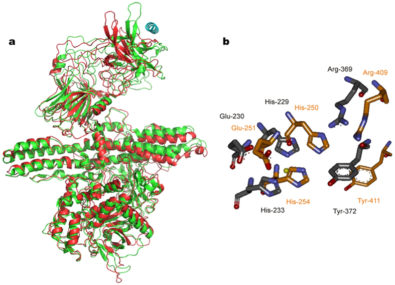
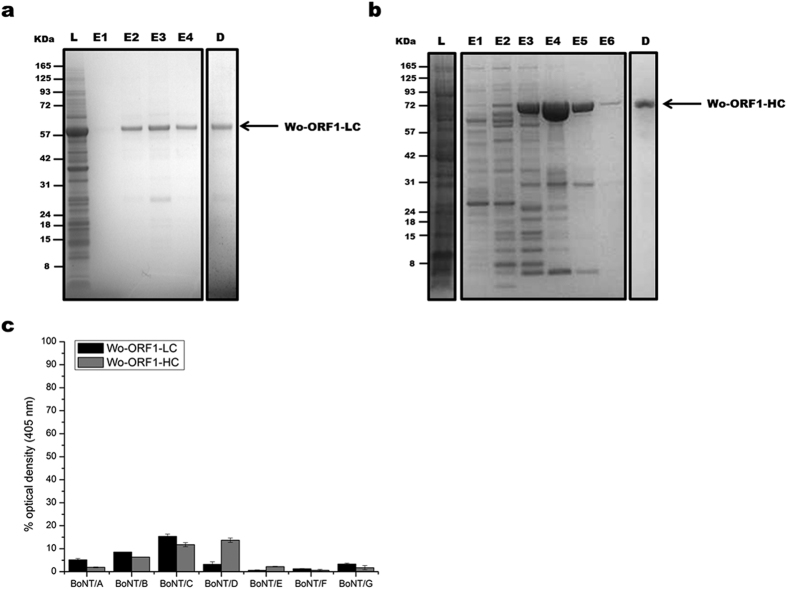
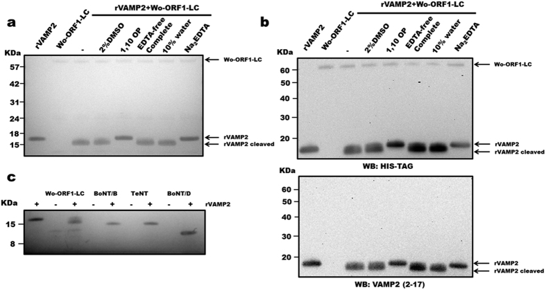
The genome of Weissella oryzae SG25T was recently sequenced and a botulinum neurotoxin (BoNT) like gene was identified by bioinformatics methods. The typical three-domains organization of BoNTs with a N-terminal metalloprotease domain, a translocation and a cell binding domains could be identified. The BoNT family of neurotoxins is rapidly growing, but this was the first indication of the possible expression of a BoNT toxin outside the Clostridium genus. We performed molecular modeling and dynamics simulations showing that the 50 kDa N-terminal domain folds very similarly to the metalloprotease domain of BoNT/B, whilst the binding part is different. However, neither the recombinant metalloprotease nor the binding domains showed cross-reactivity with the standard antisera that define the seven serotypes of BoNTs. We found that the purified Weissella metalloprotease cleaves VAMP at a single site untouched by the other VAMP-specific BoNTs. This site is a unique Trp-Trp peptide bond located within the juxtamembrane segment of VAMP which is essential for neurotransmitter release. Therefore, the present study identifies the first non-Clostridial BoNT-like metalloprotease that cleaves VAMP at a novel and relevant site and we propose to label it BoNT/Wo.


Figures

Figure 1
Figure 1. Molecular models of BoNT/B and of the Weisella oryzae open reading frame 1 and comparison of their catalytic sites.
(a) Superposition of the Cα chain trace of BoNT/B (green) to the model of Weissella oryzae (red, NCBI gi: 653854119) after molecular dynamics minimization. The structure of the N- and the middle domains, LC and HN respectively, are very similar whilst major differences are observed in the area of the C-terminal domain, HC, where the binding to the polysialoganglioside and synaptotagmin receptor takes place. The α-helix on the top right position (in blue) is the segment 40–60 of synaptotagmin which is involved in BoNT/B binding. (b) Arrangement of active site amino acids of BoNT/B (carbon atoms in grey) and comparison with the predicted configuration of corresponding residues of Wo-ORF1 (carbon atoms in orange). In addition to the presence of the zinc-binding motif residues of metalloproteases, His-Glu-X-X-His, also the Arg and Tyr residues (residues 369 and 372 of BoNT/B) of the second shell of zinc coordination are present and show the same configuration. The yellow sphere represents the active site zinc atom.
Figure 2
Figure 2. Purification of recombinant Wo-ORF1 proteins and serotyping by indirect ELISA.
Panels (a,b) show, respectively, the purification and characterization of Wo-ORF1-LC and of Wo-ORF1-HC, as recombinant proteins. Full-length Wo-ORF-LC or Wo-ORF1-HC (arrows) were expressed in competent E. coli cells. Total lysates “L” were purified with an affinity HisTrap HP column. The eluted fractions “E”, followed by progressive numbers, were pooled and dialysed overnight “D”. SDS-PAGE gel was stained with SimplyBlue™ SafeStain. These images are representative of three independent expression and purifications experiments. (c) The seven BoNT serotypes or Wo-ORF1-LC or Wo-ORF1-HC were incubated with BoNT serotype specific polyclonal antisera provided by the CDC, Atlanta. The ELISA test was developed using ABTS. The mean optical density at 405 nm of the average of three independent sets of experiments each one consisting of triplicates. The values obtained with the standard sera (anti serotype A, BoNT/A; anti serotype B, BoNT/B; etc.) were taken as 100% and the values obtained with the Weissella proteins expressed as percentages. Bars represent S.D. values.
Figure 3
Figure 3. Metalloproteolytic activity of Wo-ORF1-LC and comparison with those of Clostridial neurotoxins.
(a) 1 μg of rVAMP2 was incubated with 1 μg of recombinant Wo-ORF1-LC in 50 mM Na2HPO4, pH 7.4 in the presence or absence of different inhibitors or their vehicles, as indicated. SDS-PAGE gel was stained with SimplyBlue™ SafeStain. (b) The same samples described in panel (a), were immunoblotted using an anti-His tag antibody (top panel) or an antibody recognizing the N-terminal segment of VAMP2 (bottom panel). (c) 1 μg of rVAMP2 was incubated with 1 μg of recombinant Wo-ORF1-LC or BoNT/B, /D and TeNT in 50 mM Na2HPO4 buffer, pH 7.4, as indicated. SDS-PAGE gel was stained with SimplyBlue™ SafeStain. All figures are representative at least of three independent experiments.
Figure 4
Figure 4. Identification of the cleavage site using mass spectrometry analysis.
(a) MS spectrum acquired in reflector mode of the peptide at m/z = 1063.69 Da, which is generated upon incubation of rVAMP2 with Wo-ORF1-LC in the absence of EDTA. (b) Annotated MS/MS spectrum of the peptide at m/z = 1063.69 Da. b and y ion series are almost complete and indicate that the peptide sequence is WKNLKMMI (theoretical monoisotopic mass 1063.56 Da; 122 ppm mass difference). The mass spectrometry analysis was performed twice and representative spectra are shown. (c) Localization and relative length of the SNARE domain, the juxtamembrane region and the transmembrane (TM) domain of VAMP with the cleavage sites of the proteases used in Fig. 3c.

References

    1. Johnson E. A. & Montecucco C. Botulism. Handb Clin Neurol 91, 333–368 (2008). - PubMed
    1. Hill K. K. & Smith T. J. Genetic diversity within Clostridium botulinum serotypes, botulinum neurotoxin gene clusters and toxin subtypes. Curr Top Microbiol Immunol 364, 1–20 (2013). - PubMed
    1. Centers for Disease Control and Prevention, D. o. H. a. H. S. Vol. 77 61083–61115 (Fed. Regist., 2012). - PubMed
    1. Pantano S. & Montecucco C. The blockade of the neurotransmitter release apparatus by botulinum neurotoxins. Cell Mol Life Sci. 71, 793–811 (2014). - PMC - PubMed
    1. Südhof T. C. & Rizo J. Synaptic vesicle exocytosis. Cold Spring Harb Perspect Biol. 3 (2011). - PMC - PubMed

Publication types

LinkOut - more resources